

Protein deficiency induces alterations in the distribution of T-cell subsets in experimental pulmonary tuberculosis
- PMID: 9488377
- PMCID: PMC107997
- DOI: 10.1128/IAI.66.3.927-931.1998
Protein deficiency induces alterations in the distribution of T-cell subsets in experimental pulmonary tuberculosis
Abstract
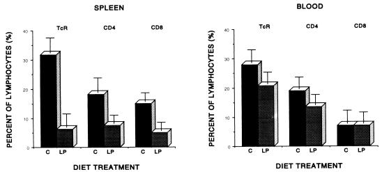
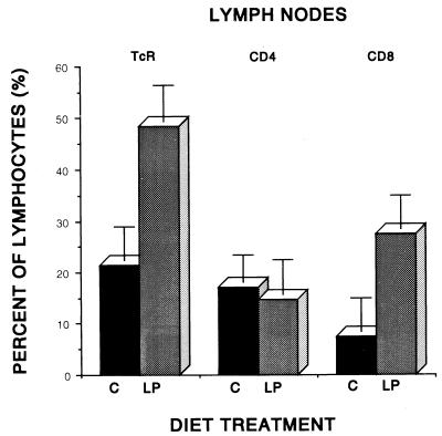
Previous research has suggested that dietary protein deficiency alters resistance to experimental pulmonary tuberculosis, in part, by affecting the distribution and trafficking of antigen-reactive T cells. In this study, guinea pigs were maintained on either a protein-deficient (10% ovalbumin) or control (30% ovalbumin) diet and infected 4 to 6 weeks later with a low dose of virulent Mycobacterium tuberculosis H37Rv by the respiratory route. Monoclonal antibodies directed against the CD4 or CD8 markers on guinea pig lymphocytes were used in a flow cytofluorometric assay to determine the proportion of each subset in the peripheral circulation, spleen, and bronchotracheal lymph nodes at 4 weeks after infection. In uninfected guinea pigs, only the spleen exhibited an effect of diet on T-cell distribution, with small but consistent reductions in the proportions of both CD4 and CD8 T lymphocytes. However, following infection, protein deficiency exerted a profound effect on T-cell distribution. Malnourished, tuberculous guinea pigs harbored only 20 and 60% of the T cells (as a proportion of total lymphoid cells) found in the spleen and blood, respectively, of their well-nourished counterparts. Normal relative proportions of CD4 and CD8 cells were observed, however. In striking contrast, the bronchotracheal lymph nodes of protein-deprived guinea pigs with tuberculosis contained more than twice the numbers of T cells of control guinea pigs, and the normal CD4-to-CD8 ratio was reversed. Peripheral T-cell function, as measured by the delayed hypersensitivity skin test to tuberculin, and antigen-induced lymphoproliferation in vitro were markedly suppressed in protein-malnourished animals. Conversely, purified protein derivative-induced (but not concanavalin A-induced) proliferation was significantly enhanced in cultures of lymph node cells from protein-deprived tuberculous animals. Taken together, these results suggest that immunological abnormalities and loss of antimycobacterial resistance in the lungs of protein-deficient guinea pigs may be explained, in part, by sequestration of antigen-reactive T cells in the lymph nodes draining the site of infection.
Figures
References
-
- Baker D, Healy D G, Verghese S, Schäfer H, Turk J L. Phenotypic analysis of guinea pig Langerhans cells with antibodies directed against leucocyte surface antigens. Int Arch Allergy Appl Immunol. 1988;86:350–358. - PubMed
-
- Ellner J J, Wallis R S. Immunologic aspects of mycobacterial infections. Rev Infect Dis. 1989;11:S455–S459. - PubMed
Publication types
MeSH terms
Substances
Grants and funding
LinkOut - more resources
Full Text Sources
Research Materials
