. 1998 Mar;66(3):966-73.
doi: 10.1128/IAI.66.3.966-973.1998.

MP1 encodes an abundant and highly antigenic cell wall mannoprotein in the pathogenic fungus Penicillium marneffei

Affiliations

MP1 encodes an abundant and highly antigenic cell wall mannoprotein in the pathogenic fungus Penicillium marneffei

L Cao et al. Infect Immun. 1998 Mar.

Abstract

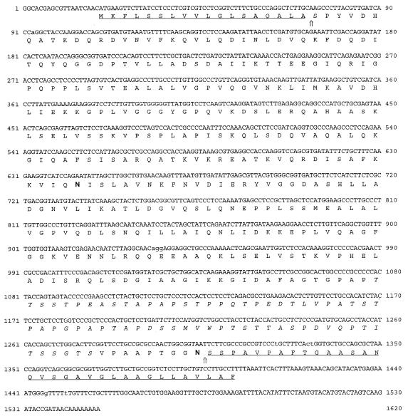
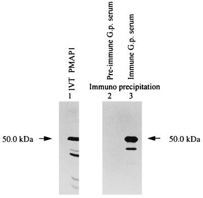
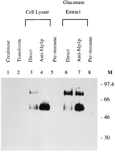
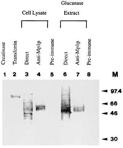
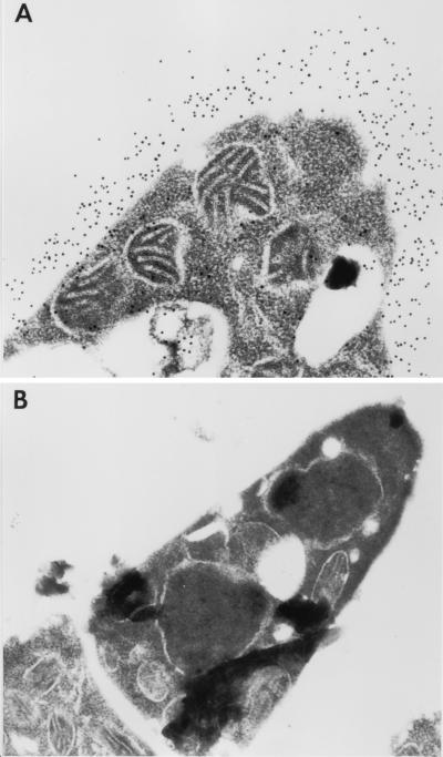
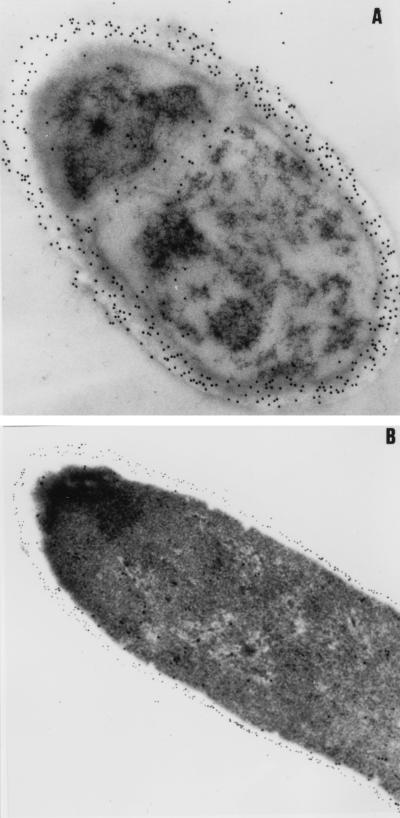
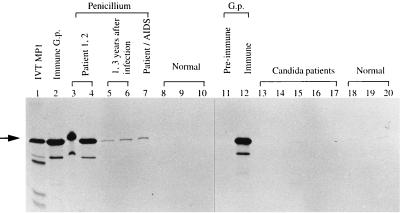
We cloned the MP1 gene, which encodes an abundant antigenic cell wall mannoprotein from the dimorphic pathogenic fungus Penicillium marneffei. MP1 is a unique gene without homologs in sequence databases. It codes for a protein, Mp1p, of 462 amino acid residues, with a few sequence features that are present in several cell wall proteins of Saccharomyces cerevisiae and Candida albicans. It contains two putative N glycosylation sites, a serine- and threonine-rich region for O glycosylation, a signal peptide, and a putative glycosylphosphatidylinositol attachment signal sequence. Specific anti-Mp1p antibody was generated with recombinant Mp1p protein purified from Escherichia coli to allow further characterization of Mp1p. Western blot analysis with anti-Mp1p antibody revealed that Mp1p has predominant bands with molecular masses of 58 and 90 kDa and that it belongs to a group of cell wall proteins that can be readily removed from yeast cell surfaces by glucanase digestion. In addition, Mp1p is an abundant yeast glycoprotein and has high affinity for concanavalin A, a characteristic indicative of a mannoprotein. Furthermore, ultrastructural analysis with immunogold staining indicated that Mp1p is present in the cell walls of the yeast, hyphae, and conidia of P. marneffei. Finally, it was observed that infected patients develop a specific antibody response against Mp1p, suggesting that this protein represents a good cell surface target for host humoral immunity.


Figures

FIG. 1
FIG. 1
DNA and amino acid sequences of Mp1p. MP1 cDNA contains a single open reading frame encoding 462 amino acid residues with a predicted molecular mass of 47.8 kDa. The N-terminal cleavable signal peptide of 18 amino acids and the C-terminal cleavable GPI signal peptide of 33 amino acids are underlined. A 67-amino-acid serine- and threonine-rich region is in italics. Two potential N glycosylation sites, at positions 209 and 429, are indicated by boldface type.
FIG. 2
FIG. 2
In vitro translation of MP1. MP1 was in vitro translated with Promega’s rabbit reticulocyte lysate, and a major band of 50 kDa can be detected (lane 1). Mp1p was immunoprecipitated with guinea pig (G.p.) immune serum against P. marneffei cells (lane 3) but not with preimmune serum (lane 2).
FIG. 3
FIG. 3
Mp1p is present in the glucanase extract fraction of the P. marneffei yeast cell wall. Both the cell lysate and glucanase extract of P. marneffei yeast cells were made from the same cell pellet. They were resuspended in the same final volume so that equivalent amounts of lysate or extract could be loaded onto the gel. Western blot analysis was carried out with rabbit anti-Mp1p antibody. Mp1p was detected in both the total yeast cell lysate (lane 3) and the cell wall glucanase extract (lane 6). Two protein bands, of 58 and 90 kDa, were seen on the Western blot. They were specifically immunoprecipitated with guinea pig anti-Mp1p antibody (lanes 4 and 7) but not with preimmune serum (lanes 5 and 8). Lanes 1 and 2 show two control proteins, creatinase as a nonglycosylated protein and transferrin as a glycosylated protein. Neither reacts with rabbit anti-Mp1p antibody. M, molecular mass (in kilodaltons).
FIG. 4
FIG. 4
Mp1p is a mannoprotein. All samples were loaded in the same order as in Fig. 3. ConA was used because it has high affinity for mannoproteins. M, molecular mass (in kilodaltons).
FIG. 5
FIG. 5
Immunoelectron microscopy of P. marneffei yeast cells stained with rabbit anti-Mp1p antibody. (A) P. marneffei yeast cells stained with rabbit anti-Mp1p antibody. Immunogold particles were specifically located throughout the entire thickness of the yeast cell wall. (B) Negative control stained with preimmune rabbit serum. Magnification, ×17,000.
FIG. 6
FIG. 6
Immunoelectron microscopy of P. marneffei conidia (A) and hyphae (B) stained with rabbit anti-Mp1p antibody.
FIG. 7
FIG. 7
Specific immunoprecipitation of Mp1p by penicilliosis patient sera. A major band of 50 kDa (arrow) was the in vitro-translated (IVT) full-length Mp1p. Mp1p can be immunoprecipitated by guinea pig (G.p.) immune serum (lanes 2 and 12) but not preimmune serum (lane 11). Mp1p protein was immunoprecipitated by sera from two immunocompetent patients with acute penicilliosis (lanes 3 and 4), from two people at 1 and 3 years after recovery from penicilliosis (lanes 5 and 6), and from an AIDS-penicilliosis patient (lane 7). Controls were sera from healthy blood donors (lanes 8 to 10 and 18 to 20) and from patients infected with C. albicans (lanes 13 to 17).

Similar articles

Cited by

References

    1. Bailey D A, Feldmann P J F, Bovey M, Gow N A R, Brown A J P. The Candida albicans HYR1 gene, which is activated in response to hyphal development, belongs to a gene family encoding yeast cell wall proteins. J Bacteriol. 1996;178:5353–5360. - PMC - PubMed
    1. Caras I W, Weddell G N. Signal peptide for protein secretion directing glycophospholipid membrane anchor attachment. Science. 1989;243:1196–1198. - PubMed
    1. Casadevall A. Antibody immunity and invasive fungal infections. Infect Immun. 1995;63:4211–4218. - PMC - PubMed
    1. Cassone A, Conti S, De Bernardis F, Polonelli L. Antibodies, killer toxins and antifungal immunoprotection: a lesson from nature? Immunol Today. 1997;18:164–169. - PubMed
    1. Cogliati M, Roverselli A, Boelaert J R, Taramelli D, Lombardi L, Viviani M A. Development of an in vitro macrophage system to assess Penicillium marneffei growth and susceptibility to nitric oxide. Infect Immun. 1997;65:279–284. - PMC - PubMed

Publication types

Associated data

LinkOut - more resources