. 1998 Mar;66(3):1076-81.
doi: 10.1128/IAI.66.3.1076-1081.1998.

Chimeric clostridial cytotoxins: identification of the N-terminal region involved in protein substrate recognition

Affiliations

Chimeric clostridial cytotoxins: identification of the N-terminal region involved in protein substrate recognition

F Hofmann et al. Infect Immun. 1998 Mar.

Abstract

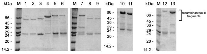
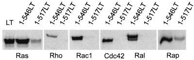
Clostridium sordellii lethal toxin is a member of the family of large clostridial cytotoxins that glucosylate small GTPases. In contrast to Clostridium difficile toxins A and B, which exclusively modify Rho subfamily proteins, C. sordellii lethal toxin also glucosylates Ras subfamily proteins. By deletion analysis and construction of chimeric fusion proteins of C. sordellii lethal toxin and C. difficile toxin B, we localized the enzyme activity of the lethal toxin to the N terminus of the holotoxin and identified the region involved in protein substrate specificity. The toxin fragment of the N-terminal 546 amino acid residues of C. sordellii lethal toxin glucosylated Rho and Ras subfamily proteins, as the holotoxin did. Deletion of a further 30 amino acid residues from the C terminus of this active fragment drastically reduced glucotransferase activity and blocked glucohydrolase activity. Exchange of amino acid residues 364 through 516 of lethal toxin for those in the active toxin B fragment (1 to 546) allowed glucosylation of Ras subfamily proteins. In contrast, the chimera with amino acids 1 to 364 from toxin B, 365 to 468 from lethal toxin, and 469 to 546 from toxin B exhibited markedly reduced modification of Ras subfamily proteins, whereas modification of Rac and Cdc42 was hardly changed. The data indicate that the region of amino acid residues 364 through 516 primarily defines the substrate specificity of C. sordellii lethal toxin.


Figures

FIG. 1
FIG. 1
Purification of recombinant toxin chimeras. The N-terminal toxin chimeras were constructed as GST fusion proteins, expressed in E. coli, and purified by affinity chromatography and subsequent thrombin cleavage. Lanes 1 to 3 and 8 to 9, toxin chimeras 1-516LT517-546ToxB (lane 1), 1-134ToxB135-516LT517-546ToxB (lane 2), 1-134LT135-516ToxB517-546LT (lane 3), 1-364ToxB365-516LT517-546ToxB (lane 8), and 1-364LT365-516ToxB517-546LT (lane 9) after thrombin cleavage of the fusion proteins. Lanes 4 to 7 and 10 to 13, GST fusion proteins of 1-468LT469-546ToxB (lane 4), 1-468ToxB469-546LT (lane 5), 1-271ToxB272-546LT (lane 6), 1-271LT272-546ToxB (lane 7), 1-546LT (lane 10), 1-517LT (lane 11), 1-364ToxB365-468LT469-546ToxB (lane 12), and 1-364LT365-468ToxB469-546LT (lane 13). Two micrograms of protein was loaded on lanes 1 to 3 and lanes 5, 6, 8, and 12; 4 μg of protein was loaded on lanes 4, 7, 10, 11, and 13. The band at 26 kDa represents GST, which does not interfere with the assays.
FIG. 2
FIG. 2
Protein substrate specificity of glucosylation by LT, 1-546LT and 1-517LT. Recombinant Rho, Rac1, Cdc42, Ras, Ral, and Rap (1 μg of each) were glucosylated by LT (100 nM) and 1-546LT and 1-517LT (100 nM each) in the presence of [14C]UDP-glucose for 30 min. Then, labeled proteins were analyzed by SDS-PAGE and phosphorimaging (shown).
FIG. 3
FIG. 3
Time course of the glucosylation of Ras by LT and by the N-terminal toxin fragments 1-546LT and 1-517LT. Ras (1 μg) was incubated with LT (1 nM; ▴) or purified N-terminal toxin fragments 1-546LT (1 nM; ▪) and 1-517LT (1 μM; •), respectively, in the presence of [14C]UDP-glucose (10 μM) for the indicated times (t). Then, labeled proteins were analyzed by SDS-PAGE and phosphorimaging. Time points represent the averages of three experiments.
FIG. 4
FIG. 4
Cosubstrate specificity of glucosylation by LT and 1-546LT. Recombinant Ras (1 μg) was incubated with LT and 1-546LT in the presence of [14C]UDP-glucose (UDP-Glc), [14C]UDP-galactose (UDP-Gal), and [14C]UDP-N-acetylglucosamine (UDP-GlcNAc) for 30 min. Thereafter, labeled proteins were analyzed by SDS-PAGE and phosphorimaging (shown).
FIG. 5
FIG. 5
Time course of glucohydrolase activity of ToxB, LT, and recombinant toxin fragments. (A) LT (100 nM), 1-546LT (100 nM), and 1-517LT (1 μM) were incubated with 20 μM [14C]UDP-glucose and 100 μM UDP-glucose in a total volume of 20 μl. At the indicated time points (t), 1.5-μl samples were taken and analyzed by thin-layer chromatography and phosphorimaging. Time points represent the averages of three experiments. (B) ToxB (100 nM), 1-546ToxB (100 nM), and 1-516ToxB (1 μM) were incubated with 20 μM [14C]UDP-glucose and 100 μM UDP-glucose in a total volume of 20 μl. At the indicated time points, 1.5-μl samples were taken and analyzed by thin-layer chromatography and phosphorimaging. Glucohydrolase activity is given as picomoles of hydrolyzed UDP-glucose per total UDP-glucose (180 pmol).
FIG. 6
FIG. 6
Glucosylation patterns of recombinant GTPases by LT and ToxB fragments and chimeric toxin constructs. Recombinant Rho, Rac, Cdc42, Ras, Ral, and Rap (1 μg of each) were glucosylated by the indicated toxin fragments or chimeric constructs (100 mM each) in the presence of [14C]UDP-glucose for 30 min. Then, labeled proteins were analyzed by SDS-PAGE and phosphorimaging (shown). (A) Glucosylation patterns of 1-546LT (lane 1), 1-271ToxB272-546LT (lane 2), 1-546ToxB (lane 3), and 1-271LT272-546ToxB (lane 4). (B) Glucosylation patterns of 1-546LT (lane 1), 1-468LT469-546ToxB (lane 2), 1-546ToxB (lane 3), and 1-468ToxB469-546LT (lane 4). (C) Glucosylation patterns of 1-546LT (lane 1), 1-134ToxB135-516LT517-546ToxB (lane 2), 1-516LT517-546ToxB (lane 3), 1-546ToxB (lane 4), and 1-134LT135-516ToxB517-546LT (lane 5). (D) Glucosylation patterns of 1-546LT (lane 1), 1-364ToxB365-516LT517-546ToxB (lane 2), and 1-364LT365-516ToxB517-546LT (lane 3). (E) Glucosylation patterns of 1-546LT (lane 1), 1-364ToxB365-468LT469-546ToxB (lane 2), 1-546ToxB (lane 3), and 1-364LT365-468ToxB469-546LT (lane 4). The diagrams at the right of each panel show summaries of the chimeric constructs in the corresponding lanes. The numbers at the right of the diagrams are amino acid residues.
FIG. 7
FIG. 7
Summary of LT-ToxB chimeric constructs and their substrate specificities. ToxB parts of chimeras are shown in black and LT parts are shown in white. Symbols: ++, marked labeling; (+), poor labeling; ↑, increase in labeling compared to control LT or ToxB; ↓, decrease in labeling compared to control LT or ToxB; –, no labeling. The hatched part of the top bar represents the region that is essential for glucosylation of Ras subfamily proteins by LT. The numbers represent amino acid residues. Data are from Fig. 6. From top to bottom, the bars represent 1-546ToxB, 1-271LT272-546ToxB, 1-468ToxB469-546LT, 1-134LT135-516ToxB517-546LT, 1-364LT365-516ToxB517-546LT, 1-364LT365-468ToxB469-546LT, 1-546LT, 1-271ToxB272-546LT, 1-516LT517-546ToxB, 1-134ToxB135-516LT517-546ToxB, 1-364ToxB365-516LT517-546ToxB, 1-364ToxB365-468LT469-546ToxB, and 1-468LT469-546ToxB.

References

    1. Aktories K. Rho proteins: targets for bacterial toxins. Trends Microbiol. 1997;5:282–288. - PubMed
    1. Baldacini O, Girardot R, Green G A, Rihn B, Monteil H. Comparative study of immunological properties and cytotoxic effects of Clostridium difficile toxin B and Clostridium sordelii toxin L. Toxicon. 1992;30:129–140. - PubMed
    1. Barroso L A, Moncrief J S, Lyerly D M, Wilkins T D. Mutagenesis of the Clostridium difficile toxin B gene and effect on cytotoxic activity. Microb Pathog. 1994;16:297–303. - PubMed
    1. Bette P, Oksche A, Mauler F, Eichel-Streiber C, Popoff M R, Habermann E. A comparative biochemical, pharmacological and immunological study of Clostridium novyi α-toxin, C. difficile toxin B and C. sordellii lethal toxin. Toxicon. 1991;29:877–887. - PubMed
    1. Bongaerts G P A, Lyerly D M. Role of toxins A and B in the pathogenesis of Clostridium difficile disease. Microb Pathog. 1994;17:1–12. - PubMed

Publication types

MeSH terms

LinkOut - more resources