

Chemokine production by a human alveolar epithelial cell line in response to Mycobacterium tuberculosis
- PMID: 9488404
- PMCID: PMC108024
- DOI: 10.1128/IAI.66.3.1121-1126.1998
Chemokine production by a human alveolar epithelial cell line in response to Mycobacterium tuberculosis
Abstract
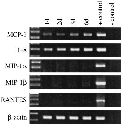
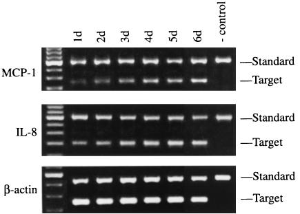
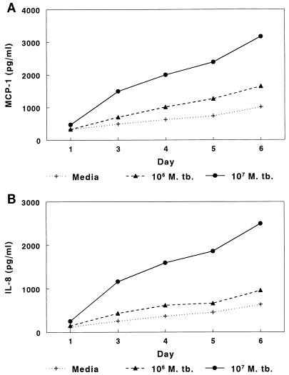
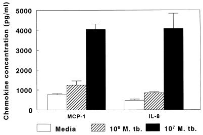
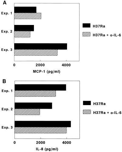
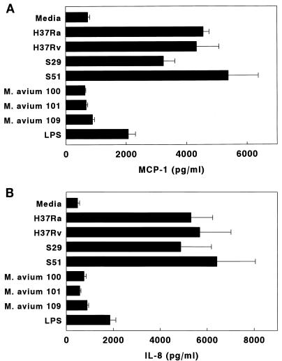
To investigate the role of chemokines during the initial local response to Mycobacterium tuberculosis in the human lung, we studied chemokine production by the human alveolar epithelial cell line A549 after infection with M. tuberculosis. M. tuberculosis-infected A549 cells produced mRNAs and protein for monocyte chemotactic protein-1 (MCP-1) and interleukin-8 (IL-8) but not mRNAs for macrophage inflammatory protein 1alpha (MIP-1alpha), MIP-1beta, and RANTES. Chemokine production in response to M. tuberculosis was not dependent on production of tumor necrosis factor alpha, IL-1beta, or IL-6. Two virulent clinical M. tuberculosis isolates, the virulent laboratory strain H37Rv, and the avirulent strain H37Ra elicited production of comparable concentrations of MCP-1 and IL-8, whereas killed M. tuberculosis and three Mycobacterium avium strains did not. The three virulent M. tuberculosis strains grew more rapidly than the avirulent M. tuberculosis strain in the alveolar epithelial cell line, and the three M. avium strains did not grow intracellularly. These findings suggest that intracellular growth is necessary for mycobacteria to elicit production of MCP-1 and IL-8 by alveolar epithelial cells but that virulence and the rate of intracellular growth do not correlate with chemokine production. Alveolar epithelial cells may contribute to the local inflammatory response in human tuberculosis by producing chemokines which attract monocytes, lymphocytes, and polymorphonuclear cells.
Figures

References
-
- Antony V B, Godbey S W, Kunkel S L, Hott J W, Hartman D L, Burdick M D, Strieter R M. Recruitment of inflammatory cells to the pleural space. Chemotactic cytokines, IL-8, and monocyte chemotactic peptide-1 in human pleural fluids. J Immunol. 1993;151:7216–7223. - PubMed
-
- Barnes P F, Chatterjee D, Abrams J S, Lu S, Wang E, Yamamura M, Brennan P J, Modlin R L. Cytokine production induced by Mycobacterium tuberculosis lipoarabinomannan. Relationship to chemical structure. J Immunol. 1992;149:541–547. - PubMed
Publication types
MeSH terms
Substances
Grants and funding
LinkOut - more resources
Full Text Sources
Other Literature Sources
Research Materials
Miscellaneous
