. 1998 Mar;66(3):1225-32.
doi: 10.1128/IAI.66.3.1225-1232.1998.

Mouse beta-defensin 1 is a salt-sensitive antimicrobial peptide present in epithelia of the lung and urogenital tract

Affiliations

Mouse beta-defensin 1 is a salt-sensitive antimicrobial peptide present in epithelia of the lung and urogenital tract

R Bals et al. Infect Immun. 1998 Mar.

Abstract

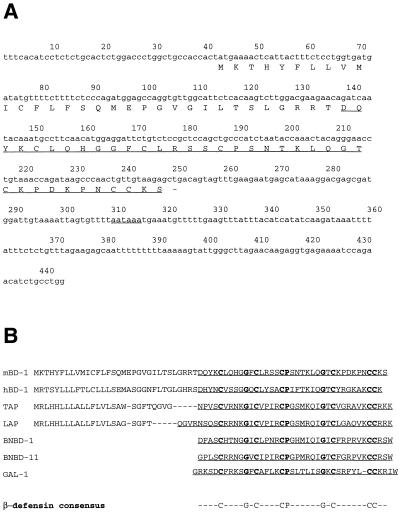
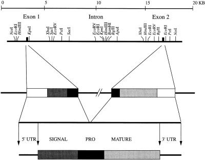
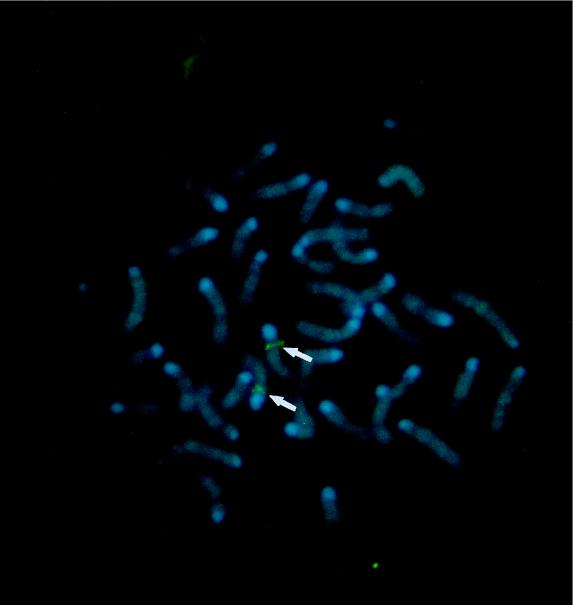
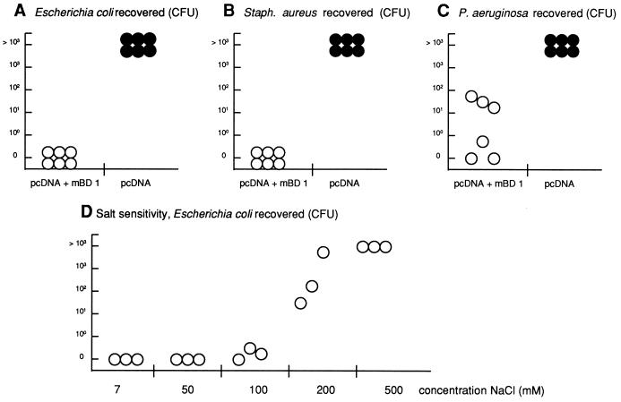
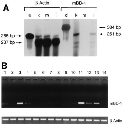
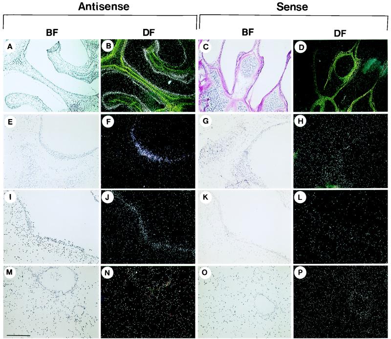
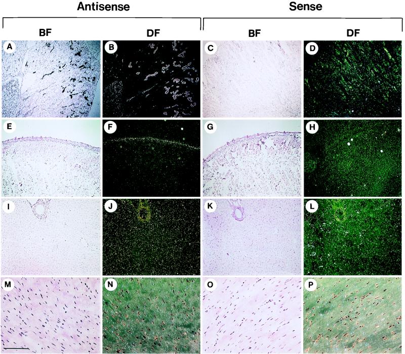
One component of host defense at mucosal surfaces appears to be epithelium-derived peptides with antimicrobial activity called defensins. Human beta-defensin 1 (hBD-1) represents the first member of the beta-defensin family isolated from humans and has been implicated in the pathogenesis of cystic fibrosis. We describe in this report the isolation and characterization of a murine homolog of hBD-1 called mouse beta-defensin 1 (mBD-1). The predicted amino acid sequence shows the hallmark features of other known epithelial beta-defensins, including the ordered array of six cysteine residues. Analysis of a genomic clone of mBD-1 revealed two exons separated by a 15-kb intron. By use of fluorescence in situ hybridization, the mBD-1 gene was localized at the proximal portion of chromosome 8, the site where mouse alpha-defensins are found. Lysates from cells transfected with the mBD-1 cDNA showed antibacterial activity against gram-positive and gram-negative bacteria. mBD-1 transcripts were found in kidney, liver, and female reproductive organ tissues. In the airways, mBD-1 is expressed diffusely throughout the epithelial cells of the large proximal airways with less expression in the small distal airways and no expression in alveolar cells. The present study demonstrates that a beta-defensin potentially homologous to human beta-defensin 1 is present in the respiratory system and other mucosal surfaces in mice.


Figures

FIG. 1
FIG. 1
cDNA and peptide sequences of mBD-1. (A) cDNA and deduced amino acid sequences of mBD-1. The first underline indicates the putative mature peptide; the dash represents the termination codon; the second underline indicates the polyadenylation signal. (B) Comparison of the putative prepropeptide sequences of mBD-1, hBD-1, TAP, and LAP, as well as the peptide sequences of bovine neutrophil β-defensins 1 and 11 (BNBD-1 and BNBD-11, respectively) and gallinacin 1 (GAL-1). The bottom line presents the consensus sequence of β-defensins.
FIG. 2
FIG. 2
Structure of the gene for mBD1. Restriction map of the gene for mBD-1 together with a schematic drawing of the gene, the cDNA, and the predicted structure of the prepropeptide. The gene is represented schematically with the following individual components: 5′ untranslated region (5′ UTR), open box; signal sequence, shaded box; interrupted prosequence (PRO), black box; mature peptide, shaded box; 3′ untranslated region (3′ UTR), open box.
FIG. 3
FIG. 3
Chromosomal FISH of an mBD-1 probe to normal metaphase chromosomes derived from mouse embryo fibroblast cells. Chromosome 8 shows a positive green signal at its proximal portion (arrows), corresponding to band 8A4.
FIG. 4
FIG. 4
Results of antibacterial activity assays. (A to C) Antibacterial liquid broth coincubation assay. E. coli (A), S. aureus (B), and P. aeruginosa (C) were added (103 to 104 CFU) to 50 μl of lysates from cells transfected with a vector containing the full-length cDNA of mBD-1 (empty circles) or with the vector alone (filled circles) and analyzed for microbial killing. Each circle represents results from an individual transfection experiment. (D) Salt sensitivity of the antimicrobial activity of mBD-1. Extracts from mBD-1-transfected cells were incubated with 5 × 10⁴ CFU of E. coli D31 in 10 mM phosphate buffer (pH 7.4) and the indicated concentrations of NaCl. After incubation at 37°C for 1 h, serial dilutions were plated and colonies were counted the following day.
FIG. 5
FIG. 5
Analysis of mBD-1 expression by RNase protection analysis and RT-PCR (A) Measurement of mBD-1 expression by RNase protection analysis. Total RNA from the kidney (k), skeletal muscle (m), and whole lung (i.e., lung and trachea) (l) was investigated. Hybridization to a labeled riboprobe against β-actin (a) and mBD-1 (d) transcripts was performed a separate tube. (B) Detection of mBD-1 expression in various mouse tissues by nested RT-PCR. Poly(A)+ RNA was isolated from mouse tissues and reverse transcribed, and the cDNAs were amplified by using two pairs of mBD-1-specific primers. A single 250-bp band was amplified by using gene-specific primers. As a control, β-actin cDNA was amplified by using specific primers. Lanes: 1, trachea, 2, lung (lung parenchyma without cartilaginous airways); 3, tongue; 4, esophagus; 5, small bowel; 6, large bowel; 7, gall bladder; 8, pancreas; 9, skeletal muscle; 10, heart; 11, fallopian tube; 12, ovary; 13, vagina; 14, brain.
FIG. 6
FIG. 6
Detection of transcripts encoding mBD-1 in the mouse respiratory tract. Antisense and sense probes were used to detect the tissue distribution of mBD-1 expression. Representative sections in dark and bright fields (DF and BF, respectively) from the nose (A to D), trachea (E to H), large bronchioles (I to L), and terminal bronchioles and lung parenchyma (M to P) are shown. Bars: A to D, 0.7 mm; E to P, 270 μm.
FIG. 7
FIG. 7
Detection of transcripts encoding mBD-1 in extrapulmonary organs. Antisense and sense probes were used to detect the tissue distribution of mBD-1 expression. Representative sections in dark and bright fields (DF and BF, respectively) from the kidney (A to D), tongue (E to H), liver (I to L), and heart muscle (M to P) are shown. Bars: A to H, 0.7 mm; I to L, 270 μm; M to P, 130 μm.

References

    1. Bensch K W, Raida M, Magert H-J, Schulz-Knappe P, Forssmann W G. hBD-1: a novel β-defensin from human plasma. FEBS Lett. 1995;368:331–335. - PubMed
    1. Diamond G, Jones D E, Bevins C L. Airway epithelial cells are the site of expression of a mammalian antimicrobial peptide gene. Proc Natl Acad Sci USA. 1993;90:4596–4600. - PMC - PubMed
    1. Diamond G, Russell J P, Bevins C L. Inducible expression of an antibiotic peptide gene in lipopolysaccharide-challenged tracheal epithelial cells. Proc Natl Acad Sci USA. 1996;93:5156–5160. - PMC - PubMed
    1. Diamond G, Zasloff M, Eck H, Brasseur M, Maloy W L, Bevins C L. Tracheal antimicrobial peptide, a cysteine-rich peptide from mammalian tracheal mucosa: peptide isolation and cloning of cDNA. Proc Natl Acad Sci USA. 1991;88:3952–3956. - PMC - PubMed
    1. Eisenhauer P B, Harwig S S L, Lehrer R I. Cryptdins: antimicrobial defensins of the murine small intestine. Infect Immun. 1992;60:3556–3565. - PMC - PubMed

Publication types