

doi: 10.1128/IAI.66.3.1237-1243.1998.
M-cell surface beta1 integrin expression and invasin-mediated targeting of Yersinia pseudotuberculosis to mouse Peyer's patch M cells
Affiliations
- PMID: 9488419
- PMCID: PMC108039
- DOI: 10.1128/IAI.66.3.1237-1243.1998
Item in Clipboard
M-cell surface beta1 integrin expression and invasin-mediated targeting of Yersinia pseudotuberculosis to mouse Peyer's patch M cells
Infect Immun.
1998 Mar.
Abstract
Quantitative analysis of Yersinia pseudotuberculosis infection of murine gut loops revealed that significantly more wild-type bacteria associated with Peyer's patch M cells than with dome enterocytes or goblet cells. An invasin-deficient mutant was significantly attenuated for M-cell invasion, while beta1 integrin expression was demonstrated in the apical membranes of M cells but not enterocytes. M-cell targeting by Yersinia pseudotuberculosis in vivo may, therefore, be mediated primarily by the interaction of invasin with cell surface beta1 integrins.
Figures

CLSM images of mouse Peyer’s patch FAE incubated for 120 min with Y. pseudotuberculosis YPIII/pIB1 and dual stained for M cells (red) and bacteria (green). (a) Surface view. Several bacteria are adherent to M cells, and a single bacterium (arrow) is adherent to an enterocyte. At depths of 2 μm (b) and 6 μm (c), large numbers of bacteria are located within the M cells. (d) Invaded bacteria are present 16 μm below the FAE surface, at which depth the cell type initially entered cannot be determined. Bar, 10 μm.
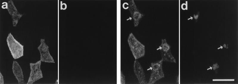
CLSM images of mouse Peyer’s patch FAE incubated for 60 min with Y. pseudotuberculosis YPIII/pIB1 and dual stained for M cells (a and c) and bacteria (b and d). (a and b) Surface views. (c and d) Images at 1 μm in depth. Bacteria are absent from the surface of this region of FAE (a and b). Bacterial invasion (d [arrows]) is accompanied by a redistribution of UEA1 binding sites in the subapical region of the cells, observed on a confocal optical section as rings of UEA1 staining around the invading bacteria (c [arrows]). Bar, 10 μm.
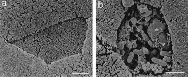
SEM images of mouse Peyer’s patch FAE incubated for 60 min with Y. pseudotuberculosis YPIII/pIB1. Both images depict a central M cell surrounded by enterocytes (and part of a brush cell in panel b). The M cell in panel a lacks adherent bacteria and exhibits a normal surface morphology. In contrast, the association of large numbers of bacteria with the M cell in panel b is accompanied by disruption of the M-cell surface. Bars, 2 μm.
CLSM images of mouse Peyer’s patch FAE dual stained for M cells (a) and β1 integrins (b and c). (a and b) Surface views. UEA1-stained M cells express β1 integrins in their apical membranes (stained cells in panels a and b), whereas β1 integrin expression is absent from the apical membranes of enterocytes (unstained cells in panels a and b). (c) At a depth of 2 μm, both M cells and enterocytes express β1 integrins in their lateral membranes. Bar, 10 μm.
References
-
- Autenrieth I B, Firsching R. Penetration of M cells and destruction of Peyer’s patches by Yersinia enterocolitica: an ultrastructural and histological study. J Med Microbiol. 1996;44:285–294. - PubMed
-
- Beaulieu J-F. Differential expression of the VLA family of integrins along the crypt-villus axis in the human small intestine. J Cell Sci. 1992;102:427–436. - PubMed
-
- Bergelson J M, Finberg R W. Integrins as receptors for virus attachment and cell entry. Trends Microbiol. 1993;1:287–288. - PubMed
-
- Bliska J B, Falkow S. Interplay between determinants of cellular entry and cellular disruption in the enteropathogenic Yersinia. Curr Opin Infect Dis. 1994;7:323–328.
Publication types
MeSH terms
Substances
LinkOut - more resources
Full Text Sources
Other Literature Sources
