. 1998 Mar;18(3):1137-46.
doi: 10.1128/MCB.18.3.1137.

Activity of a trypanosome metacyclic variant surface glycoprotein gene promoter is dependent upon life cycle stage and chromosomal context

Affiliations

Activity of a trypanosome metacyclic variant surface glycoprotein gene promoter is dependent upon life cycle stage and chromosomal context

S V Graham et al. Mol Cell Biol. 1998 Mar.

Abstract

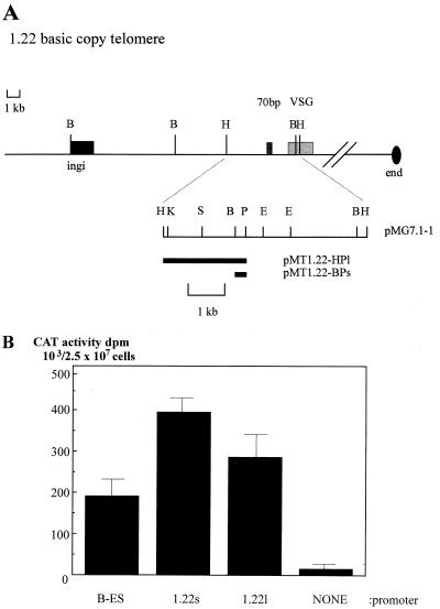
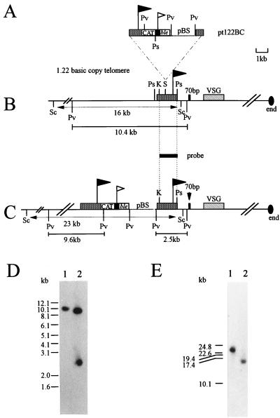
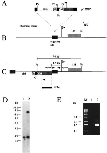
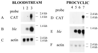
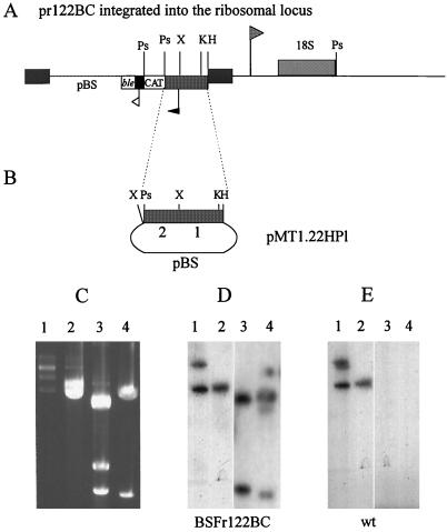
African trypanosomes evade the mammalian host immune response by antigenic variation, the continual switching of their variant surface glycoprotein (VSG) coat. VSG is first expressed at the metacyclic stage in the tsetse fly as a preadaptation to life in the mammalian bloodstream. In the metacyclic stage, a specific subset (<28; 1 to 2%) of VSG genes, located at the telomeres of the largest trypanosome chromosomes, are activated by a system very different from that used for bloodstream VSG genes. Previously we showed that a metacyclic VSG (M-VSG) gene promoter was subject to life cycle stage-specific control of transcription initiation, a situation unique in Kinetoplastida, where all other genes are regulated, at least partly, posttranscriptionally (S. V. Graham and J. D. Barry, Mol. Cell. Biol. 15:5945-5956, 1985). However, while nuclear run-on analysis had shown that the ILTat 1.22 M-VSG gene promoter was transcriptionally silent in bloodstream trypanosomes, it was highly active when tested in bloodstream-form transient transfection. Reasoning that chromosomal context may contribute to repression of M-VSG gene expression, here we have integrated the 1.22 promoter, linked to a chloramphenicol acetyltransferase (CAT) reporter gene, back into its endogenous telomere or into a chromosomal internal position, the nontranscribed spacer region of ribosomal DNA, in both bloodstream and procyclic trypanosomes. Northern blot analysis and CAT activity assays show that in the bloodstream, the promoter is transcriptionally inactive at the telomere but highly active at the chromosome-internal position. In contrast, it is inactive in both locations in procyclic trypanosomes. Both promoter sequence and chromosomal location are implicated in life cycle stage-specific transcriptional regulation of M-VSG gene expression.


Figures

FIG. 1
FIG. 1
Activity of the 1.22 M-VSG gene promoter in transient transfection in bloodstream forms. (A) Map of the 1.22 basic copy telomere (28) and the clone pMG7.1-1 derived from its 3′ end (15). Horizontal black bars show the extent of the short and long promoter-containing fragments cloned in pMT122-BPs and pMT122-HPl. Abbreviations: B, BamHI; H, HindIII; K, KpnI; S, SalI; P, PstI; E, EcoRI. Hatched box, VSG gene region; stippled box, 70-bp repeat region; black box, ingi retroposon sequence; oval, end of the telomere. (B) Short (1.22s) and long (1.22l) versions of the 1.22 M-VSG gene promoter region were tested for the ability to drive expression of a CAT reporter gene, flanked by actin RNA processing signals (35), in transient transfection in bloodstream trypanosomes. Promoter regions tested were as follows: B-ES, the 221 bloodstream expression site promoter (73); 1.22s, the insert in pMT1.22-BPs; 1.22l, a KpnI/PstI fragment derived from the insert in pMT1.22-HPl; and NONE, the CAT gene flanked by actin RNA processing signals but with no promoter upstream. Values are means and deviations from the means for eight experiments.
FIG. 2
FIG. 2
Insertion of plasmid pt122BC into the 1.22 endogenous expression telomere. (A) Structure of plasmid pt122BC which has been linearized at the SalI site within the 1.22 promoter region. Abbreviations: Pv, PvuII; Ps, PstI; pBS, pBluescript sequences; CAT, CAT reporter gene; ble, selectable marker gene encoding phleomycin resistance. Stippled boxes, 1.22 promoter region; black box, procyclin/PARP promoter region; open boxes, marker genes; black flag; 1.22 promoter; white flag, procyclin/PARP promoter; dotted line, pBluescript sequence. (B) Partial map of the 1.22 basic copy telomere showing the targeting site, a SalI (S) restriction enzyme site. Also shown are the PvuII and SacI fragments containing the promoter. Abbreviations: K, KpnI; S, SalI; Ps, PstI; Pv, PvuII; Sc, SacI. Stippled box, the 1.22 promoter-containing region; black box, 70-bp repeat region; hatched box, VSG coding region; black flag, 1.22 promoter; oval, end of the telomere. (C) Result of targeting pt122BC into the 1.22 telomere. The new PvuII and SacI fragments, generated by the insertion, which contain the promoter are shown. The horizontal black bar between panels B and C represents the KpnI/PstI fragment used as a probe in hybridizations in panels D and E. (D) Southern blot analysis of PvuII-digested genomic DNA, fractionated on a 0.6% agarose gel, from wild-type trypanosomes (track 1) and from BSFtelo122BC trypanosomes (track 2). (E) Southern blot analysis of SacI-digested genomic DNA fractionated on a FIGE gel of DNA isolated from wild-type trypanosomes (track 1) and from BSFtelo122BC trypanosomes (track 2). Both Southern blots were hybridized with the KpnI/PstI promoter probe shown above panel C in 5× SSC at 65°C and washed to 0.1× SSC at 65°C.
FIG. 3
FIG. 3
Insertion of plasmid pr122BC into the ribosomal nontranscribed spacer region. (A) Structure of plasmid pr122BC linearized at the unique NotI site within the ribosomal locus targeting fragment. Abbreviations: Ps, PstI; Pv, PvuII; pBS, pBluescript sequences; CAT, CAT reporter gene; ble, selectable marker gene encoding phleomycin resistance. Dark stippled boxes, ribosomal locus targeting sequence; light stippled box, 1.22 promoter region; black box, procyclin/PARP promoter region; open boxes, marker genes; black flag, 1.22 promoter; white flag, procyclin/PARP promoter; dotted line, pBluescript sequence. (B) Partial map of the ribosomal locus showing the targeting site, containing a unique NotI (N) restriction enzyme site. Dark stippled box, targeting region; cross-hatched box, 18S coding region; cross-hatched flag, ribosomal locus promoter; Ps, PstI. (C) Result of integrating pr122BC into the ribosomal nontranscribed spacer region. Note that pr122BC is designed to insert in reverse orientation with respect to the ribosomal transcription unit. The horizontal black bar beneath the map in panel C represents the 1.6-kb KpnI/PstI 1.22 promoter fragment used as a probe in hybridizations in panel D. The size of the fragment expected when this probe is hybridized to PstI-digested genomic DNA stably transformed with pr122BC is shown above the map in panel C. Horizontal arrows indicate the approximate location of the primers (Mprom and Rprom) used in the PCR shown in panel E to amplify the region between the inserted plasmid and the ribosomal promoter. The size of the expected amplified fragment is shown. (D) Southern blot analysis of genomic DNA cut with PstI and fractionated on a 0.6% agarose gel isolated from track 1 (wild-type trypanosomes and track 2 (BSFribo122BC trypanosomes). The blot was hybridized with the 1.6-kb KpnI/PstI 1.22 promoter region probe shown below panel C in 5× SSC at 65°C and washed to 0.1× SSC at 65°C. (E) Linkage of the 1.22 promoter region and the ribosomal locus promoter tested by PCR. The primers Mprom (5′) and Rprom (3′) (C) were used in an amplification reaction with PstI-cut genomic DNA isolated from wild-type trypanosomes (track 1) and BSFribo122BC trypanosomes (track 2). Track M, 1-kb marker ladder. PCR products were separated by agarose gel (1.5%) electrophoresis, and the gel was stained with ethidium bromide (0.5 μg/ml).
FIG. 4
FIG. 4
Northern blot analysis of reporter gene expression driven by the 1.22 promoter in stably transformed trypanosomes. (A to C) Assay with bloodstream stable transformants. Total RNA was isolated from cultured bloodstream-form cells of the wild type (track 1), BSFtelo122BC (track 2), and BSFribo122BC (track 3). (D to F) Assay with procyclic stable transformants. Total RNA was isolated from cultured bloodstream-form cells of the wild type (track 4), PFtelo122BC (track 5), PFribo122BC (track 6), and from cells transiently transfected with p5′parpCAT3′parp and p5′parpble3′parp, constructs where either the CAT or ble gene was expressed from a procyclin/PARP promoter (track 7). RNA was fractionated on a denaturing formaldehyde gel, blotted onto a nylon membrane, and hybridized sequentially with the 32P-labeled probes shown. Hybridization was in 3× SSC–50% formamide at 42°C. The ble and actin probes were labeled by random priming, and the CAT probe was a 32P-labeled in vitro-transcribed CAT antisense probe. Blots were washed to 0.5× SSC at 65°C. Following each hybridization, the probe was removed by boiling in 0.5× SSC and the filter was autoradiographed to check that no residual hybridization remained.
FIG. 5
FIG. 5
Nuclear run-on analysis of transcription initiation of the CAT gene inserted in the ribosomal locus in stably transformed bloodstream cells. (A) Partial map of the nontranscribed spacer region of rDNA with plasmid pr122BC inserted. (B) Restriction map of plasmid pMT1.22-HPl from which was derived the 1.22 promoter region driving CAT reporter gene expression in pr122BC. The three DNA fragments which should result from digesting plasmid pMT1.22-HPl with HindIII and XbaI are labeled 1, 2, and pBS. Abbreviations: H, HindIII; K, KpnI; X, XbaI; P, PstI; pBS, pBluescript sequence. Dark grey box, 1.22 promoter region; light grey box, 18S rRNA gene; open box, CAT gene; open flag, ribosomal promoter; black flag, 1.22 promoter. (C) Ethidium bromide-stained gel of a PstI digest of pR4, an rDNA repeat unit (39) (track 1), pTbαβ-T1, an αβ-tubulin repeat unit (63), digested with HindIII (track 2), pMT1.22-HPl digested with HindIII/XbaI (track 3), and p5′parpCAT3′parp digested with HindIII/PstI (track 4). (D) Result of hybridizing a Southern blot of the gel in panel C with a 32P-labeled nascent transcript probe from nuclei isolated from BSFribo122BC trypanosomes, cloned line r10. (E) A blot very similar to that used in panel D, hybridized with a 32P-labeled nascent transcript probe from nuclei isolated from wild-type (wt) trypanosomes. To avoid excessive signal, there is 1/10 the amount of DNA loaded in tracks 1 (pR4) as in tracks 2 to 4. Tracks 3 and 4 of the Southern blots in panels D and E have been subjected to a longer exposure than tracks 1 and 2, but the exposure of the Southern blot in panel D, tracks 3 and 4, is the same as in panel E, tracks 3 and 4. Hybridizations were in 3× SSC at 55°C, and blots were washed to 0.1× SSC 65°C.

References

    1. Alarcon C M, Son H J, Hall T, Donelson J E. A monocistronic transcript for a trypanosome variant surface glycoprotein. Mol Cell Biol. 1994;14:5579–5591. - PMC - PubMed
    1. Auffray C, Rougeon F. Purification of mouse immunoglobulin heavy-chain messenger RNAs from total myeloma tumour RNA. Eur J Biochem. 1980;107:303–314. - PubMed
    1. Barry J D. The relative significance of mechanisms of antigenic variation in African trypanosomes. Parasitol Today. 1997;13:212–218. - PubMed
    1. Barry J D, Graham S V, Matthews K R, Shiels P G, Shonekan O A. Stage-specific mechanisms for activation and expression of variant surface glycoprotein genes in Trypanosoma brucei. Biochem Soc Trans. 1990;18:708–710. - PubMed
    1. Ben Amar M F, Pays A, Tebabi P, Dero B, Seebeck T, Steinert M, Pays E. Structure and transcription of the actin gene of Trypanosoma brucei. Mol Cell Biol. 1988;8:2166–2176. - PMC - PubMed

Publication types

MeSH terms

Substances

LinkOut - more resources