. 1998 Mar;18(3):1201-12.
doi: 10.1128/MCB.18.3.1201.

Gal4p-mediated chromatin remodeling depends on binding site position in nucleosomes but does not require DNA replication

Affiliations

Gal4p-mediated chromatin remodeling depends on binding site position in nucleosomes but does not require DNA replication

M Xu et al. Mol Cell Biol. 1998 Mar.

Abstract

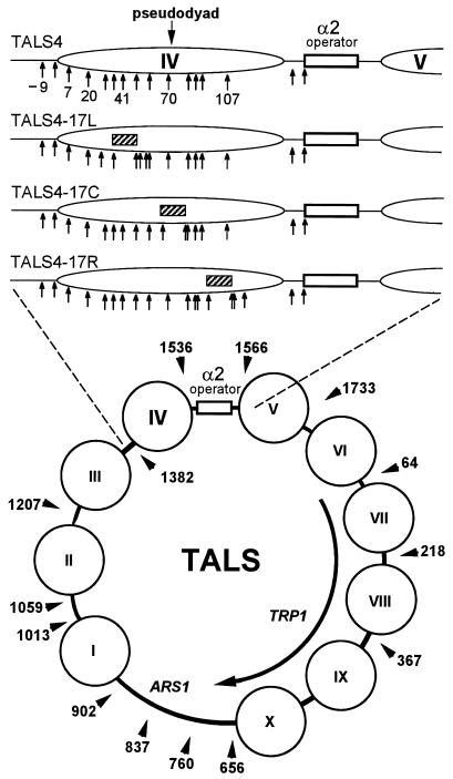
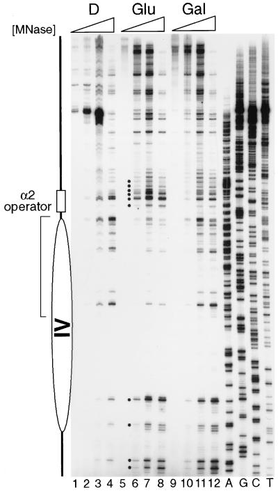
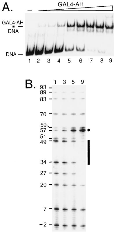
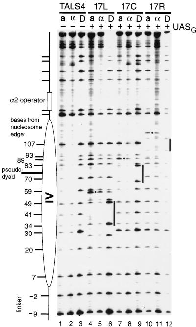
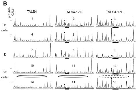
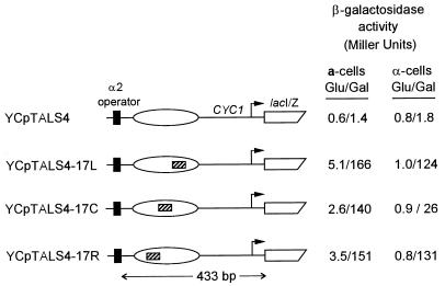
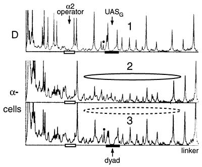
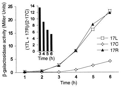
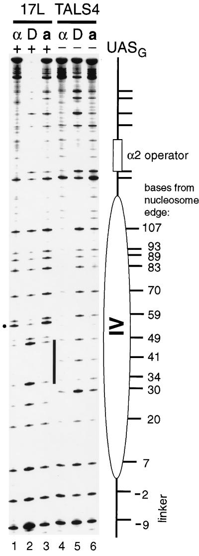
Biochemical studies have demonstrated decreased binding of various proteins to DNA in nucleosome cores as their cognate sites are moved from the edge of the nucleosome to the pseudodyad (center). However, to date no study has addressed whether this structural characteristic of nucleosomes modulates the function of a transcription factor in living cells, where processes of DNA replication and chromatin modification or remodeling could significantly affect factor binding. Using a sensitive, high-resolution methyltransferase assay, we have monitored the ability of Gal4p in vivo to interact with a nucleosome at positions that are known to be inaccessible in nucleosome cores in vitro. Gal4p efficiently bound a single cognate site (UASG) centered at 41 bp from the edge of a positioned nucleosome, perturbing chromatin structure and inducing transcription. DNA binding and chromatin perturbation accompanying this interaction also occurred in the presence of hydroxyurea, indicating that DNA replication is not necessary for Gal4p-mediated nucleosome disruption. These data extend previous studies, which demonstrated DNA replication-independent chromatin remodeling, by showing that a single dimer of Gal4p, without the benefit of cooperative interactions that occur at complex wild-type promoters, is competent for invasion of a preestablished nucleosome. When the UASG was localized at the nucleosomal pseudodyad, relative occupancy by Gal4p, nucleosome disruption, and transcriptional activation were substantially compromised. Therefore, despite the increased nucleosome binding capability of Gal4p in cells, the precise translational position of a factor binding site in one nucleosome in an array can affect the ability of a transcriptional regulator to overcome the repressive influence of chromatin.


Figures

FIG. 1
FIG. 1
Chromatin structure of the TALS4 minichromosome and its derivatives in α-cells. The lower diagram shows the locations of preferential cleavages by MNase (arrowheads), in m.u., as determined at low resolution (10 to 20 bp) by indirect end labeling, and inferred positions of nucleosomes (circles) in TALS minichromosomes (55). The α2 operator is indicated by the open rectangle. The upper diagram shows a to-scale enlargement of the region from m.u. 1347 to 1619 (left to right) from the minichromosome TALS4 and its derivatives. This region was amplified by PCR and cloned in the reverse orientation to construct the lacIZ reporter plasmids of Fig. 6 and 7. The position of nucleosome IV (ellipse) (m.u. 1375 to 1520), determined by mapping of MNase cutting sites at high resolution (59) (Fig. 2), and locations of target CG sites (arrows) relative to the left edge (arbitrarily assigned position 1) of the nucleosome are given. A 32-bp sequence (see Materials and Methods) containing a near-consensus 17-bp binding site for Gal4p (UASG; hatched bar) was inserted at three different positions into TALS4 (no UASG) to create the three UASG-containing plasmids, TALS4-17L, TALS4-17C, and TALS4-17R. In α-cells, nucleosome IV is precisely positioned next to the α2 operator, incorporating UASG at three different translational positions, centered 41 bp from the left (17L) and right (17R) edges or at the center (17C; pseudodyad), of the nucleosome.
FIG. 2
FIG. 2
Primer extension analysis of MNase-cut sites in TALS4-17L. Nuclei (lanes 5 to 12) and protein-free DNA (D) (lanes 1 to 4) from α-cells containing TALS4-17L and pRS426-GAL4 grown in glucose (Glu) (lanes 5 to 8) or galactose (Gal) (lanes 9 to 12) were isolated and treated with increasing concentrations of MNase as follows: 0.05, 0.25, 0.5, and 1 U/ml, respectively, in lanes 1 to 4; 0, 2.5, 5, and 10 U/ml in lanes 5 to 8; and 0, 1.25, 2.5, and 5 U/ml in lanes 9 to 12. The higher concentrations used to digest cells from glucose were to compensate for increased cell numbers. The dots mark cleavage sites that are enhanced in glucose-grown cells, and the bracket delineates those sites that are protected relative to control DNA. MNase cleavages and the inferred nucleosome position (ellipse) are as those observed in the propositus TALS plasmid by Shimizu et al. (59). Loss of protection due to binding of activated Gal4p, indicating disruption of the nucleosome, is observed when cells are grown in galactose (40, 63).
FIG. 3
FIG. 3
In vitro SssI footprinting of Gal4-AH bound to a single site. (A) Mobility shift gel showing resolution of naked DNA probe containing sequences from m.u. 1267 to 1508 of TALS4-17L (DNA) from the complex of probe bound by Gal4-AH (Gal4-AH · DNA). The concentrations of Gal4-AH monomer added to each sample and the resultant percent occupancies (quantified with a phosphorimager) of the probe are as follows: lane 1, 0 nM; lane 2, 0.21 nM, 7.1%; lane 3, 0.42 nM, 10%; lane 4, 0.85 nM, 17%; lane 5, 1.7 nM, 59%; lane 6, 3.3 nM, 70%; lane 7, 6.6 nM, 90%; lane 8, 13.3 nM, 90%; and lane 9, 27.5 nM, 93%. (B) Duplicate binding reaction mixtures were incubated with purified SssI DNA MTase, and selected reaction mixtures (numerals indicated above gel correspond to samples in panel A) were treated to identify methylated cytosines as described previously (31). The signal intensity of a given band corresponds directly to the amount of methylation at that cytosine. UASG is indicated by the bar, and the location of each CG site in bases from the left edge of the positioned nucleosome in α-cells is given to facilitate comparison to data in Fig. 4, 5, and 9. Protection against SssI of two CG sites, one in each UASG half-site (sites 34 and 49), as well as sites just outside UASG (sites 27 and 51), and an accompanying enhancement of methylation (marked by dot) occur with increasing concentrations of Gal4-AH. As the 17-bp UASG is contained in the same 32-bp sequence within each of the plasmids, these sites can be used to assess protections and enhancement of SssI methylation upon Gal4p binding within chromatin in vivo. The corresponding positions of CG sites from the left edge of nucleosome IV within TALS4-17C and TALS4-17R can be obtained by adding 32 and 64 bp, respectively, to each of the above locations.
FIG. 4
FIG. 4
Remodeling of a positioned nucleosome by Gal4p binding is dependent on the position of UASG. The chromatin structures in living yeast cells grown in galactose of the four different plasmids depicted in Fig. 1, TALS4 (lanes 1 to 3), TALS4-17L (lanes 4 to 6), TALS4-17C (lanes 7 to 9), and TALS4-17R (lanes 10 to 12), were analyzed in the presence of pRS426-GAL4, which overexpresses Gal4p from the wild-type GAL4 promoter. Protein-free DNA for each plasmid (D) was methylated by SssI in vitro to identify target CG sequences and site preferences of the enzyme (lanes 3, 6, 9, and 12). For analysis of chromatin, DNA was rapidly isolated from isogenic, SssI-expressing a-cells (lanes 1, 4, 7, and 10) and α-cells (lanes 2, 5, 8, and 11), and modified cytosines were identified as described previously (31). The number of nucleotides that the C residue in each target CG site is away from the operator-distal edge (i.e., left edge) of the positioned nucleosome in TALS4 in α-cells is indicated. Where present, each UASG is marked by a bar at the immediate right of the samples that were methylated in vitro (D), and the position of the hypermethylated residue next to each binding site is indicated by a dot.
FIG. 5
FIG. 5
Chromatin remodeling and Gal4p occupancy in a- and α-cells at endogenous and overexpressed levels of Gal4p. (A) TALS4-17C (+UASG) and TALS4 (−UASG) were introduced into SssI+ a- and α-cells containing endogenous levels of Gal4p (−pRS426-GAL4) (i.e., transformed with pRS426 vector only) or overexpressing Gal4p (+pRS426-GAL4) and probed with SssI MTase by growing the cells in galactose. Positions of CG sites were identified in protein-free DNA (D) as indicated in the legend to Fig. 4. Endogenous levels of Gal4p in a-cells led to marked disruption of chromatin in the region of nucleosome IV (compare lanes 7 and 8 to lane 10), whereas no remodeling was visible in α-cells (compare lanes 3 and 5 to lane 4). Overexpression of Gal4p increased disruption of nucleosome IV in α-cells only slightly at sites 107, 89, and 30 (compare lanes 1 and 2 to lanes 3 and 5). Note that overexpression of Gal4p in cells harboring TALS4 (no UASG) had no effect on chromatin structure (compare lanes 9 and 10). (B) Scans of in vivo SssI probing data. Phosphorimager scans from an experiment completely independent from that presented in panel A are shown, analyzing disruption of TALS4, TALS4-17C, and TALS4-17L in a- and α-cells grown in galactose containing endogenous (transformed with pRS426 vector) (scans 1 to 3 and 10 to 12) or overexpressed (transformed with pRS426-GAL4) (scans 4 to 6 and 13 to 15) levels of Gal4p. UASG is indicated by filled bars, and the site that becomes hypermethylated upon Gal4p binding is marked by dots. The CG within the α2 operator is located at the leftmost side of each scan, and the linker is at the right. Perturbation of control a-cell chromatin, seen as increases in methylation at many CG sites in the region (except sites within UASG that are protected by bound Gal4p in the presence of pRS426-GAL4), occurs equally well in TALS4-17C and TALS4-17L (compare scans 2 and 3 to scan 1 and compare scans 5 and 6 to scan 4). The precisely positioned nucleosome in TALS4 and TALS4-17C in α-cells is depicted as a solid ellipse. Perturbation of this nucleosome in α-cells in TALS4-17L (compare scans 12 and 10 and compare scans 15 and 13) is indicated by the dashed ellipse. Endogenous levels of Gal4p efficiently remodel UASG-containing a-cell chromatin (compare scans 2 and 3 to scan 1) but disrupt chromatin in α-cells only when UASG is removed from the pseudodyad in TALS4-17L (i.e., lack of chromatin perturbation in scan 11 versus scan 10 but clear disruption in scan 12 versus scan 10 or 11). Note that the level of disruption in TALS4-17L at endogenous levels of Gal4p (scan 12) is greater than that in TALS4-17C even when Gal4p is overexpressed (scan 14).
FIG. 5
FIG. 5
Chromatin remodeling and Gal4p occupancy in a- and α-cells at endogenous and overexpressed levels of Gal4p. (A) TALS4-17C (+UASG) and TALS4 (−UASG) were introduced into SssI+ a- and α-cells containing endogenous levels of Gal4p (−pRS426-GAL4) (i.e., transformed with pRS426 vector only) or overexpressing Gal4p (+pRS426-GAL4) and probed with SssI MTase by growing the cells in galactose. Positions of CG sites were identified in protein-free DNA (D) as indicated in the legend to Fig. 4. Endogenous levels of Gal4p in a-cells led to marked disruption of chromatin in the region of nucleosome IV (compare lanes 7 and 8 to lane 10), whereas no remodeling was visible in α-cells (compare lanes 3 and 5 to lane 4). Overexpression of Gal4p increased disruption of nucleosome IV in α-cells only slightly at sites 107, 89, and 30 (compare lanes 1 and 2 to lanes 3 and 5). Note that overexpression of Gal4p in cells harboring TALS4 (no UASG) had no effect on chromatin structure (compare lanes 9 and 10). (B) Scans of in vivo SssI probing data. Phosphorimager scans from an experiment completely independent from that presented in panel A are shown, analyzing disruption of TALS4, TALS4-17C, and TALS4-17L in a- and α-cells grown in galactose containing endogenous (transformed with pRS426 vector) (scans 1 to 3 and 10 to 12) or overexpressed (transformed with pRS426-GAL4) (scans 4 to 6 and 13 to 15) levels of Gal4p. UASG is indicated by filled bars, and the site that becomes hypermethylated upon Gal4p binding is marked by dots. The CG within the α2 operator is located at the leftmost side of each scan, and the linker is at the right. Perturbation of control a-cell chromatin, seen as increases in methylation at many CG sites in the region (except sites within UASG that are protected by bound Gal4p in the presence of pRS426-GAL4), occurs equally well in TALS4-17C and TALS4-17L (compare scans 2 and 3 to scan 1 and compare scans 5 and 6 to scan 4). The precisely positioned nucleosome in TALS4 and TALS4-17C in α-cells is depicted as a solid ellipse. Perturbation of this nucleosome in α-cells in TALS4-17L (compare scans 12 and 10 and compare scans 15 and 13) is indicated by the dashed ellipse. Endogenous levels of Gal4p efficiently remodel UASG-containing a-cell chromatin (compare scans 2 and 3 to scan 1) but disrupt chromatin in α-cells only when UASG is removed from the pseudodyad in TALS4-17L (i.e., lack of chromatin perturbation in scan 11 versus scan 10 but clear disruption in scan 12 versus scan 10 or 11). Note that the level of disruption in TALS4-17L at endogenous levels of Gal4p (scan 12) is greater than that in TALS4-17C even when Gal4p is overexpressed (scan 14).
FIG. 6
FIG. 6
The location of UASG in a positioned nucleosome modulates transcriptional activation by Gal4p. The region indicated in Fig. 1 encompassing the α2 operator and nucleosome (ellipse) from TALS4 and its derivatives (UASG is indicated by hatched bar) were subcloned upstream of lacIZ to create the four indicated YCp constructs. Reporter plasmids were transformed into cells expressing wild-type levels of Gal4p, and levels of β-galactosidase activity were determined after growth in medium containing glucose (Glu) or galactose (Gal). The expression levels of β-galactosidase shown are the averages obtained from two different yeast transformants from two representative, independent experiments that included all of the samples. In addition, although the absolute levels of expression varied slightly between experiments, the fold inductions from glucose to galactose were very similar for YCpTALS4-17C versus YCpTALS4-17L and YCpTALS4-17R in α-cells for two independent transformants in two additional experiments.
FIG. 7
FIG. 7
A nucleosome is positioned next to the α2 operator in the lacIZ reporter plasmids in α-cells. Linear phosphorimager scans of SssI methylation of YCpTALS4 (no UASG) and YCpTALS4-17C (UASG is demarcated by the filled bar) in SssI-expressing cells which express endogenous levels of Gal4p are shown. The open bar indicates the α2 operator. Cells were grown in galactose and then processed to identify methylated cytosines. For YCpTALS4 (scan 2), note the protection by the nucleosome (solid ellipse) (compare scans 2 and 1). Insertion of UASG (i.e., YCpTALS4-17C [scan 3]) leads to partial disruption of the nucleosome (depicted by the dashed ellipse) in α-cells (compare scans 3 and 2). The increase in signal at the hypermethylated site (marked by the dot) also indicates partial occupancy of UASG by Gal4p.
FIG. 8
FIG. 8
Transcriptional activation of the lacIZ reporter plasmids in α-cells at limiting levels of Gal4p expression. Cells containing YCpTALS4, YCpTALS4-17C (17C), YCpTALS4-17L (17L), and YCpTALS4-17R (17R) were initially cultured in medium containing glucose then transferred to galactose and assayed at 1-h intervals for expression of β-galactosidase. The activity of YCpTALS4 lacking UASG remained below 0.1 Miller unit at all time points. The inset shows the relative activities of 17L and 17R versus 17C at several time points.
FIG. 9
FIG. 9
Gal4p binds 41 bp from the edge of a positioned nucleosome and causes disruption in the absence of DNA replication. Cells overexpressing Gal4p and harboring TALS4-17L (lanes 1–3; UASG is indicated by the bar at right of lane 3) or TALS4 (lanes 4 to 6; no UASG) were initially grown in glucose-containing medium to repress synthesis of SssI MTase and Gal4p and were then transferred to galactose medium that also contained hydroxyurea. Following the induction of Gal4p and SssI expression, identification of lower-strand cytosines methylated in chromatin in vivo was performed as described (31). Positions of CG sites were identified in protein-free DNA (lanes D) as indicated in the legend to Fig. 4. Note the increase in methylation in chromatin of both cell types in the presence of UASG (for α-cells, compare lanes 1 and 4; for a-cells, compare lanes 3 and 6), which is indicative of chromatin remodeling.

References

    1. Almer A, Rudolph H, Hinner A, Hörz W. Removal of positioned nucleosomes from the yeast PHO5 promoter upon PHO5 induction releases additional upstream activating DNA elements. EMBO J. 1986;5:2689–2696. - PMC - PubMed
    1. Archer T K, Cordingley M G, Marsaud V, Richard-Foy H, Hager G L. Steroid transactivation at a promoter organized in a specifically-positioned array of nucleosomes. In: Gustafsson J A, Eriksson H, Carlstedt-Duke J, editors. Steroid/thyroid hormone receptor family and gene regulation. Berlin, Germany: Berkhauser Verlag, AG; 1989. pp. 221–238.
    1. Bajwa W, Torchia T E, Hopper J E. Yeast regulatory gene GAL3: carbon regulation; UASGal elements in common with GAL1, GAL2, GAL7, GAL10, GAL80, and MEL1; encoded protein strikingly similar to yeast and Escherichia coli galactokinases. Mol Cell Biol. 1988;8:3439–3447. - PMC - PubMed
    1. Barton M C, Emerson B M. Regulated expression of the β-globin gene locus in synthetic nuclei. Genes Dev. 1994;8:2453–2465. - PubMed
    1. Becker P B, Wu C. Cell-free system for assembly of transcriptionally repressed chromatin from Drosophila embryos. Mol Cell Biol. 1992;12:2241–2249. - PMC - PubMed

Publication types

LinkOut - more resources