. 1998 Mar;18(3):1236-47.
doi: 10.1128/MCB.18.3.1236.

Overexpression of the nucleoporin CAN/NUP214 induces growth arrest, nucleocytoplasmic transport defects, and apoptosis

Affiliations

Overexpression of the nucleoporin CAN/NUP214 induces growth arrest, nucleocytoplasmic transport defects, and apoptosis

J Boer et al. Mol Cell Biol. 1998 Mar.

Abstract

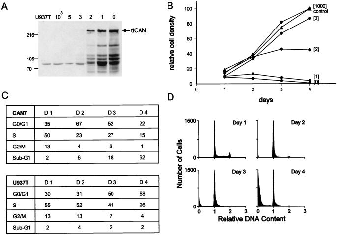
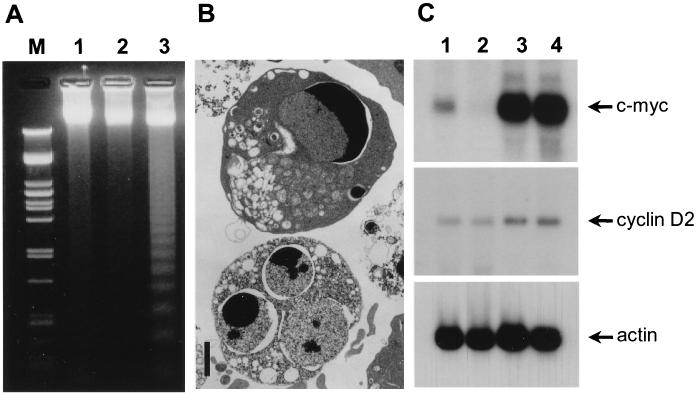
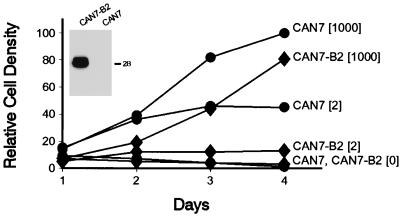
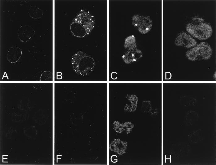
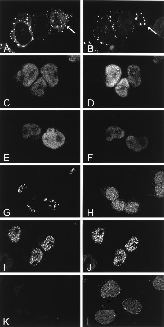
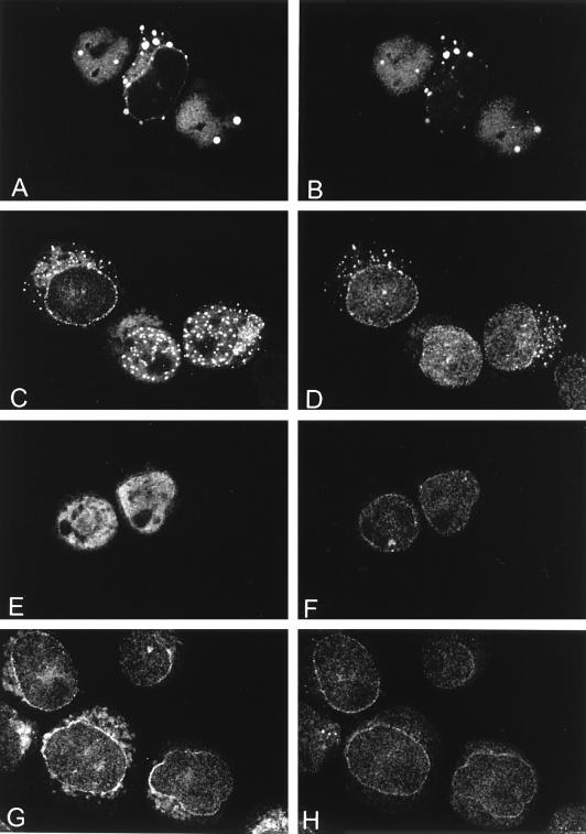
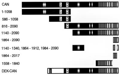
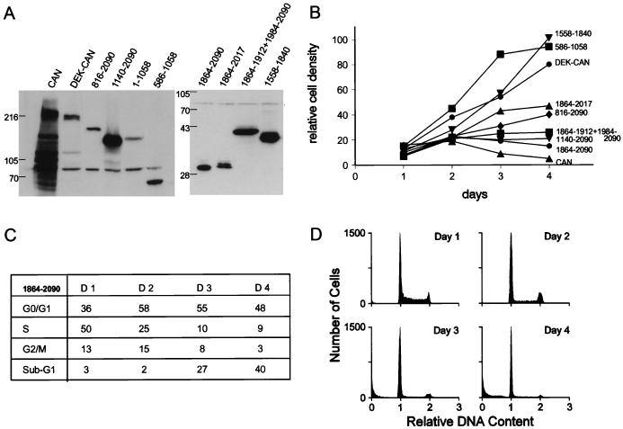
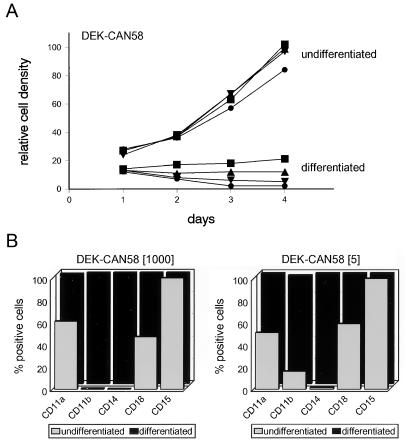
The human CAN gene was first identified as a target of t(6;9)(p23;q34), associated with acute myeloid leukemia and myelodysplastic syndrome, which results in the expression of a DEK-CAN fusion gene. CAN, also called NUP214, is a nuclear pore complex (NPC) protein that contains multiple FG-peptide sequence motifs. It interacts at the NPC with at least two other proteins, the nucleoporin NUP88 and hCRM1 (exportin 1), which was recently shown to function as a nuclear export receptor. Depletion of CAN in knockout mouse embryonic cells results in cell cycle arrest in G2, followed by inhibition of nuclear protein import and a block of mRNA export. We overexpressed CAN and DEK-CAN in U937 myeloid precursor cells. DEK-CAN expression did not interfere with terminal myeloid differentiation of U937 cells, whereas CAN-overexpressing cells arrested in G0, accumulated mRNA in their nuclei, and died in an apoptotic manner. Interestingly, we found that hCRM1 and import factor p97/importin beta colocalized with the ectopically expressed CAN protein, resulting in depletion of both factors from the NPC. Overexpression of the C-terminal FG-repeat region of CAN, which contains the binding site for hCRM1, caused sequestering of hCRM1 in the nucleoplasm and was sufficient to inhibit cell growth and to induce apoptosis. These results confirm that CAN plays a crucial role in nucleocytoplasmic transport and imply an essential role for hCRM1 in cell growth and survival.


Figures

FIG. 1
FIG. 1
CAN-overexpressing cells are growth inhibited. (A) Inducible expression of HA1-tagged CAN in CAN7 cells grown for 24 h in the presence of 1,000, 5, 3, 2, 1, and 0 ng of tetracycline per ml, as indicated above the lanes, was assayed by Western blot analysis of the sodium dodecyl sulfate (SDS)–6% polyacrylamide gel. Induced parental U937T cells serve as a negative control. Each lane contains lysate from 5 × 10⁵ cells. The blot was probed with the anti-HA1 monoclonal antibody 12CA5. The arrow indicates ttCAN protein. Lysates of cells expressing large amounts of ttCAN protein show specific truncated products, which do not seem to affect the results. The sizes of molecular mass standards, run in an adjacent lane, are indicated on the left in kilodaltons. (B) Growth curves of induced CAN7 cells (•) and U937T control cells (▴). Cultures were maintained in medium containing the indicated tetracycline concentrations, and viability was measured daily by a nonradioactive proliferation assay. The relative density was calculated as a percentage of the density of uninduced cells on day 4. Mean values of triplicate determinations are plotted; the standard deviations were below 10%. This experiment is one of three that all gave similar results. (C) The cell cycle phase distribution of induced CAN7 cells (upper panel) and U937T cells (lower panel) at 1, 2, 3, and 4 days after tetracycline withdrawal was calculated from flow cytometric measurements of the DNA content. (D) Flow cytometry profiles showing DNA fluorescence of propidium iodide-stained CAN7 cell nuclei at 1, 2, 3, and 4 days after withdrawal of tetracycline. This is a representative experiment of three, all of which gave similar results.
FIG. 2
FIG. 2
Overexpression of CAN induces G0 apoptosis in U937T cells. (A) Agarose gel electrophoresis of DNA from CAN7 cells cultured for 3 days in the absence of tetracycline (lane 3) shows internucleosomal DNA cleavage, whereas DNA from the parent U937T cells grown in the presence (lane 1) or absence (lane 2) of 1 μg of tetracycline per ml remains unfragmented. PstI-digested λ DNA served as a molecular weight marker (lane M). (B) Electron micrographs showing apoptotic CAN-overexpressing CAN7 cells after 3 days of induction. Bar, 2 μm. (C) Northern blot analysis of 15 μg of RNA isolated from the total culture (lane 1) and the FACS-sorted diploid-cell fraction (lane 2) of CAN7 cells induced for 40 h, compared to the total cultures of uninduced CAN7 cells (lane 3) and induced parental U937T cells (lane 4). The amounts of c-myc (2.4 kb; top panel), and cyclin D2 (6.0 kb; middle panel) mRNA were compared to the levels of actin mRNA (2.0 kb; bottom panel).
FIG. 3
FIG. 3
Bcl-xL coexpression does not rescue CAN-overexpressing cells. Growth curves of a representative Bcl-xL-overexpressing CAN7 clone, CAN7-B2 (⧫), compared to CAN7 (•), both grown in 1,000, 2, and 0 ng of tetracycline per ml for 4 days. Mean relative density values of triplicate cultures are plotted against time; the standard deviations were below 10%. This experiment is one of three, all of which gave similar results. Inset: Western blot of an SDS–9% polyacrylamide gel containing lysate from 5 × 10⁵ cells per lane, probed with a mouse monoclonal antibody to Bcl-xL. The 29-kDa doublet represents the Bcl-xL protein. The position of the 28-kDa molecular mass standard is indicated on the right.
FIG. 4
FIG. 4
Polyadenylated RNA export defect in CAN-overexpressing cells. (A to C) Confocal images of endogenous CAN expression in U937T cells (A) and overexpressed CAN in CAN7 cells 20 h (B) and 48 h (C) after induction; the cells were stained with the anti-CAN antiserum αCNC. (D) Overexpression of HA-CAN1864-2090 in U937T cells stained with the anti-HA antibody 12CA5. (E to H) Subcellular localization of polyadenylated RNA in control U937T cells (E) and CAN7 cells after 48 h (F) and 60 h (G) of induction and in HA-CAN 1864-2090 cells after 60 h of induction (H) analyzed by in situ hybridization with an FITC-conjugated oligo(dT)50 probe.
FIG. 5
FIG. 5
Colocalization of full-length CAN and CAN mutants with hCRM1. The subcellular distribution of hCRM1 and CAN proteins after 2 days of induction is shown. (A, C, E, G, I, and K) Confocal microscopy showing immunodetection of induced CAN and mutant proteins by an anti-HA1 monoclonal antibody followed by a goat anti-mouse Texas red-conjugated secondary antibody. (B, D, F, H, J, and L) Distribution of endogenous hCRM1 in the same cells stained with the anti-hCRM1 antiserum and a goat anti-rabbit FITC-linked antibody. hCRM1 colocalized with full-length CAN (A and B [cells indicated by arrows]), with the C-terminal FG repeat regions of CAN, CAN 1864–2090 (C and D) and CAN (1140–1340, 1864–1912, 1984–2090) (E and F), and with DEK-CAN (I and J). In contrast, hCRM1 did not colocalize with the more N-terminally located FG repeat region of CAN, represented by CAN 1558–1840 (G and H); it showed normal distribution in the nuclear membrane and the nucleoplasm, similar to that of induced U937T cells (K and L).
FIG. 6
FIG. 6
Double immunostaining of CAN and mutants with p97/importin β. The subcellular distribution of p97 and CAN proteins after 2 days of induction is shown. (A, C, E, and G) Confocal images of indirect immunofluorescence with the anti-CAN polyclonal antiserum anti-CNC, detected with a Texas red-conjugated goat anti-rabbit antibody. (B, D, F, and H) Confocal images of the same cells immunostained with an antibody to p97 (MAb3E9), detected with an FITC-conjugated goat anti-mouse antibody. Endogenous p97 colocalized with overexpressed CAN in the nuclear membrane and in the cytoplasmic and nucleoplasmic speckles (A and B). In cells overexpressing CAN 816–2090, only the nuclear membrane and the cytoplasmic speckles showed colocalization of the CAN mutant with p97 (C and D). Cells overexpressing CAN 1140–2090 showed normal p97 localization in the nuclear membrane (E and F), comparable to cells expressing endogenous levels of CAN (G and H).
FIG. 7
FIG. 7
Overview of CAN deletion mutants. Black bars represent CAN and CAN mutant proteins; numbers on the left represent amino acid boundaries. Predicted structural motifs are represented as follows: vertical lines, FG repeats; diamonds, FXF repeats; LZ, coiled-coil 1 and adjacent leucine zipper; AH, coiled-coil 2. Horizontal stripes indicate an acidic region in the DEK sequence (white bar).
FIG. 8
FIG. 8
The C terminus of CAN is sufficient to inhibit cell growth. (A) Western blot analysis of SDS–6% polyacrylamide (left panel) and SDS–10% polyacrylamide (right panel) gels with lysates from 5 × 10⁵ induced cells expressing the indicated CAN mutants and DEK-CAN. CAN (1140–1340, 1864–1912, 1984–2090) is abbreviated to CAN 1864−1912+1984−290. Blots were probed as described for Fig. 1A. (B) Growth curves of cells overexpressing selected CAN mutants and DEK-CAN grown in the absence of tetracycline for 4 days. Data shown are the mean values of triplicate cultures from one experiment of at least three independent experiments that gave similar results. (C) Cell cycle phase distribution of induced CAN 1864–2090 cells. (D) Quantitation of the DNA content by flow cytometric analysis in CAN 1864–2090 cells, overexpressing the hCRM1-binding domain, on days 1, 2, 3, and 4 after withdrawal of tetracycline.
FIG. 9
FIG. 9
Differentiation antigen expression in U937T cells expressing DEK-CAN. (A) Growth curves of DEK-CAN58 cells cultured in medium containing 1,000 (▪), 10 (▵), 5 (▾), or 0 (•) ng of tetracycline per ml, seeded at 105 cells/ml in the presence (differentiated) or absence (undifferentiated) of 1 ng of TGFβ1 per ml and 250 ng of D3 per ml. The cells were cultured in normal medium containing the indicated tetracycline concentrations for 3 days prior to the induction of differentiation. (B) DEK-CAN58 cells cultured in 1,000 ng of tetracycline per ml (no DEK-CAN expression) and 5 ng of tetracycline per ml (partial DEK-CAN induction) were differentiated for 5 days in medium containing TGFβ1 and D3 (differentiated; black bars). Undifferentiated cells cultured in 1,000 and 5 ng of tetracycline per ml are also shown (undifferentiated; gray bars). Expression of the indicated differentation antigens (CD11a, CD11b, CD14, CD15, and CD18) was evaluated by cytofluorimetry with specific monoclonal antibodies. Results are expressed as percentages of antigen-positive cells.

References

    1. Adam E J H, Adam S A. Identification of cytosolic factors required for nuclear location sequence-mediated binding to the nuclear envelope. J Cell Biol. 1994;125:547–555. - PMC - PubMed
    1. Adam S A, Gerace L. Cytosolic proteins that specifically bind nuclear localization signals are receptors for nuclear import. Cell. 1991;66:837–847. - PubMed
    1. Adam S A, Marr R S, Gerace L. Nuclear protein import in permeabilized mammalian cells requires soluble cytoplasmic factors. J Cell Biol. 1990;111:807–816. - PMC - PubMed
    1. Amberg D C, Goldstein A L, Cole C N. Isolation and characterization of RAT1: an essential gene of Saccharomyces cerevisiae required for the efficient nucleocytoplasmic trafficking of mRNA. Genes Dev. 1992;6:1173–1189. - PubMed
    1. Boise L H, González-Garcia M, Postema C E, Ding L, Lindsten T, Turka L A, Mao X, Nuñez G, Thompson C B. bcl-x, a bcl-2-related gene that functions as a dominant regulator of apoptotic cell death. Cell. 1993;74:597–608. - PubMed

Publication types

MeSH terms