. 1998 Mar;18(3):1248-56.
doi: 10.1128/MCB.18.3.1248.

Two members of the Tcf family implicated in Wnt/beta-catenin signaling during embryogenesis in the mouse

Affiliations

Two members of the Tcf family implicated in Wnt/beta-catenin signaling during embryogenesis in the mouse

V Korinek et al. Mol Cell Biol. 1998 Mar.

Abstract

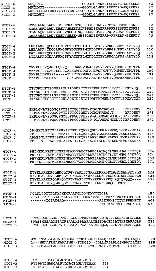
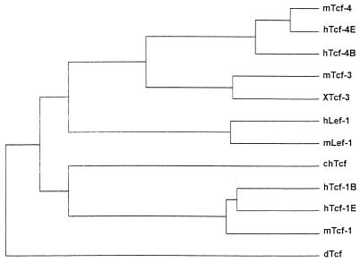
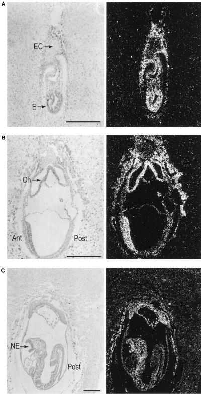
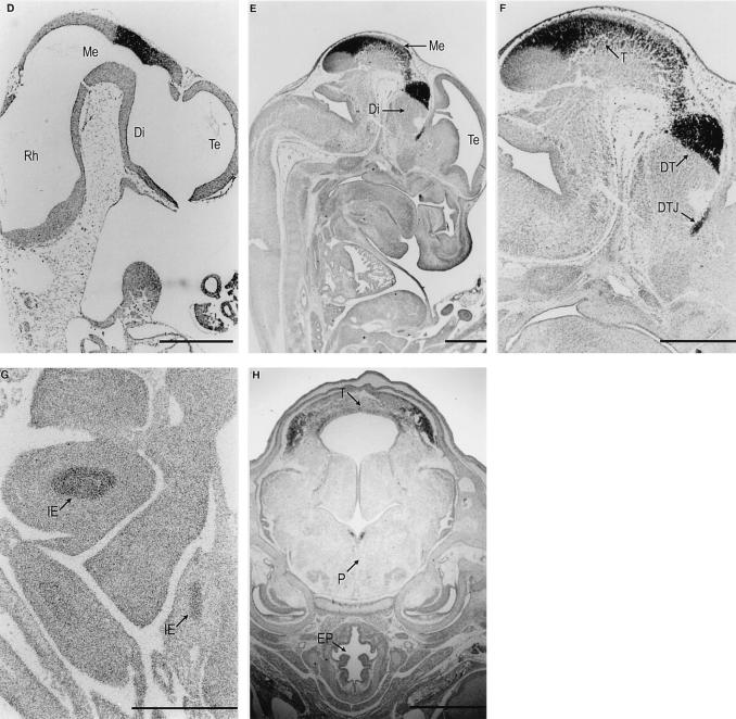
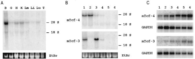
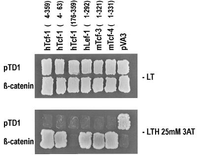
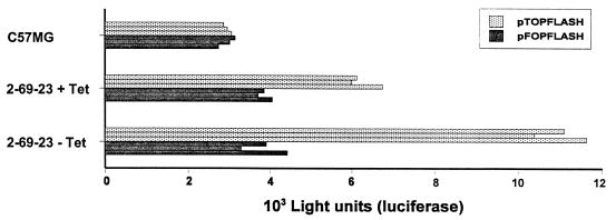
Tcf transcription factors interact with beta-catenin and Armadillo to mediate Wnt/Wingless signaling. We now report the characterization of genes encoding two murine members of the Tcf family, mTcf-3 and mTcf-4. mTcf-3 mRNA is ubiquitously present in embryonic day 6.5 (E6.5) mouse embryos but gradually disappears over the next 3 to 4 days. mTcf-4 expression occurs first at E10.5 and is restricted to di- and mesencephalon and the intestinal epithelium during embryogenesis. The mTcf-3 and mTcf-4 proteins bind a canonical Tcf DNA motif and can complex with the transcriptional coactivator beta-catenin. Overexpression of Wnt-1 in a mammary epithelial cell line leads to the formation of a nuclear complex between beta-catenin and Tcf proteins and to Tcf reporter gene transcription. These data demonstrate a direct link between Wnt stimulation and beta-catenin/Tcf transcriptional activation and imply a role for mTcf-3 and -4 in early Wnt-driven developmental decisions in the mouse embryo.


Figures

FIG. 1
FIG. 1
Sequence comparison of mTcf-4, hTcf-4, mTcf-3, and XTcf-3. The highly conserved N-terminal β-catenin interaction domain and the HMG box region are underlined. hTcf-4 is 98% identical to mTcf-4 at the amino acid level; the overall match between mTcf-3 and XTcf-3 is 75%; in particular, they align in the region C terminal to the HMG box (65% identity), which provides a unique signature for individual Tcf factors.
FIG. 2
FIG. 2
Phylogenetic relationship among members of the Tcf/Lef gene family based on aligned amino acid sequences. Relative branch lengths are drawn proportional to the number of inferred substitutions per site.
FIG. 3
FIG. 3
In situ hybridization analysis of mTcf-3 (A to C) and mTcf-4 (D to H) expression on murine embryo sections. mTcf-3 was detected in E6.5 (A), E7.5 (B), and E8.5 (C) embryos. Bright-field (left) and the accompanying dark-field (right) pictures of the longitudinal sections are shown. Embryo proper (E), ectoplacental cone (EC), chorion (Ch), and neuroectoderm (NE) are indicated. mTcf-4 expression was first observed at E10.5 (D) in the central nervous system. The expression remains restricted at E13.5 (E). (F) Detail of the head region; (G) detail of the intestine at E13.5; (D to G) parasagittal sections; (H) coronal section of the head at E16.5. Rhombencephalon (Rh), mesencephalon (Me), diencephalon (Di), telencephalon (Te), tectum (T), dorsal thalamus (DT), di-telencephalic junction (DTJ), intestinal epithelium (IE), pons (P), and esophagus (EP) are indicated. The bar in panel G represents 0.1 mm; bars in other panels represent 0.3 mm.
FIG. 3
FIG. 3
In situ hybridization analysis of mTcf-3 (A to C) and mTcf-4 (D to H) expression on murine embryo sections. mTcf-3 was detected in E6.5 (A), E7.5 (B), and E8.5 (C) embryos. Bright-field (left) and the accompanying dark-field (right) pictures of the longitudinal sections are shown. Embryo proper (E), ectoplacental cone (EC), chorion (Ch), and neuroectoderm (NE) are indicated. mTcf-4 expression was first observed at E10.5 (D) in the central nervous system. The expression remains restricted at E13.5 (E). (F) Detail of the head region; (G) detail of the intestine at E13.5; (D to G) parasagittal sections; (H) coronal section of the head at E16.5. Rhombencephalon (Rh), mesencephalon (Me), diencephalon (Di), telencephalon (Te), tectum (T), dorsal thalamus (DT), di-telencephalic junction (DTJ), intestinal epithelium (IE), pons (P), and esophagus (EP) are indicated. The bar in panel G represents 0.1 mm; bars in other panels represent 0.3 mm.
FIG. 4
FIG. 4
Northern blot analysis of mTcf-4 and mTcf-3 expression. (A) Tissue-specific mTcf-4 expression in mouse embryo at E18.5. The analysis was performed on total RNA isolated from brain (B), gut (G), heart (H), kidney (K), limbs (Lm), liver (Li), lung (Lu), and thymus (T). (B) mTcf-4 and mTcf-3 expression in C57MG cells (lane 1), PC12 cells (lane 2), embryonic stem cells (lane 3), 3T3 fibroblasts (lane 4), RBL-5 T-lymphocyte cells (lane 5), and NS-1 B lymphocyte cells (lane 6). The positions of 18S and 28S rRNAs are shown. EtBr, ethidium bromide stain. (C) Northern blot analysis of poly(A)+ RNA isolated from different parts of the intestine. Lanes: 1, duodenum; 2, jejunum; 3, ileum; 4, cecum; 5, proximal colon; 6, distal colon. GAPDH, control hybridization with a GAPDH probe.
FIG. 5
FIG. 5
Two-hybrid mating assay for the interaction of Tcfs and β-catenin. Baits (indicated above the lanes; pVA3 encodes p53) and preys were transformed in MATa and MATα yeast strains, respectively, and mated with each other. As shown by growth on selective plates (lower panel) or β-galactosidase assay (not shown), all Tcfs (but not the N-terminally truncated hTcf-1 or simian virus 40 large T) interact specifically with β-catenin and not with a control p53 protein (pVA3). All matings grow on nonselective plates (upper panel). −LT, medium lack Leu and Trp; −LTH 25mM 3AT, medium lacking Leu, Trp, and His and containing 25 mM 3-aminotriazole.
FIG. 6
FIG. 6
Inducible expression of Wnt-1 in C57MG cells. (A) Northern blot analysis of Wnt-1 RNA in C57MG cells (clone 2-69-23) carrying a Wnt-1 transgene under the control of a tetracycline (Tet)-repressible promoter. Poly(A)+ RNA was isolated from cells cultured in the presence of tetracycline (50 ng/ml) or for 24 h in the absence of the drug. The Wnt-1 transcript was detected by blot hybridization after gel electrophoresis using a 32P-labeled probe for Wnt-1. A longer exposure detects the presence of the major Wnt-1 transcript in tetracycline-treated cells. Wnt-1 expression was quantitated with the model 300A computing densitometer (Molecular Dynamics), and basal levels were found to be 50 times lower than the derepressed levels (data not shown). A probe for neomycin was used to confirm the presence of equal levels of RNA in each lane (not shown). (B) Western blot analysis of 2-69-23 cells. Equal amounts of Triton X-100-soluble protein from cells cultured in the presence of tetracycline (50 ng/ml) or for 24 h in the absence of the drug were analyzed by immunoblotting with anti-Wnt-1 antibody.
FIG. 7
FIG. 7
Wnt-1 induces a Tcf–β-catenin complex in C57MG cells, as determined by a gel retardation assay performed with nuclear extracts from 2-69-23 cells growing in the presence or absence (Wnt-1 induction) of tetracycline (Tet; 50 ng/ml). Samples in lanes 1 and 5 were incubated under standard conditions. Anti-β-catenin antibody (0.2 μg) was added to the samples in lanes 2, 6, and 9. A control antibody (human CD4; 0.2 μg) was added to the samples in lanes 3 and 7. Anti-Tcf-4 antibody (affinity purified, 0.2 μg) was added to samples in lanes 8 and 9. Samples in lanes 4 and 10 were incubated with a probe mutated in the Tcf binding site.
FIG. 8
FIG. 8
Wnt-1 activates Tcf reporter construct in C57MG cells. C57MG cells and 2-69-23 cells growing in the presence or absence of tetracycline (Tet) were transfected with 1 μg of the indicated luciferase reporter construct and 1 μg of plasmid pCATCONTROL as an internal control. Cells were harvested after 24 h, and luciferase and CAT values were determined. All experiments were done in triplicate; triplicate values are given.

References

    1. Aberle H, Bauer A, Stappert J, Kispert A, Kemler R. β-Catenin is a target for the ubiquitin-proteasome pathway. EMBO J. 1997;16:3797–3804. - PMC - PubMed
    1. Barker, N. Unpublished data.
    1. Behrens J, von Kries J P, Kuehl M, Bruhn L, Wedlich D, Grosschedl R, Birchmeier W. Functional interaction of β-catenin with the transcription factor LEF-1. Nature. 1996;382:638–642. - PubMed
    1. Brown A M, Wildin R S, Prendergast T J, Varmus H E. A retrovirus vector expressing the putative mammary oncogen int-1 causes partial transformation of a mammary epithelial cell line. Cell. 1986;46:1001–1009. - PubMed
    1. Brunner E, Peter O, Schweizer L, Basler K. Pangolin encodes a Lef-1 homologue that acts downstream of Armadillo to transduce the Wingless signal in Drosophila. Nature. 1997;385:829–833. - PubMed

Publication types

MeSH terms

Substances

Associated data