. 1998 Mar;18(3):1322-30.
doi: 10.1128/MCB.18.3.1322.

ETS-core binding factor: a common composite motif in antigen receptor gene enhancers

Affiliations

ETS-core binding factor: a common composite motif in antigen receptor gene enhancers

B Erman et al. Mol Cell Biol. 1998 Mar.

Abstract

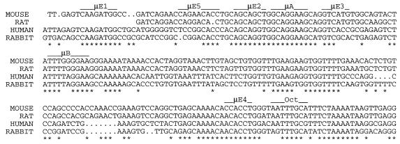
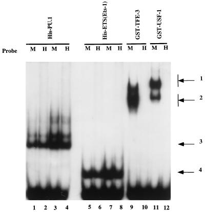
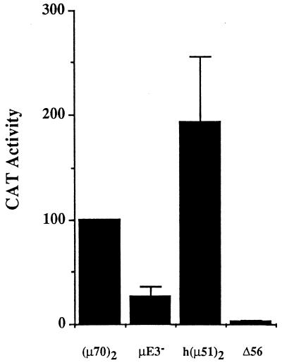
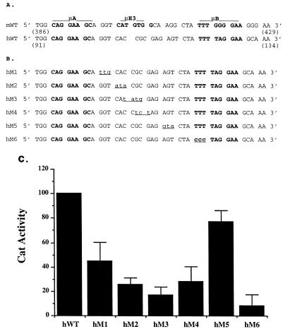
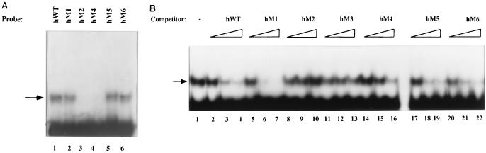
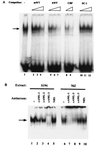
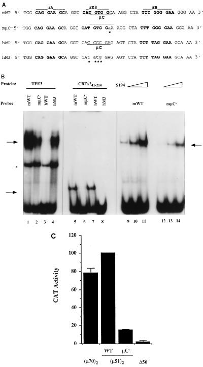
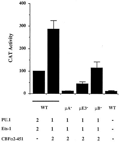
A tripartite domain of the murine immunoglobulin mu heavy-chain enhancer contains the muA and muB elements that bind ETS proteins and the muE3 element that binds leucine zipper-containing basic helix-loop-helix (bHLH-zip) factors. Analysis of the corresponding region of the human mu enhancer revealed high conservation of the muA and muB motifs but a striking absence of the muE3 element. Instead of bHLH-zip proteins, we found that the human enhancer bound core binding factor (CBF) between the muA and mu elements; CBF binding was shown to be a common feature of both murine and human enhancers. Furthermore, mutant enhancers that bound prototypic bHLH-zip proteins but not CBF did not activate transcription in B cells, and conversely, CBF transactivated the murine enhancer in nonlymphoid cells. Taking these data together with the earlier analysis of T-cell-specific enhancers, we propose that ETS-CBF is a common composite element in the regulation of antigen receptor genes. In addition, these studies identify the first B-cell target of CBF, a protein that has been implicated in the development of childhood pre-B-cell leukemias.


Figures

FIG. 1
FIG. 1
Alignment of IgH μ enhancer sequences. Sequences of IgH μ enhancer from four different species (GenBank accession no. V01523 for mouse, M13799 for rat, K01901 for human, and X13700 for rabbit) were aligned by using the Pileup program in the Wisconsin package version 8.1 (Genetics Computer Group, Madison, Wis.). Elements containing previously identified recognition motifs are overlined, and positions where the nucleotides from all species are identical are indicated by asterisks.
FIG. 2
FIG. 2
DNA binding analysis of PU.1, Ets-1, TFE3, and USF1 to the murine and human enhancers. The human (H) and murine (M) μ enhancer probes were used in binding assays with the following: lanes 1 and 2, His-PU.1 (80 ng); lanes 3 and 4, His-PU.1 (160 ng); lanes 5 and 6, Ets-1(ETS) (100 ng); lanes 7 and 8, Ets-1(ETS) (200 ng); lanes 9 and 10, GST-TFE3 (50 ng); and lanes 11 and 12, GST-USF1 (40 ng). Arrows: 1 and 2, USF and TFE3 binding to the murine probe only; 3, PU.1-DNA complex; 4, Ets-1(ETS)–DNA complex. EMSAs were performed as described previously (5).
FIG. 3
FIG. 3
The minimal murine and human IgH μ enhancers activate transcription comparably. Reporter plasmids (5 μg) containing dimeric murine μ70 [(μ70)2], murine μE3 mutant (μE3), and human [h(μ51)2] enhancers were transfected into S194 plasma cell lines, and CAT assays were performed by ELISA as described in Materials and Methods. CAT enzyme activity is shown on the y axis as the percentage of the amount of CAT enzyme obtained with the dimeric murine μ70 reporter. Results show the averages of at least two transfections carried out in duplicate. Δ56 refers to an enhancerless reporter. Error bars indicate the average deviations of the data.
FIG. 4
FIG. 4
Identification of an element in the human IgH enhancer in the region corresponding to the murine μE3 motif. (A) Comparison of the murine and human enhancers indicating the absence of a μE3 motif in the human enhancer. The μA, μE3, and μB motifs are indicated in boldface, and the nucleotide numbers of the murine and human enhancers are from references and , respectively. (B) Sequences of a panel of mutants in the human enhancer (hM1 to hM6) corresponding to the region spanning the murine μE3 motif. The altered sequence in each mutant is indicated in lowercase and underlined. (C) Transcriptional activities of the minimal human mutant enhancers. Reporter plasmids (5 μg) containing dimeric wild-type hWT and mutant (hM1 to hM6) enhancers were transfected into S194 plasma cell lines, and CAT assays were performed by ELISA as described in Materials and Methods. CAT enzyme activity is shown on the y axis as the percentage of the activity of the reporter plasmid containing the hWT enhancer. Results shown are the averages of at least two transfections carried out in duplicate. Error bars indicate the average deviations of the data.
FIG. 5
FIG. 5
DNA binding of PU.1 and Ets-1 to the human enhancer mutants. EMSAs were carried out with bacterially expressed and purified His-Ets-1(ETS) (lanes 1 to 6) and His-PU.1 (lanes 7 to 12) proteins and hWT and mutant enhancer probes as indicated above the lanes. The mutant probes are numbered as in Fig. 4B. Specific nucleoprotein complexes are indicated by arrows 1 [His-Ets-1(ETS)–DNA] and 2 (His-PU.1–DNA). A lower-mobility complex in the lanes with the PU.1 protein is due to the double occupancy of the μB and μA sites.
FIG. 6
FIG. 6
CBFα2 binds the human IgH μ enhancer in vitro. (A) EMSAs were carried out with bacterially expressed and purified CBFα241-190, which contains the DNA binding Runt domain of CBFα2, and hWT and mutant enhancer probes as indicated. Specific nucleoprotein complexes in lanes 1, 2, 5, and 6 are indicated by an arrow. (B) In vitro competition assays with CBFα241-190 bound to the human WT probe. EMSAs were carried out as described in the text, with 25-, 125-, and 250-fold molar excesses of competitor DNA fragments indicated by triangles. Competitor DNA was excised as dimeric fragments from reporter plasmids used for transfections in Fig. 4C and contain wild-type or mutated human enhancer sequences as indicated.
FIG. 7
FIG. 7
CBFα2 binds the murine IgH μ enhancer in vitro. EMSAs were carried out with bacterially expressed and purified CBFα241-190, which contains the DNA binding Runt domain of CBFα2, and the mWT μ enhancer and μA, μE3, and μB mutant enhancer probes as indicated.
FIG. 8
FIG. 8
The murine and human μ enhancers bind CBF in B-cell nuclear extracts. (A) In vitro competition assays with CBF bound to a high-affinity consensus binding probe. EMSAs were carried out with 20 μg of S194 nuclear extracts with 32P-labeled CBF oligonucleotide DNA probes (50,000 cpm), in the presence of no competitor DNA (lane 1), 5-, 25-, and 133-fold molar excesses of murine μ enhancer DNA (lanes 2 to 4) and human μ enhancer DNA (lanes 5 to 7), and 8- and 33-fold molar excesses of self (lanes 8 and 9) and 16-, 32-, and 66-fold molar excesses of nonspecific (lanes 10 to 12) competitor oligonucleotides, indicated by triangles. Specific nucleoprotein complexes are indicated by an arrow. The nonspecific competitor is a high-affinity binding site for Ets-1 (28). (B) Supershift EMSAs were carried out with 20 μg of S194 plasma cell and 27 μg of 70Z pre-B-cell extracts and a CBF high-affinity consensus binding site probe. Lanes 1 to 5 show complexes formed by the incubation of S194 extracts with CBF probes followed by no antiserum (lane 1), antiserum specific for AML-1, -2, and -3 (lanes 2, 3, and 4, respectively), and normal rabbit serum (NRS) (lane 5). Lanes 6 to 10 show complexes formed by the incubation of 70Z extracts with CBF probes followed by no antiserum (lane 6), antiserum specific for AML-1, -2, and -3 (lanes 7, 8, and 9, respectively), and normal rabbit serum (lane 10). Specific complexes are indicated by an arrow on the left.
FIG. 9
FIG. 9
CBFα2 binding correlates with minimal μ enhancer activity in B-cell lines. (A) Sequences of murine and human wild-type enhancers compared to sequences of two mutants in the murine (mμC) and human (hM3) enhancers. The overlapping μE3 (bHLH-zip protein binding) and μC (CBF binding) sites in the murine enhancer are overlined and underlined, respectively. The murine μC mutation alters the single nonoverlapping base between the two motifs. This position in the CBFα2 consensus binding site has been shown to be important for binding (19). The μC motif in the human motif is also underlined and is shown to be mutated in the hM3 mutation, which introduces an E-box motif into the human enhancer sequence that is absent in the wild-type sequence. (B) EMSAs were carried out with bacterially expressed and purified GST-TFE3, CBFα241-214, and S194 plasma cell extracts and the indicated murine and human probes. Lanes 1 to 4 show complexes formed by the incubation of approximately 50 ng of GST-TFE3 with the indicated probes. Specific complexes in lanes 1, 2, and 4 are indicated by the top arrow on the left, and a nonspecific complex is indicated by an asterisk. Lanes 5 to 8 show complexes formed by the incubation of 100 ng of CBFα241-214 and the indicated probes. Specific nucleoprotein complexes in lanes 5 and 7 are indicated by the bottom arrow on the left. Lanes 9 to 11 and 12 to 14 show complexes formed by the incubation of 4, 8, and 16 μg of S194 extracts, respectively, with the mWT and mμC probes. The μE3 binding complex is indicated by the arrow on the right. (C) Transcriptional activity of the murine enhancer. Reporter plasmids (5 μg) containing dimeric murine wild-type [WT or (μ70)2] and mutant (μC) enhancers or no enhancer (Δ56) were transfected into the M12 B-cell line, and CAT assays were performed by ELISA as described in the Materials and Methods. CAT enzyme activity is shown on the y axis as the percentage of the activity of the reporter plasmid containing the wild-type murine (μ70)2 enhancer. Results shown are the averages of at least two transfections carried out in duplicate. Error bars indicate the average deviations of the data.
FIG. 10
FIG. 10
CBFα2 activates the IgH μ enhancer in nonlymphoid cells in cooperation with Ets-1. HeLa cells were transfected with reporter plasmids containing the (μ70)2 enhancer along with expression plasmids for PU.1 (2 μg) and Ets-1 (2 μg) (bar 1) and for PU.1 (1 μg), Ets-1 (1 μg), and CBFα2 (2 μg) (bar 2) or with an empty pEVRF2 expression plasmid as carrier DNA (bar 6). The transcription activation abilities of PU.1 (1 μg), Ets-1 (1 μg), and CBFα2 (2 μg) were also tested by cotransfecting these reporter plasmids with binding site mutant versions of the (μ70)2 enhancer, μA (bar 3), μE3 (bar 4), and μB (bar 5). Cells were harvested 2 days after being transfected, and CAT analysis was performed by ELISA. Results shown are the averages of at least two transfections carried out in duplicate. Error bars indicate the average deviations of the data.

References

    1. Cameron S, Taylor D S, TePas E C, Speck N A, Mathet-Prevot B. Identification of a critical regulatory site in the human interleukin-3 promoter by in vivo footprinting. Blood. 1994;83:2851–2859. - PubMed
    1. Carvajal, I., and R. Sen. Unpublished observations.
    1. Chen J, Young F, Bottaro A, Stewart V, Smith R K, Alt F. Mutations of the intronic IgH enhancer and its flanking sequences differentially affect accessibility of the JH locus. EMBO J. 1993;12:4635–4645. - PMC - PubMed
    1. Crute B, Lewis A, Wu Z, Bushweller J, Speck N. Biochemical and biophysical properties of the core-binding factor alpha2 (AML1) DNA-binding domain. J Biol Chem. 1996;271:26251–26260. - PubMed
    1. Ephrussi A, Church G M, Tonegawa S, Gilbert W. B lineage specific interactions of an immunoglobulin enhancer with cellular factors in vivo. Science. 1985;227:134–140. - PubMed

Publication types

MeSH terms