. 1998 Mar;18(3):1349-58.
doi: 10.1128/MCB.18.3.1349.

Repression of GCN5 histone acetyltransferase activity via bromodomain-mediated binding and phosphorylation by the Ku-DNA-dependent protein kinase complex

Affiliations

Repression of GCN5 histone acetyltransferase activity via bromodomain-mediated binding and phosphorylation by the Ku-DNA-dependent protein kinase complex

N A Barlev et al. Mol Cell Biol. 1998 Mar.

Abstract

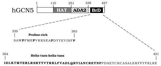
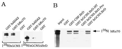
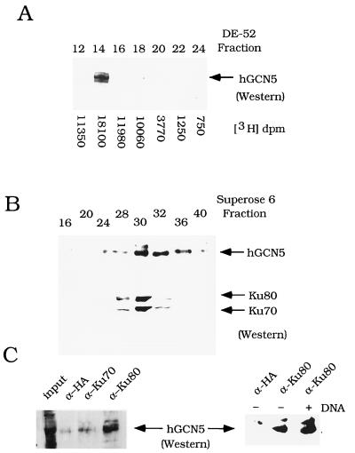
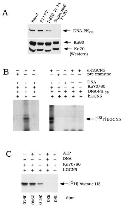
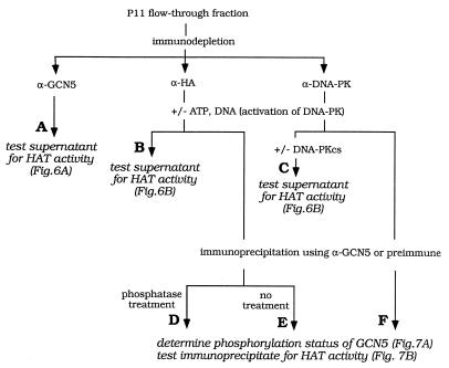
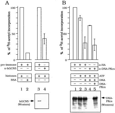
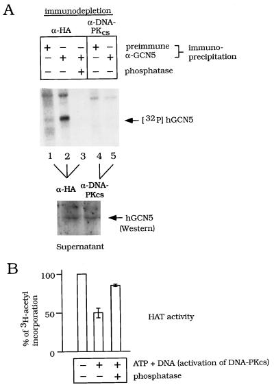
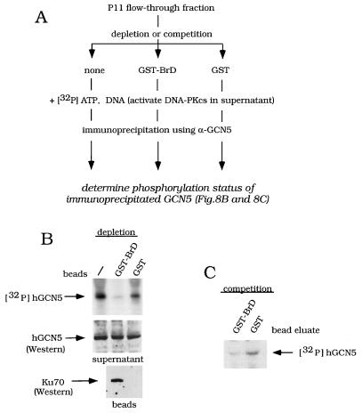
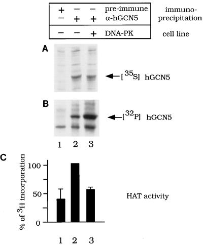
GCN5, a putative transcriptional adapter in humans and yeast, possesses histone acetyltransferase (HAT) activity which has been linked to GCN5's role in transcriptional activation in yeast. In this report, we demonstrate a functional interaction between human GCN5 (hGCN5) and the DNA-dependent protein kinase (DNA-PK) holoenzyme. Yeast two-hybrid screening detected an interaction between the bromodomain of hGCN5 and the p70 subunit of the human Ku heterodimer (p70-p80), which is the DNA-binding component of DNA-PK. Interaction between intact hGCN5 and Ku70 was shown biochemically using recombinant proteins and by coimmunoprecipitation of endogenous proteins following chromatography of HeLa nuclear extracts. We demonstrate that the catalytic subunit of DNA-PK phosphorylates hGCN5 both in vivo and in vitro and, moreover, that the phosphorylation inhibits the HAT activity of hGCN5. These findings suggest a possible regulatory mechanism of HAT activity.


Figures

FIG. 1
FIG. 1
hGCN5 domain structure. The domains of GCN5 are aa 1 to 110 (nonessential function) (14), aa 110 to 251 (the HAT domain) (16), aa 251 to 338 (the ADA2 interaction domain) (14), and aa 338 to 427 (the BrD motif) (53). Sequences within the BrD are aa 339 to 363 (the proline-rich sequence [hBrD.Pro]) and aa 364 to 421 (the helix-turn-helix-turn motif [hBrD.HT]). The prolines and helix-turn-helix-turn region are shown in boldface.
FIG. 2
FIG. 2
In vitro interactions between hGCN5 and Ku70. (A) hGCN5 binding to GST-hKu70. Equal amounts of GST-hADA2, GST-hKu70, or GST beads were incubated with either 35S-labeled hGCN5 or hGCN5ΔBrD translated in vitro. After washing, the material remaining on beads was eluted with 20 mM reduced glutathione and subjected to SDS-PAGE and autoradiography. (B) hKu70 binding to GST-BrDs. Equal amounts of GST, GST-CBP.BrD, GST-hGCN5.BrD.HT, GST-hGCN5.BrD.Pro, GST-hGCN5.BrD, and GST beads were incubated with in vitro-translated 35S-labeled hKu70. The material bound to beads was treated the same as for panel A. hKu70 input represented 50% of the amount used in each reaction. See Table I, footnote a, for explanation of GST fusion protein abbreviations.
FIG. 3
FIG. 3
Association of hGCN5 and Ku70/80 in HeLa nuclear extract. (A) Cofractionation of hGCN5 and HAT activity over DE-52 ion-exchange chromatography. Western blot analysis was performed on DE-52 column fractions, using α-hGCN5 polyclonal serum. The arrow designates the position of hGCN5 protein. HAT activity was quantitated by liquid scintillation counting, and the values are shown for each fraction. (B) Cofractionation of Ku70/80 and hGCN5 over Superose 6 sizing chromotography. Western blot analysis was performed on Superose 6 column fractions. Upper panel, hGCN5 immunoblot; lower panel, Ku70/80 immunoblot. The column was calibrated with size marker proteins: dextran blue (2,000 kDa) fraction 15; thyroglobulin (669 kDa) fraction 24; ferritin (440 kDa) fraction 28; aldolase (158 kDa) fraction 32. (C) Coimmunoprecipitation analysis of Ku and hGCN5. Western blot analysis of hGCN5 was performed on immunoprecipitates from fraction 14 after DE-52 column chromatography (A). The monoclonal antibodies used in immunoprecipitation were α-HA epitope, α-Ku70, and α-Ku80. The input lane (left panel) represents 15% of the material used in each reaction. The right panel shows the effect of adding sonicated salmon sperm DNA. Silver staining of immunoprecipitates with anti-Ku80 or anti-HA serum indicated no differences in the overall pattern of proteins bound by each antiserum (data not shown).
FIG. 4
FIG. 4
Effect of Ku/DNA-PKcs on recombinant hGCN5. (A) Western blot analysis of DNA-PKcs in sequential column fractions. Input, HeLa nuclear extract; P11 FT, FT from the P11 column; DE-52 Fr.14, fraction 14 (0.3 M KCl elution) (Fig. 3A); Superose 6 Fr.30, fraction 30 (Fig. 3B). Equal volumes of fractions described above were subjected to SDS-PAGE and transferred onto nitrocellulose. Blots were incubated with either DNA-PKcs monoclonal antibody (upper panel) or a mixture of Ku70 and p80 monoclonal antibodies (lower panel). (B) Phosphorylation of hGCN5 by DNA-PKcs in vitro. Recombinant hGCN5 was incubated with sonicated salmon sperm DNA, purified Ku70/80, and purified DNA-PKcs, as indicated. In the control experiment in the left panel, recombinant hGCN5 was not added to the lane marked −. [γ-32P]ATP was added and, following the kinase reaction, samples were immunoprecipitated with α-hGCN5 or preimmune serum and subjected to SDS-PAGE and autoradiography. (C) HAT activity assay of phosphorylated hGCN5. Recombinant hGCN5 and purified DNA-PKcs were incubated with ATP, sonicated salmon sperm DNA, and Ku70/80, as indicated. Following the HAT assay, to visualize 3H-histones, samples were subjected to SDS-PAGE and fluorography. One-fourth of the sample volume was quantitated by liquid scintillation counting, and these values are shown below the gel.
FIG. 5
FIG. 5
Outline of experiments testing the effect of DNA-PK on endogenous hGCN5. HeLa nuclear extract was fractionated on a P11 column, and the FT was tested for hGCN5-dependent HAT activity (column A) as well as DNA-PKcs-dependent effects on hGCN5’s HAT activity (columns B and C) and DNA-PKcs-dependent phosphorylation of hGCN5 (columns D to F). The experimental results are shown in Fig. 6 and 7.
FIG. 6
FIG. 6
Effect of immunodepletion of endogenous hGCN5 or DNA-PKcs on HAT activity in HeLa cell extracts. (A) HAT activity of endogenous hGCN5. P11 FT fraction was preincubated with preimmune (lanes 1 to 3) or hGCN5 (lane 4) antiserum. Proteins bound to antibodies were depleted by incubation with protein G beads followed by centrifugation. Supernatants were incubated with either BSA (lane 2) or free histones (lanes 1, 3, and 4) and subjected to HAT assay; 100% activity represented the incorporation of the 3H-acetyl moiety (dpm) mediated by the P11 FT fraction after mock immunodepletion with preimmune antiserum (lanes 1 and 3). Activities of other samples were calculated as a percentage of the mock-treated values. The standard deviations are based on results of two independent experiments. (B) Effect of DNA-PKcs on HAT activity in P11 FT. P11 FT was preincubated with HA-epitope monoclonal antibody (lanes 1 to 3) or DNA-PKcs monoclonal antibody (lanes 4 and 5) and immunodepleted as for panel A. Prior to the HAT assay, immunodepleted samples were incubated with ATP (lanes 2 to 5) and nonspecific DNA (lanes 3 to 5). Purified DNA-PKcs was added to the DNA-PKcs-immunodepleted sample (lane 5). Following the kinase reaction, samples were subjected to HAT assay; 100% activity represented the incorporation of 3H-acetyl moiety (dpm) mediated by the P11 FT fraction after mock immunodepletion with HA epitope monoclonal antibodies (lane 1). Activities of other samples were calculated as a percentage of the mock-treated values. The standard deviations are based on results of three independent experiments.
FIG. 7
FIG. 7
Effect of immunodepletion of DNA-PKcs and phosphatase treatment on phosphorylation and HAT activity of endogenous hGCN5. (A) Effect on phosphorylation of hGCN5. The P11 FT was preincubated with HA epitope monoclonal antibody (lanes 1 to 3) or DNA-PKcs monoclonal antibody (lanes 4 and 5) and immunodepleted as for Fig. 6A. The kinase reaction was done in the immunodepleted samples in the presence of [γ-32P]ATP and sonicated salmon sperm DNA. Following the kinase reaction, hGCN5 was immunoprecipitated and phosphatase was added to the reaction in lane 3, as indicated. The phosphorylation status of hGCN5 was determined by SDS-PAGE and autoradiography (upper panel). To show that the amount of hGCN5 input in any of the lanes in the upper panel was the same following immunodepletion by α-HA (lanes 1 to 3) or α-DNA-PKcs (lanes 4 and 5), hGCN5 input was analyzed by Western blotting (lower panel). (B) Effect of phosphatase treatment on HAT activity of hGCN5. The P11 FT was preincubated with HA epitope monoclonal antibody as for panel A. The kinase reaction was performed with nonradiolabeled ATP and sonicated salmon sperm DNA where indicated. Following the kinase reaction, samples were immunoprecipitated with α-hGCN5, and HAT activity was determined; 100% activity represented the incorporation of 3H-acetyl moiety (dpm) mediated by the P11 FT fraction without activation of DNA-PKcs (lane 1). Activities of other samples were calculated as a percentage of the mock-treated values. Standard deviations are based on results of three independent experiments.
FIG. 8
FIG. 8
Effect of BrD affinity depletion or affinity competition on hGCN5 phosphorylation in the P11 FT fraction. (A) Outline of experiments testing effect of GST-BrD on hGCN5 phosphorylation. P11 column FT was tested for Ku70-dependent effects on hGCN5 phosphorylation, following either affinity depletion of Ku70 or affinity competition for Ku70, using GST-BrD. The experimental results are shown in panels B (depletion) and C (competition). (B) Effect of affinity depletion of Ku70 on phosphorylation of hGCN5. The P11 FT was either not treated (−) or incubated with GST-BrD beads or GST beads. Proteins bound to the beads were depleted by centrifugation. Upper panel, [γ-32P]ATP and sonicated salmon sperm DNA were added to the supernatants to activate DNA-PKcs. Following the kinase reaction, samples were immunoprecipitated using α-hGCN5 and subjected to SDS-PAGE and autoradiography. Middle and lower panels, following bead depletion, either the supernatants were assayed for hGCN5 (middle) or the beads were assayed for Ku70 (lower), using Western analysis. (C) Effect of affinity competition for Ku70 on phosphorylation of hGCN5. GST-BrD and GST were eluted from the glutathione-Sepharose beads, using reduced glutathione. The P11 FT was incubated with the bead-eluted GST-BrD or GST, and a kinase assay was performed as for panel B.
FIG. 9
FIG. 9
Phosphorylation levels and HAT activity of hGCN5 in DNA-PKcs+ and DNA-PKcs glioblastoma cells. MO59K cells (DNA-PK+) (lane 3) or MO59J cells (DNA-PK) (lanes 1 and 2) were transfected with hGCN5 expressed from the cytomegalovirus promoter. The cells were lysed, and samples were immunoprecipitated with hGCN5 antiserum (lanes 2 and 3) or preimmune serum as a control (lane 1). Transfected cells were labeled with either [35S]Met (A) to determine hGCN5 expression levels in the two cell lines or 32Pi (B) to detect the levels of hGCN5 phosphorylation. (C) Histogram of the HAT activity detected in immunoprecipitates from unlabeled cells that were similarly transfected with hGCN5. Standard deviations are based on results of two independent experiments. The high background of HAT activity in lane 1 derived from a combination of nonspecific precipitation of HAT activity by the protein G-Sepharose beads, as well as by the preimmune serum (data not shown). Total levels of incorporation of 32Pi into proteins from the two cell lines were comparable, as judged by SDS-PAGE analysis (data not shown).

References

    1. Abmayr S M, Workman J L. Preparation of nuclear and cytoplasmic extracts from mammalian cells, units 12.1.1–12.1.9. In: Ausubel F M, et al., editors. Current protocols in molecular biology. New York, N.Y: John Wiley & Sons; 1993. - PubMed
    1. Anderson C W. DNA damage and the DNA-activated protein kinase. Trends Biochem Sci. 1993;18:433–437. - PubMed
    1. Arany Z, Sellers W R, Livingston D M, Eckner R. E1A-associated p300 and CREB-associated CBP belong to a conserved family of coactivators. Cell. 1994;77:799–800. . (Letter.) - PubMed
    1. Bannister A, Gottlieb T, Kouzarides T, Jackson S. c-Jun is phosphorylated by the DNA-dependent protein kinase in vitro; definition of the minimal kinase recognition motif. Nucleic Acids Res. 1993;21:1289–1295. - PMC - PubMed
    1. Bannister A, Kouzarides T. The CBP co-activator is a histone acetyltransferase. Nature. 1996;384:641–643. - PubMed

Publication types

MeSH terms

LinkOut - more resources