. 1998 Mar;18(3):1379-87.
doi: 10.1128/MCB.18.3.1379.

Regulation of the p85/p110 phosphatidylinositol 3'-kinase: stabilization and inhibition of the p110alpha catalytic subunit by the p85 regulatory subunit

Affiliations

Regulation of the p85/p110 phosphatidylinositol 3'-kinase: stabilization and inhibition of the p110alpha catalytic subunit by the p85 regulatory subunit

J Yu et al. Mol Cell Biol. 1998 Mar.

Abstract

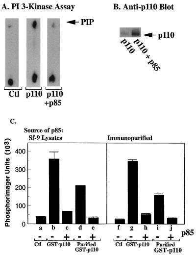
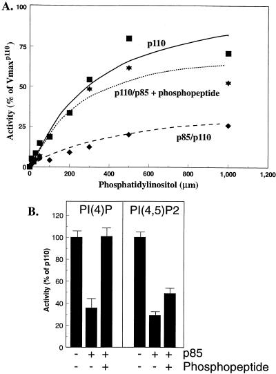
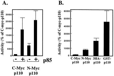
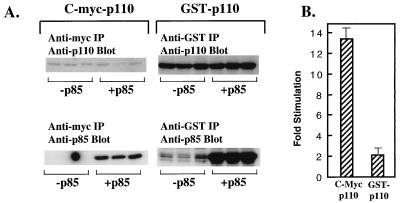
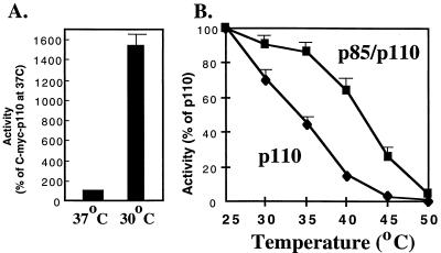
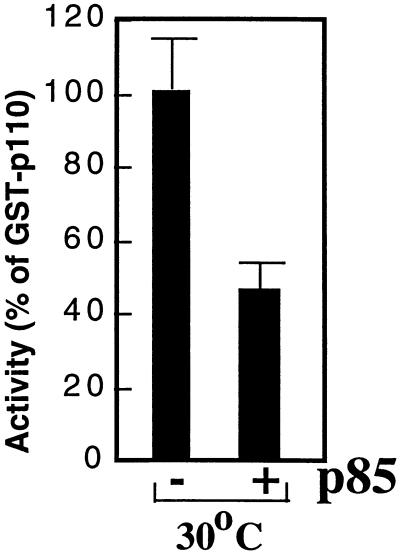
We propose a novel model for the regulation of the p85/pl10alpha phosphatidylinositol 3'-kinase. In insect cells, the p110alpha catalytic subunit is active as a monomer but its activity is decreased by coexpression with the p85 regulatory subunit. Similarly, the lipid kinase activity of recombinant glutathione S-transferase (GST)-p110alpha is reduced by 65 to 85% upon in vitro reconstitution with p85. Incubation of p110alpha/p85 dimers with phosphotyrosyl peptides restored activity, but only to the level of monomeric p110alpha. These data show that the binding of phosphoproteins to the SH2 domains of p85 activates the p85/p110alpha dimers by inducing a transition from an inhibited to a disinhibited state. In contrast, monomeric p110 had little activity in HEK 293T cells, and its activity was increased 15- to 20-fold by coexpression with p85. However, this apparent requirement for p85 was eliminated by the addition of a bulky tag to the N terminus of p110alpha or by the growth of the HEK 293T cells at 30 degrees C. These nonspecific interventions mimicked the effects of p85 on p110alpha, suggesting that the regulatory subunit acts by stabilizing the overall conformation of the catalytic subunit rather than by inducing a specific activated conformation. This stabilization was directly demonstrated in metabolically labeled HEK 293T cells, in which p85 increased the half-life of p110. Furthermore, p85 protected p110 from thermal inactivation in vitro. Importantly, when we examined the effect of p85 on GST-p110alpha in mammalian cells at 30 degrees C, culture conditions that stabilize the catalytic subunit and that are similar to the conditions used for insect cells, we found that p85 inhibited p110alpha. Thus, we have experimentally distinguished two effects of p85 on p110alpha: conformational stabilization of the catalytic subunit and inhibition of its lipid kinase activity. Our data reconcile the apparent conflict between previous studies of insect versus mammalian cells and show that p110alpha is both stabilized and inhibited by dimerization with p85.


Figures

FIG. 1
FIG. 1
Expression of p110α in Sf-9 cells. (A) Control Sf-9 cells or p110α baculovirus-infected Sf-9 cells were additionally infected without the p85 baculovirus or not infected. The cells were lysed, and PI 3′-kinase activities in the lysate were measured. (B) Western blot of parallel samples with anti-p110α antibody. (C) Left panel: control Sf-9 lysates, lysates from cells expressing GST-p110α, or glutathione-Sepharose-purified GST-p110α was mixed with lysates from cells expressing p85 as indicated. Lipid kinase activity was then measured. Right panel: control Sf-9 lysates, lysates from cells expressing GST-p110α, or glutathione-Sepharose-purified GST-p110α was mixed with immunopurified p85 as indicated, and PI 3′-kinase activities were measured. All determinations were made in triplicate, and the data are the means ± standard errors of the means from three experiments.
FIG. 2
FIG. 2
Lipid kinase activities of p110α monomers and p85/p110α dimers. (A) N-myc p110α monomers were incubated for 30 min at 4°C in the absence or presence of p85 and then incubated for an additional 60 min at 4°C in the absence or presence of 1 μM bisphosphopeptide. The mixtures were assayed in the presence of 0 to 1,000 μM PI and 1 mM ATP. After 10 min at 22°C, the lipids were extracted and analyzed as described in Materials and Methods. All determinations were performed in duplicate, and the data are the means from three separate experiments. Curves represent the best Michaelis-Menten fit and were generated with Kaleidograph software. (B) N-myc-p110 monomers or p85/N-myc-p110 dimers were incubated in the absence or presence of phosphopeptide as described above. The samples were assayed with sonicated mixtures of PI,PS and either PI[4]P or PI[4,5]P2 as described in Materials and Methods. The data are the means ± standard errors of the means for two (PIP) or three (PIP2) experiments.
FIG. 3
FIG. 3
Inhibition of p110α by p85-S608A. (A) Expression of wild-type p85 and p85-S608A in Sf-9 lysates was measured by blotting with anti-p85 antibody. (B) Wild-type p85 and p85-S608A were immobilized on anti-p85–protein A-Sepharose beads, incubated with lysates from cells expressing N-myc-p110α, washed, and assayed for lipid kinase activity. Lipid kinase activity bound to wild-type p85 was defined as 100%. (C) Lysates from Sf-9 cells expressing N-myc-p110α were incubated in the absence or presence of wild-type p85 or p85-S608A and then assayed for lipid kinase activity (expressed as a percentage of lipid kinase activity in the absence of p85). (D) Lysates from Sf-9 cells expressing N-myc-p110α were incubated in the absence or presence of immunopurified wild-type p85 or p85-S608A and then assayed for lipid kinase activity (expressed as in panel C). (E) Sf-9 cells were infected with N-myc-p110α alone or were coinfected with wild-type p85 or p85-S608A. Lipid kinase activity in the cell lysates was determined (expressed as in panel C). Inset: p110α expression was determined by blotting with anti-p110α antibody. (F) Lysates containing N-myc-p110α and wild-type p85 or p85-S608A were incubated in the absence or presence of bisphosphopeptide (1 μM) for 1 h, and lipid kinase activity was determined. Activation was expressed as fold stimulation over activity in the absence of phosphopeptide. All determinations were made in duplicate or triplicate, and the data are the means ± standard errors of the means of four separate experiments.
FIG. 4
FIG. 4
Inhibition of p110α by dephosphorylated p85. (A) Forty-eight hours after infection with p85 baculovirus, Sf-9 cells were labeled with [32P]orthophosphate for 4 h. The cells were lysed, treated with recombinant protein phosphatase 1 (0.5 μg) or not treated, and immunoprecipitated with anti-p85–protein A–Sepharose beads. Proteins were eluted and separated by SDS-PAGE, and the dried gel was visualized by autoradiography and quantitated with a Molecular Dynamics PhosphorImager. (B) Lysates from Sf-9 cells expressing p85 were treated in the absence or presence of recombinant protein phosphatase 1 (0.5 μg). p85 was purified by absorption on anti-p85–protein A–Sepharose beads and mixed with p110α, and lipid kinase activity was determined. All determinations were made in triplicate, and the data are the means ± standard errors of the means of two separate experiments.
FIG. 5
FIG. 5
Activity of epitope-tagged p110α: comparison of N-terminal versus C-terminal tags. (A) HEK 293T cells were transfected with expression vectors for C-myc- or N-myc-p110α (30 μg) plus control vector or an expression vector for p85 (30 μg). Forty-eight hours after transfection the cells were lysed, and anti-myc immunoprecipitates were prepared. The immune complexes were assayed for lipid kinase activity. Parallel samples were eluted, separated by SDS-PAGE, and blotted sequentially with anti-p110α and anti-p85 antibodies. PI 3′-kinase specific activity was calculated relative to p110α expression, as measured by blotting. Data from different experiments were normalized to the activity of C-myc-p110α in each experiment. All determinations were performed in triplicate, and the data are the means ± standard errors of the means from five separate experiments. (B) HEK 293T cells were transfected with 30 (C-myc-p110α, N-myc-p110α, and GST-p110α) or 45 μg (3HA-p110α) of DNA. The cells were lysed after 48 h, and anti-myc, anti-HA, and anti-GST immunoprecipitates were prepared. Lipid and protein determinations and data normalization were performed as described for panel A. All determinations were performed in triplicate, and the data are the means ± standard errors of the means from four separate experiments.
FIG. 6
FIG. 6
Effect of p85 on C-myc-p110α versus GST-p110α. HEK 293T cells were transfected with expression vectors for C-myc p110α or GST-p110α in the presence of a control vector or the expression vector for p85. (A) Anti-myc or anti-GST immunoprecipitates (IP) were blotted with anti-p110 antibody (top lanes) or anti-p85 antibody (lower lanes). (B) Protein and lipid kinase activities in anti-myc or anti-GST immunoprecipitates were calculated as described in the legend for Fig. 5A. The data are expressed as the fold stimulation for each construct in the presence versus the absence of p85. All determinations were performed in triplicate, and the data are the means ± standard errors of the means from two separate experiments.
FIG. 7
FIG. 7
Stabilization of p110 by p85 in mammalian cells. HEK 293T cells were transfected with vectors for C-myc-p110 in the absence or presence of p85. Thirty-six hours after transfection the cells were labeled with [35S]methionine overnight and then chased for the indicated times in medium containing 10× methionine. The cells were lysed, and immunoprecipitated p110 was separated by SDS-PAGE and visualized by autoradiography. The data are representative of two separate experiments.
FIG. 8
FIG. 8
Effect of temperature on the activity of p110α. (A) HEK 293T cells were transfected with expression vectors for C-myc-p110α and then maintained for 48 h at 37 or 30°C. The cells were then lysed, and protein expression and lipid kinase activities were determined as described in the legend for Fig. 5A. All determinations were performed in triplicate, and the data are the means ± standard errors of the means (SEM) from two experiments. (B) Recombinant p110 or p85/p110 dimers were incubated at the indicated temperatures for 30 min. After being chilled on ice, the samples were assayed for lipid kinase activity at 22°C. The data are the means ± SEM from three separate experiments.
FIG. 9
FIG. 9
Inhibition of p110 by p85 in mammalian cells. HEK 293T cells were transfected with GST-p110α in the presence of control vector or p85 expression vector as indicated. After 48 h at 30°C, the cells were lysed and protein and lipid kinase activities were determined. The activities were normalized to the activity of GST-p110α. All determinations were performed in triplicate, and the data are representative of four separate experiments.

References

    1. Antonetti D A, Algenstaedt P, Kahn C R. Insulin receptor substrate 1 binds two novel splice variants of the regulatory subunit of phosphatidylinositol 3-kinase in muscle and brain. Mol Cell Biol. 1996;16:2195–2203. - PMC - PubMed
    1. Backer J M, Jr, Myers M G, Shoelson S E, Chin D J, Sun X J, Miralpeix M, Hu P, Margolis B, Skolnik E Y, Schlessinger J, White M F. The phosphatidylinositol 3′-kinase is activated by association with IRS-1 during insulin stimulation. EMBO J. 1992;11:3469–3479. - PMC - PubMed
    1. Booker G W, Gout I, Downing A K, Driscoll P C, Boyd J, Waterfield M D, Campbell I D. Solution structure and ligand-binding site of the SH3 domain of the p85α subunit of phosphatidylinositol 3-kinase. Cell. 1993;73:813–822. - PubMed
    1. Cantley L C, Auger K R, Carpenter C, Duckworth B, Graziani A, Kapeller R, Soltoff S. Oncogenes and signal transduction. Cell. 1991;64:231–302. - PubMed
    1. Carpenter C L, Auger K R, Chanudhuri M, Yoakim M, Schaffhausen B, Shoelson S, Cantley L C. Phosphoinositide 3-kinase is activated by phosphopeptides that bind to the SH2 domains of the 85-kDa subunit. J Biol Chem. 1993;268:9478–9483. - PubMed

Publication types

MeSH terms