. 1998 Mar;18(3):1489-97.
doi: 10.1128/MCB.18.3.1489.

EWS, but not EWS-FLI-1, is associated with both TFIID and RNA polymerase II: interactions between two members of the TET family, EWS and hTAFII68, and subunits of TFIID and RNA polymerase II complexes

Affiliations

EWS, but not EWS-FLI-1, is associated with both TFIID and RNA polymerase II: interactions between two members of the TET family, EWS and hTAFII68, and subunits of TFIID and RNA polymerase II complexes

A Bertolotti et al. Mol Cell Biol. 1998 Mar.

Abstract

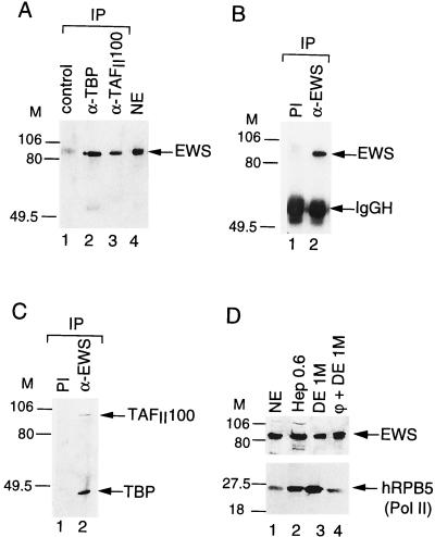
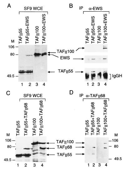
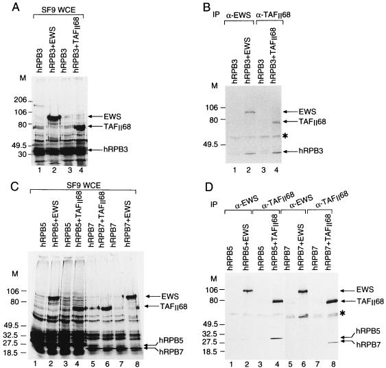
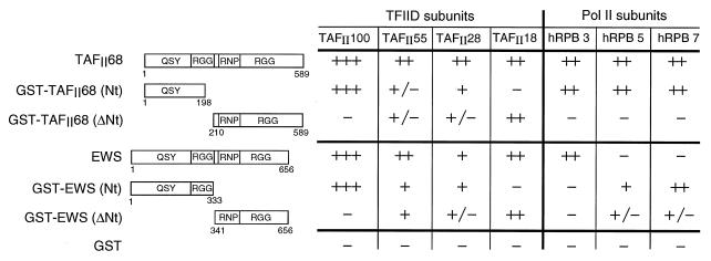
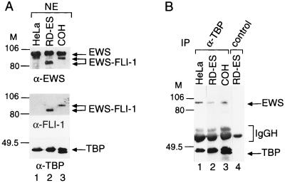
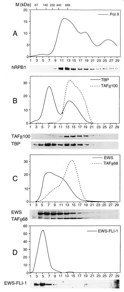
The t(11;22) chromosomal translocation specifically linked to Ewing sarcoma and primitive neuroectodermal tumor results in a chimeric molecule fusing the amino-terminus-encoding region of the EWS gene to the carboxyl-terminal DNA-binding domain encoded by the FLI-1 gene. As the function of the protein encoded by the EWS gene remains unknown, we investigated the putative role of EWS in RNA polymerase II (Pol II) transcription by comparing its activity with that of its structural homolog, hTAFII68. We demonstrate that a portion of EWS is able to associate with the basal transcription factor TFIID, which is composed of the TATA-binding protein (TBP) and TBP-associated factors (TAFIIs). In vitro binding studies revealed that both EWS and hTAFII68 interact with the same TFIID subunits, suggesting that the presence of EWS and that of hTAFII68 in the same TFIID complex may be mutually exclusive. Moreover, EWS is not exclusively associated with TFIID but, similarly to hTAFII68, is also associated with the Pol II complex. The subunits of Pol II that interact with EWS and hTAFII68 have been identified, confirming the association with the polymerase. In contrast to EWS, the tumorigenic EWS-FLI-1 fusion protein is not associated with either TFIID or Pol II in Ewing cell nuclear extracts. These observations suggest that EWS and EWS-FLI-1 may play different roles in Pol II transcription.


Figures

FIG. 1
FIG. 1
EWS is associated with TFIID and copurifies with Pol II. (A) The anti-hTBP (α-TBP) and the anti-hTAFII100 (α-TAFII100) MAbs coimmunoprecipitate EWS from a HeLa cell NE. HeLa cell NE was immunoprecipitated (IP) with either an unrelated antibody (lane 1) or a MAb raised against TBP (3G3; lane 2) or hTAFII100 (2D2; lane 3). Beads were washed and boiled, and bound proteins were analyzed by Western blotting using an antibody raised against the N-terminal domain of EWS that recognizes the endogenous EWS protein in HeLa cell NE (lane 4). M, markers in kilodaltons. (B and C) The anti-EWS antibody coimmunoprecipitates components of the TFIID complex. HeLa cell NE was immunoprecipitated with either the anti-EWS PAb (lanes 2) or the preimmune serum (PI; lanes 1). Beads were washed and boiled, and bound proteins were analyzed by Western blotting using either the anti-EWS antibody (B) or the anti-TBP MAb 3G3 together with the anti-hTAFII100 MAb 2D2 (C). In panel B, the IgG heavy chain (IgGH) is indicated. (D) EWS copurifies with Pol II. The previously described chromatographic fractions obtained during the purification of Pol II (3) were tested by Western blotting using an antibody raised against either EWS (upper panel) or the fifth-largest subunit of Pol II (hRPB5; lower panel). Hep, Heparin-Ultrogel column; DE, DEAE 5PW HPLC column; φ, Phenyl-5PW HPLC column.
FIG. 2
FIG. 2
Interactions of EWS and hTAFII68 with other components of the human TFIID complex. SF9 cells were coinfected with recombinant baculoviruses expressing hTAFII100 and hTAFII55 either individually or pairwise with EWS (A) and hTAFII68 (C) as indicated. After 44 h of infection, proteins were radiolabeled with [α-35S]methionine and [α-35S]cysteine for 4 h. WCEs were made, proteins were separated by SDS-PAGE, and gels were dried and subjected to autoradiography. M, markers in kilodaltons. (B and D) From the protein extracts, EWS and TAFII68 were immunoprecipitated (IP) with either the anti-EWS (α-EWS) PAb (B) or the anti-hTAFII68 (α-TAFII68) MAb (D) as indicated. Resin-bound proteins were analyzed by Western blotting with antibodies raised against either EWS, hTAFII100, and hTAFII55 separately (B) or hTAFII68, hTAFII100, and hTAFII55 (D). In panel B, peroxidase-conjugated goat anti-mouse IgG-IgM-specific secondary antibodies were used; in panel D, peroxidase-conjugated goat anti-mouse κ-type light-chain-specific secondary antibody was used. IgGH, IgG heavy chain.
FIG. 3
FIG. 3
Interactions between EWS or hTAFII68 and the different subunits of Pol II. (A and C) SF9 cells were coinfected with recombinant baculoviruses expressing subunits of Pol II either individually or pairwise with EWS and hTAFII68 as indicated. After 44 h of infection, proteins were radiolabeled with [α-35S]methionine and [α-35S]cysteine for 4 h. WCEs were made, proteins were separated by SDS-PAGE, and gels were dried and subjected to autoradiography. M, markers in kilodaltons. (B and D) From the WCEs, EWS or hTAFII68 was immunoprecipitated (IP) with either the anti-EWS (α-EWS) antibody or the anti-hTAFII68 (α-TAFII68) MAb as indicated. Resin-bound proteins were then analyzed by SDS-PAGE followed by autoradiography. The asterisk indicates a nonspecific protein species.
FIG. 4
FIG. 4
Mapping the domains of EWS and hTAFII68 which interact with the subunits of TFIID and Pol II. Numbers in the diagrams refer to amino acid positions in either hTAFII68 or EWS. The results of the protein-protein interaction assay, using either baculovirus-overexpressed full-length proteins or E. coli-produced GST fusion proteins, are summarized as follows: +++, ++, +, +/−, and −, strong, moderate, weak, very weak but detectable, and no interactions between the indicated proteins.
FIG. 5
FIG. 5
The oncogenic fusion protein EWS–FLI-1 does not coimmunoprecipitate with the TFIID complex in Ewing sarcoma cell lines. (A) The PAb raised against the N-terminal domain of EWS recognizes both wild-type EWS and the two different EWS–FLI-1 fusion proteins in NEs from the two Ewing sarcoma cell lines, RD-ES (lane 2) and COH (lane 3). NEs from HeLa, RD-ES and COH cells were analyzed by Western blotting using the anti-EWS (α-EWS) antibody (upper panel), the anti-FLI-1 (α-FLI-1) MAb (middle panel), and the anti-TBP (α-TBP) MAb 3G3 (lower panel). M, markers in kilodaltons. (B) NEs from the various cell lines were immunoprecipitated (IP) with the anti-TBP MAb 3G3 (lane 1 to 3). Beads were washed and boiled, and bound proteins were analyzed by Western blotting with the anti-EWS antibody and the anti-TBP MAb. The control immunoprecipitation using an unrelated MAb is shown in lane 4.
FIG. 6
FIG. 6
Sedimentation of RD-ES cell NE through a 20 to 40% glycerol gradient indicates that the endogenous EWS–FLI-1 fusion protein is present in low-molecular-mass ranges. The relative sedimentations of the largest subunit of Pol II (A), TAFII100 and TBP (B), EWS and TAFII68 together (C), and EWS–FLI-1 (D) were determined by Western blotting using antibodies raised against either the CTD of the largest subunit of Pol II (A), TAFII100 and TBP (B), or EWS and TAFII68 (C). To better visualize EWS–FLI-1 that is only weakly detected in panel C by the EWS antibody, in panel D the anti-FLI-1 antibody was used. In each panel, the upper part shows a quantification of the Western blot. Values represent the percentage of a given protein present in each fraction compared to the total amount of this protein loaded on the glycerol gradient. Positions of markers (M) of known molecular mass standards are indicated at the top of panel A.

References

    1. Acker J, de Graaff M, Cheynel I, Khazak V, Kedinger C, Vigneron M. Interactions between the human RNA polymerase II subunits. J Biol Chem. 1997;272:16815–16821. - PubMed
    1. Bailly R A, Bosselut R, Zucman J, Cormier F, Delattre O, Roussel M, Thomas G, Ghysdael J. DNA-binding and transcriptional activation properties of the EWS–FLI-1 fusion protein resulting from the t(11;22) translocation in Ewing sarcoma. Mol Cell Biol. 1994;14:3230–3241. - PMC - PubMed
    1. Bertolotti A, Lutz Y, Heard D J, Chambon P, Tora L. hTAFII68 a novel RNA/SSDNA-binding protein with homology to the pro-oncoproteins TLS/FUS and EWS is associated with both TFIID and RNA polymerase II. EMBO J. 1996;15:5022–5031. - PMC - PubMed
    1. Besse S, Vigneron M, Pichard E, Puvlon-Dutilleul F. Synthesis and maturation of viral transcripts in herpes simplex virus type 1 infected HeLa cells: the role of interchromatin granules. Gene Expr. 1995;4:143–161. - PMC - PubMed
    1. Brou C, Chaudhary S, Davidson I, Lutz Y, Wu J, Egly J M, Tora L, Chambon P. Distinct TFIID complexes mediate the effect of different transcriptional activators. EMBO J. 1993;12:489–499. - PMC - PubMed

Publication types

MeSH terms