. 1998 Mar;18(3):1611-21.
doi: 10.1128/MCB.18.3.1611.

Reinitiation of DNA synthesis and cell division in senescent human fibroblasts by microinjection of anti-p53 antibodies

Affiliations

Reinitiation of DNA synthesis and cell division in senescent human fibroblasts by microinjection of anti-p53 antibodies

V Gire et al. Mol Cell Biol. 1998 Mar.

Abstract

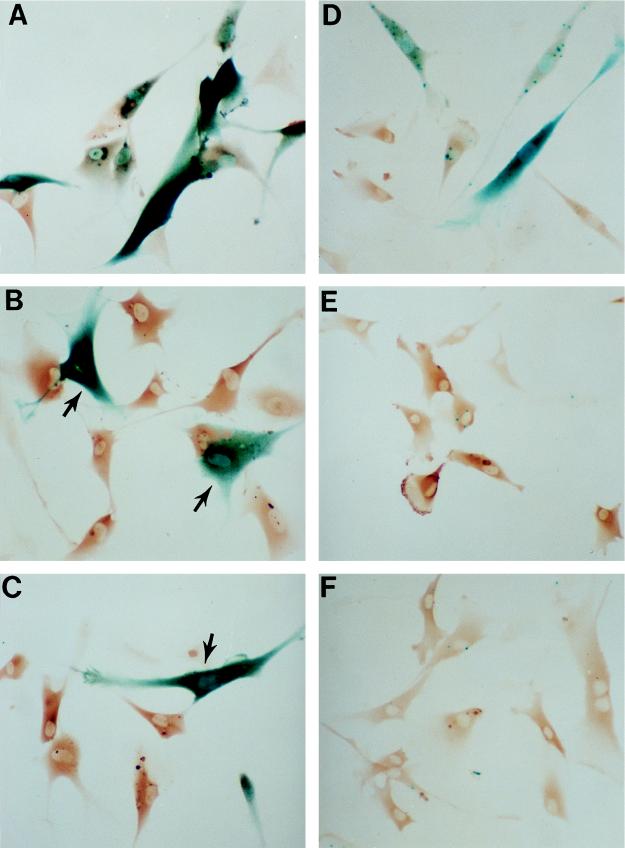
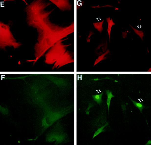
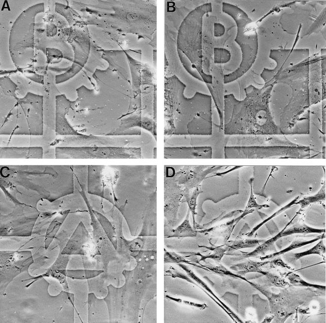
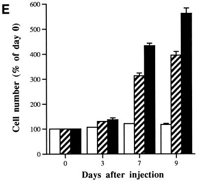
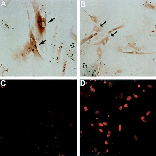
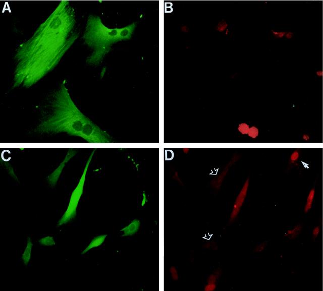
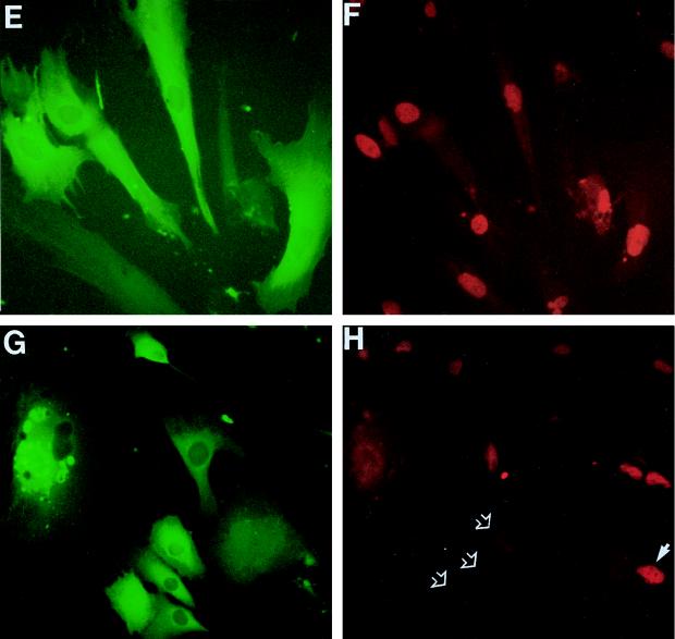
In human fibroblasts, growth arrest at the end of the normal proliferative life span (induction of senescence) is dependent on the activity of the tumor suppressor protein p53. In contrast, once senescence has been established, it is generally accepted that reinitiation of DNA synthesis requires loss of multiple suppressor pathways, for example, by expression of Simian virus 40 (SV40) large T antigen, and that even this will not induce complete cell cycle traverse. Here we have used microinjection of monoclonal antibodies to the N terminus of p53, PAb1801 and DO-1, to reinvestigate the effect of blocking p53 function in senescent human fibroblasts. Unexpectedly, we found that both antibodies induce senescent cells to reenter S phase almost as efficiently as SV40, accompanied by a reversion to the "young" morphology. Furthermore, this is followed by completion of the cell division cycle, as shown by the appearance of mitoses, and by a four- to fivefold increase in cell number 9 days after injection. Immunofluorescence analysis showed that expression of the p53-inducible cyclin/kinase inhibitor p21sdi1/WAF1 was greatly diminished by targeting p53 with either PAb1801 or DO-1 but remained high and, moreover, still p53 dependent in cells expressing SV40 T antigen. As previously observed for induction, the maintenance of fibroblast senescence therefore appears to be critically dependent on functional p53. We suggest that the previous failure to observe this by using SV40 T-antigen mutants to target p53 was most probably due to incomplete abrogation of p53 function.


Figures

FIG. 1
FIG. 1
Inhibition of p53-dependent transactivation by anti-p53 antibodies. p53 transactivation activity was stimulated in early-passage LacZ21 fibroblasts either by irradiation with 10 of UV J · m−2 (A to C) or by microinjection of the activating antibody PAb421 (D to F). Expression of the p53-dependent reporter was assessed 24 h later by β-gal histochemistry (punctate and/or diffuse blue staining). Microinjected cells were identified by immunoperoxidase detection of injected IgG (brown staining). Injection of either DO-1 (B) or PAb1801 (C) 24 h prior to UV treatment greatly reduced reporter expression compared to that with mouse IgG controls (A). Similarly, the response to PAb421 (D) was inhibited by coinjection of DO-1 (E) or PAb1801 (F). (Note that the β-gal-positive [blue] cells in panels B and C [arrows] were not microinjected.) Magnification, ×140.
FIG. 2
FIG. 2
Morphological reversion and reinitiation of DNA synthesis in senescent human fibroblasts induced by anti-p53 antibody PAb1801. Senescent HCA2 cells (corresponding to PDL65 plus 19 days in Table 1) were microinjected with control mouse IgG (B, E, and F), PAb1801 (C, G, and H), or plasmid SVori− (D) and analyzed 72 h later (all cells in the fields shown were injected). (B to D) Effects on morphology determined by phase-contrast microscopy compared to untreated young cultures (A). (E to H) Effects on DNA synthesis as revealed by BrdU labelling. Injected cells are identified by immunofluorescence of coinjected rat IgG with a rhodamine label (red) (E and G); nuclear BrdU incorporation (examples are indicated by arrows) is shown by immunofluorescence with FITC (green) (F and H). (Note that the weak cytoplasmic FITC fluorescence visible in panels F and H is due to detection of the injected mouse IgGs by the rabbit anti-mouse–FITC used to detect the BrdU.) Magnification, ×160.
FIG. 2
FIG. 2
Morphological reversion and reinitiation of DNA synthesis in senescent human fibroblasts induced by anti-p53 antibody PAb1801. Senescent HCA2 cells (corresponding to PDL65 plus 19 days in Table 1) were microinjected with control mouse IgG (B, E, and F), PAb1801 (C, G, and H), or plasmid SVori− (D) and analyzed 72 h later (all cells in the fields shown were injected). (B to D) Effects on morphology determined by phase-contrast microscopy compared to untreated young cultures (A). (E to H) Effects on DNA synthesis as revealed by BrdU labelling. Injected cells are identified by immunofluorescence of coinjected rat IgG with a rhodamine label (red) (E and G); nuclear BrdU incorporation (examples are indicated by arrows) is shown by immunofluorescence with FITC (green) (F and H). (Note that the weak cytoplasmic FITC fluorescence visible in panels F and H is due to detection of the injected mouse IgGs by the rabbit anti-mouse–FITC used to detect the BrdU.) Magnification, ×160.
FIG. 3
FIG. 3
Stimulation of cell proliferation in senescent fibroblasts by microinjection of antibody PAb1801 or DO-1. (A to D) Photomicrographs of senescent fibroblasts in zones predefined by reference to an underlying grid, before (A and C) and 7 days after (B and D) microinjection with either control IgG (A and B) or PAb1801 (C and D). (Note the increase in cell density in panel D and reversion of most cells to a young morphology.) Magnification, ×200. (E) Histogram showing increasing cell number with time after microinjection of DO-1 (solid bars) or PAb1801 (hatched bars) compared to control IgG (open bars). Injected cells and their progeny were identified by immunostaining of coinjected rat IgG, and their numbers are expressed as a percentage of the day 0 value. Values are means from at least three independent experiments ± SE.
FIG. 3
FIG. 3
Stimulation of cell proliferation in senescent fibroblasts by microinjection of antibody PAb1801 or DO-1. (A to D) Photomicrographs of senescent fibroblasts in zones predefined by reference to an underlying grid, before (A and C) and 7 days after (B and D) microinjection with either control IgG (A and B) or PAb1801 (C and D). (Note the increase in cell density in panel D and reversion of most cells to a young morphology.) Magnification, ×200. (E) Histogram showing increasing cell number with time after microinjection of DO-1 (solid bars) or PAb1801 (hatched bars) compared to control IgG (open bars). Injected cells and their progeny were identified by immunostaining of coinjected rat IgG, and their numbers are expressed as a percentage of the day 0 value. Values are means from at least three independent experiments ± SE.
FIG. 4
FIG. 4
Inhibition of p53 transactivating activity and increased nuclear content of p53 protein induced by PAb1801 in senescent fibroblasts. Senescent LacZ21 cells were microinjected with control IgG (A and C) or PAb1801 (B and D) and analyzed 72 h later. (A and B) β-gal expression assessed by X-Gal histochemistry (blue product); injected cells are identifiable by immunoperoxidase detection of coinjected rat IgG (brown). In panel A injected (solid arrows) and uninjected (open arrow) cells have similar β-gal activities. In panel B most injected cells have reverted to a young morphology and have undetectable β-gal activity (examples are shown by solid arrows). An uninjected β-gal-positive cell is also shown (open arrow). Magnification, ×130. (C and D) Nuclear p53 analyzed by immunofluorescence with antibody CM1 and a rhodamine label (red). Nearly all of the cells in these fields were injected. Note the heterogeneous nuclear p53 content in cells injected with PAb1801 (D), in contrast to the absence of detectable p53 in the control cells (C). Magnification, ×160.
FIG. 5
FIG. 5
Inhibition of p21sdi1/WAF1 expression in senescent fibroblasts by PAb1801 but not SV40. Senescent HCA2 cells (as for Fig. 1) were microinjected with control IgG (A and B), PAb1801 (C and D), plasmid SVori− plus control IgG (E and F), or SVori− plus PAb1801 (G and H) and analyzed 72 h later by double immunofluorescence. Injected cells are shown by immunofluorescence detection of coinjected rat IgG with an FITC label (green) (A, C, E, and G). p21 content is shown by immunofluorescence with a rhodamine label (red) (B, D, F, and H). Examples of nuclei lacking detectable p21 after injection with PAb1801 are indicated with open arrows in panel D; a p21-positive nucleus in a neighboring uninjected cell is also visible (solid arrow). Note that PAb1801 (D) but not SVori− (F) causes loss of detectable nuclear p21 and that the persistent elevation in SVori−-injected cells is also abolished by coinjection of PAb1801 (H) (compare injected cells [open arrows] with uninjected cell [closed arrow]). Magnification, ×160.
FIG. 5
FIG. 5
Inhibition of p21sdi1/WAF1 expression in senescent fibroblasts by PAb1801 but not SV40. Senescent HCA2 cells (as for Fig. 1) were microinjected with control IgG (A and B), PAb1801 (C and D), plasmid SVori− plus control IgG (E and F), or SVori− plus PAb1801 (G and H) and analyzed 72 h later by double immunofluorescence. Injected cells are shown by immunofluorescence detection of coinjected rat IgG with an FITC label (green) (A, C, E, and G). p21 content is shown by immunofluorescence with a rhodamine label (red) (B, D, F, and H). Examples of nuclei lacking detectable p21 after injection with PAb1801 are indicated with open arrows in panel D; a p21-positive nucleus in a neighboring uninjected cell is also visible (solid arrow). Note that PAb1801 (D) but not SVori− (F) causes loss of detectable nuclear p21 and that the persistent elevation in SVori−-injected cells is also abolished by coinjection of PAb1801 (H) (compare injected cells [open arrows] with uninjected cell [closed arrow]). Magnification, ×160.

Similar articles

Cited by

References

    1. Abarzua P, LoSardo J E, Gubler M L, Neri A. Microinjection of monoclonal antibody PAb421 into human SW480 colorectal carcinoma cells restores the transcription activation function to mutant p53. Cancer Res. 1995;55:3490–3494. - PubMed
    1. Alcorta D A, Xiong Y, Phelps D, Hannon G, Beach D, Barrett J C. Involvement of the cyclin-dependent kinase inhibitor p16 (INK4a) in replicative senescence of normal human fibroblasts. Proc Natl Acad Sci USA. 1996;93:13742–13747. - PMC - PubMed
    1. Atadja P, Wong H, Garkaavtsev I, Geillette C, Riabowol K. Increased activity of p53 in senescing fibroblasts. Proc Natl Acad Sci USA. 1995;92:8348–8352. - PMC - PubMed
    1. Bacchetti S. Telomere dynamics and telomerase activity in cell senescence and cancer. Cell Dev Biol. 1996;7:31–39.
    1. Banks L, Matlashewski G, Crawford L. Isolation of human p53 specific monoclonal antibodies and their use in the study of human p53 expression. Eur J Biochem. 1986;159:529–534. - PubMed

Publication types