. 1998 Mar;18(3):1682-91.
doi: 10.1128/MCB.18.3.1682.

Architecture of protein and DNA contacts within the TFIIIB-DNA complex

Affiliations

Architecture of protein and DNA contacts within the TFIIIB-DNA complex

T Colbert et al. Mol Cell Biol. 1998 Mar.

Abstract

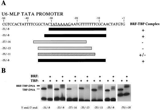
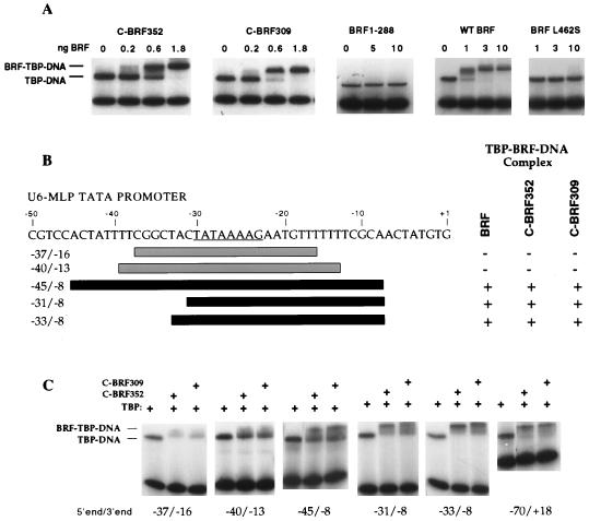
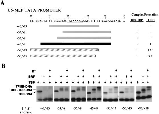
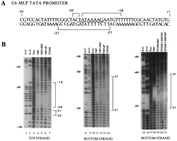
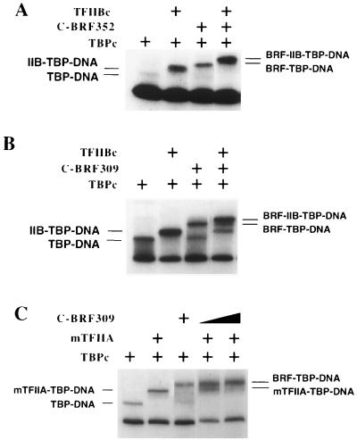
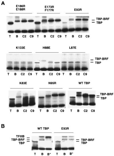
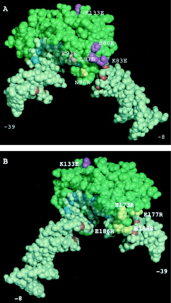
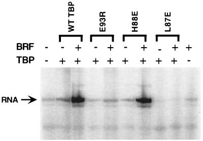
The RNA polymerase III factor TFIIIB forms a stable complex with DNA and can promote multiple rounds of initiation by polymerase. TFIIIB is composed of three subunits, the TATA binding protein (TBP), TFIIB-related factor (BRF), and B". Chemical footprinting, as well as mutagenesis of TBP, BRF, and promoter DNA, was used to probe the architecture of TFIIIB subunits bound to DNA. BRF bound to TBP-DNA through the nonconserved C-terminal region and required 15 bp downstream of the TATA box and as little as 1 bp upstream of the TATA box for stable complex formation. In contrast, formation of complete TFIIIB complexes required 15 bp both upstream and downstream of the TATA box. Hydroxyl radical footprinting of TFIIIB complexes and modeling the results to the TBP-DNA structure suggest that BRF and B" surround TBP on both faces of the TBP-DNA complex and provide an explanation for the exceptional stability of this complex. Competition for binding to TBP by BRF and either TFIIB or TFIIA suggests that BRF binds on the opposite face of the TBP-DNA complex from TFIIB and that the binding sites for TFIIA and BRF overlap. The positions of TBP mutations which are defective in binding BRF suggest that BRF binds to the top and N-terminal leg of TBP. One mutation on the N-terminal leg of TBP specifically affects the binding of the B" subunit.


Figures

FIG. 1
FIG. 1
BRF requires DNA downstream of the TATA box to form a stable BRF-TBP-DNA complex. (A) The sequence of the U6-MLP TATA promoter is shown, and the asymmetric MLP TATA box is underlined. Below the sequence, the DNA probes assayed and their abilities to bind BRF-TBP are summarized. Probes able to form a stable BRF-TBP-DNA complex are black, and probes unable to form stable complexes are gray; the hatched probe forms an unstable BRF-TBP complex. (B) Gel mobility shift assays with wild-type BRF. The probe, −70/+18, contains 40 bp of DNA both upstream and downstream of the TATA box. Reaction mixtures contained 1 ng of TBPc and 15 ng of recombinant BRF.
FIG. 2
FIG. 2
C-BRF derivatives require DNA downstream of the TATA box to form a stable complex with TBP-DNA. (A) Gel mobility shift assays using the yeast U6 promoter containing 75 bp upstream and downstream of the TATA box. The indicated amounts of C-BRF derivatives were added to reaction mixtures containing 2 ng of TBPc and the U6 promoter probe. (B) Summary of DNA probes assayed and their abilities to bind C-BRF derivatives. Probes able to form a stable BRF-TBP-DNA complex are black; probes unable to form stable complexes are gray. (C) Gel mobility shift assays with C-BRF derivatives. Reactions contain 1 ng of TBPc and either 2 ng of C-BRF352 or 4 ng of C-BRF309.
FIG. 3
FIG. 3
DNA upstream and downstream of the TATA box is required to form a stable TFIIIB-DNA complex. (A) Summary of DNA probes assayed and their abilities to bind BRF-TBP and recruit B" to form TFIIIB. Probes able to form a stable TFIIIB-DNA complex are black, and probes unable to form stable complexes are gray; the hatched probes form unstable TFIIIB complexes. (B) Gel mobility shift assays with wild-type BRF and B". Reaction mixtures contained 1 ng of TBPc, 15 ng of BRF, and 12 ng of B" as indicated.
FIG. 4
FIG. 4
Hydroxyl radical protection of TFIIIB-TBP-DNA complexes. (A) Summary of hydroxyl radical protection at U6-MLP TATA by TFIIIB. (B) Hydroxyl radical footprinting of the U6-MLP. The panel on the far right shows hydroxyl radical footprinting of the bottom strand of the U6-MLP, using either of the two C-BRF derivatives. TFIIIB indicates that TBPc, BRF, and B" were all added. The intensity of each base in each reaction was quantitated and normalized to the corresponding intensity in unbound DNA. Those bases that are reproducibly protected with the addition of TBPc, BRF, and B" are bracketed. Protection by TFIIIB was defined as a greater than 25% decrease in intensity compared with TBPc bound alone. Hydroxyl radical protection of the U6-MLP TATA nontranscribed strand with TFIIIB containing either C-BRF derivative shows similar protection to that of TFIIIB containing wild-type BRF (10a).
FIG. 5
FIG. 5
Competition assays between BRF and TFIIB and TFIIA. (A and B) Gel mobility shift assays using the U6-MLP promoter as a probe and 1 ng of TBPc with 6 ng of yeast TFIIBc (lacking residues 2 to 119) where indicated and either 2 ng of C-BRF352 or 4 ng of C-BRF309. (C) Reaction mixtures contained 1 ng of TBPc and 0.5 ng of mTFIIA where indicated and either 4 or 8 ng of C-BRF309.
FIG. 6
FIG. 6
Radical point mutations in TBP affect binding of BRF and B". (A) Gel mobility shift assay of each TBP mutant, either alone (T) or with wild-type BRF (B), C-BRF352 (C2), or C-BRF309 (C9). (B) TFIIIB complex formation of TBP mutant E93R and wild-type (WT) TBP. TBP was added alone (T), with BRF (B), or with BRF and 24 ng of B" (B").
FIG. 7
FIG. 7
Modeling of hydroxyl radical TFIIIB footprinting, DNA deletion analysis, and TBP mutagenesis to the TBP-DNA structure. (A) View of one face of the TBP-DNA complex where TFIIA binds near the N-terminal stirrup. (B) View of the opposite face of the TBP-DNA complex where TFIIB binds to the C-terminal stirrup. The conserved domain of yeast TBP is represented in green, the sugar residues of the TATA box that are protected from hydroxyl radical cleavage by TBP alone are shown in blue, and sugar residues outside of the TATA box protected from hydroxyl radical attack by TFIIIB are shown in red. TBP side chains which when mutated show decreased binding to BRF are shown in purple; TBP side chains which when mutated show no phenotype for BRF or B" binding are yellow. The pink mutation reduces binding of B" but not BRF.
FIG. 8
FIG. 8
Two TBP radical mutations fail to rescue Pol III transcription in vitro. A whole-cell extract from TBP mutant I143N was supplemented with 20 ng of BRF and 20 ng of either wild-type TBP or mutant TBP as indicated. Transcription is from the tRNA2Leu promoter.

References

    1. Bagby S, Kim S, Maldonado E, Tong K I, Reinberg D, Ikura M. Solution structure of the carboxy-terminal core domain of human TFIIB: similarity to cyclin A and interaction with TATA binding protein. Cell. 1995;82:857–867. - PubMed
    1. Baker R E, Camier S, Sentenac A, Hall B D. Gene size differentially affects the binding of yeast transcription factor tau to two intragenic regions. Proc Natl Acad Sci USA. 1987;84:8768–8772. - PMC - PubMed
    1. Barberis A, Muller C W, Harrison S C, Ptashne M. Delineation of two functional regions of transcription factor TFIIB. Proc Natl Acad Sci USA. 1993;90:5628–5632. - PMC - PubMed
    1. Bartholomew B, Durkovich D, Kassavetis G A, Geiduschek E P. Orientation and topography of RNA polymerase III in transcription complexes. Mol Cell Biol. 1993;13:942–952. - PMC - PubMed
    1. Bartholomew B, Kassavetis G A, Geiduschek E P. Two components of Saccharomyces cerevisiae transcription factor IIIB (TFIIIB) are stereospecifically located upstream of a tRNA gene and interact with the second-largest subunit of TFIIIC. Mol Cell Biol. 1991;11:5181–5189. - PMC - PubMed

Publication types

MeSH terms

LinkOut - more resources