. 1998 Mar;18(3):1725-35.
doi: 10.1128/MCB.18.3.1725.

Signaling through CD5 activates a pathway involving phosphatidylinositol 3-kinase, Vav, and Rac1 in human mature T lymphocytes

Affiliations

Signaling through CD5 activates a pathway involving phosphatidylinositol 3-kinase, Vav, and Rac1 in human mature T lymphocytes

S I Gringhuis et al. Mol Cell Biol. 1998 Mar.

Abstract

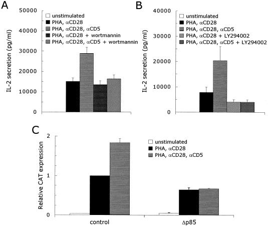
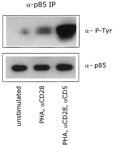
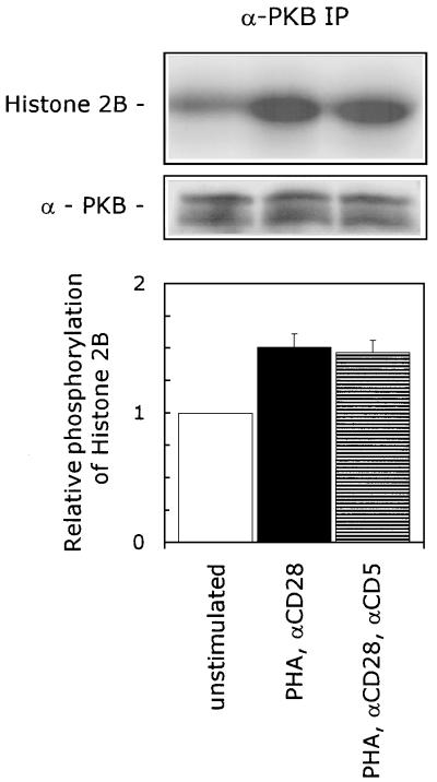
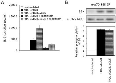
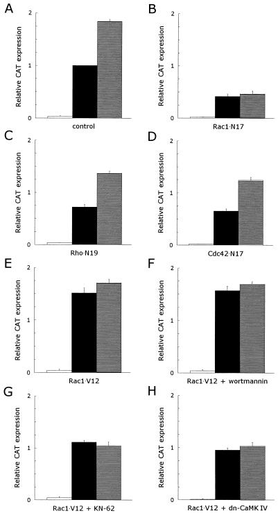
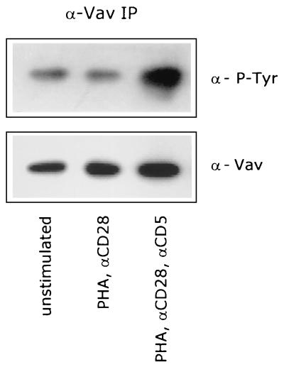
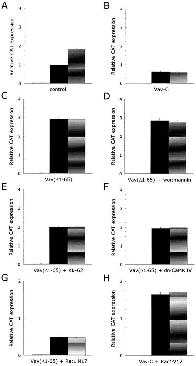
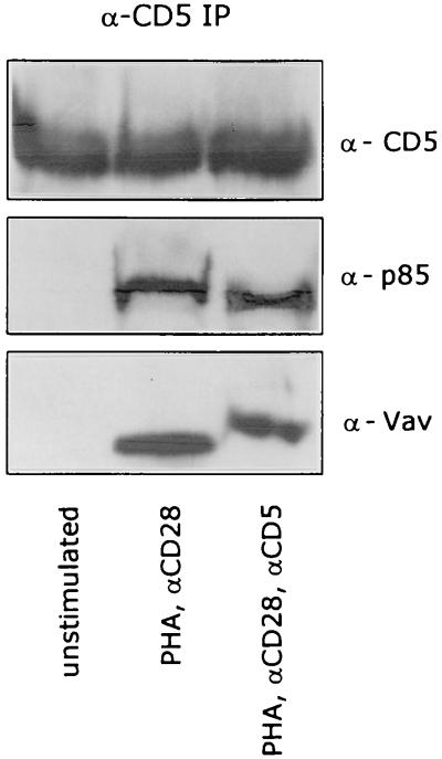
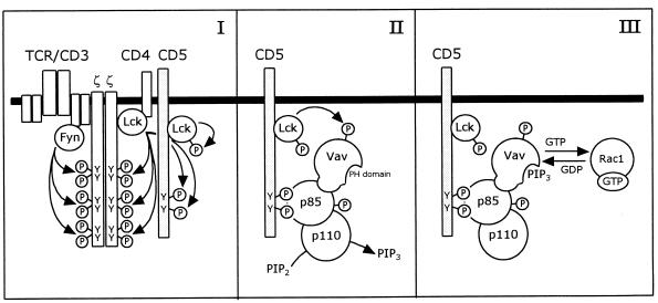
CD5 acts as a coreceptor on T lymphocytes and plays an important role in T-cell signaling and T-cell-B-cell interactions. Costimulation of T lymphocytes with anti-CD5 antibodies results in an increase of the intracellular Ca2+ levels, and subsequently in the activation of Ca2+/calmodulin-dependent (CaM) kinase type IV. In the present study, we have characterized the initial signaling pathway induced by anti-CD5 costimulation. The activation of phosphatidylinositol (PI) 3-kinase through tyrosine phosphorylation of its p85 subunit is a proximal event in the CD5-signaling pathway and leads to the activation of the lipid kinase activity of the p110 subunit. The PI 3-kinase inhibitors wortmannin and LY294002 inhibit the CD5-induced response as assessed in interleukin-2 (IL-2) secretion experiments. The expression of an inactivated Rac1 mutant (Rac1.N17) in T lymphocytes transfected with an IL-2 promoter-driven reporter construct also abrogates the response to CD5 costimulation, while the expression of a constitutively active Rac1 mutant (Rac1-V12) completely replaces the CD5 costimulatory signal. The Rac1-specific guanine nucleotide exchange factor Vav is heavily phosphorylated on tyrosine residues upon CD5 costimulation, which is a prerequisite for its activation. A role for Vav in the CD5-induced signaling pathway is further supported by the findings that the expression of a dominant negative Vav mutant (Vav-C) completely abolishes the response to CD5 costimulation while the expression of a constitutively active Vav mutant [Vav(delta1-65)] makes the CD5 costimulation signal superfluous. Wortmannin is unable to block the Vav(delta1-65)- or Rac1.V12-induced signals, indicating that both Vav and Rac1 function downstream from PI 3-kinase. Vav and Rac1 both act upstream from the CD5-induced activation of CaM kinase IV, since KN-62, an inhibitor of CaM kinases, and a dominant negative CaM kinase IV mutant block the Vav(delta1-65)-and Rac1.V12-mediated signals. We propose a model for the CD5-induced signaling pathway in which the PI 3-kinase lipid products, together with tyrosine phosphorylation, activate Vav, resulting in the activation of Rac1 by the Vav-mediated exchange of GDP for GTP.


Figures

FIG. 1
FIG. 1
Inhibition of the PI 3-kinase lipid kinase activity completely abrogates the CD5-induced upregulation of the IL-2 promoter activity in activated human T lymphocytes. (A and B) Human T lymphocytes were left unstimulated or stimulated with PHA plus anti-CD28 (αCD28) ± anti-CD5 (αCD5) in the presence or absence of 100 nM wortmannin (A) or 1 μM LY294002 (B), both inhibitors of PI 3-kinase. Cell-free supernatants were harvested after 24 h and analyzed for secreted IL-2 protein. The mean values ± SEMs for the IL-2 secretion observed in four independent experiments are shown. (C) Human T cells, prestimulated as described in Materials and Methods, were transfected with 15 μg of pCAT3e-IL-2(−319/+47) together with 15 μg of either an empty control expression plasmid (control) or the expression plasmid for a dominant negative p85 mutant (Δp85). Transfected cells were left alone for 1 h, divided into three groups, and subsequently left unstimulated or stimulated with PHA plus anti-CD28 (αCD28) or PHA plus anti-CD28 and anti-CD5 (αCD5) for 24 h. CAT expression was measured as described in Materials and Methods. The results are expressed as the relative CAT expression compared to the PHA- plus anti-CD28-induced CAT expression in the transfected control T cells, which was set at 1. The mean values ± SEMs found for the relative CAT expression in three independent experiments are shown.
FIG. 2
FIG. 2
CD5 costimulation enhances the tyrosine phosphorylation of the p85 subunit of PI 3-kinase. T cells were left unstimulated or stimulated with PHA plus anti-CD28 (αCD28) in the presence or absence of anti-CD5 (αCD5) for 5 min. p85 was immunoprecipitated from total-cell lysates, and tyrosine-phosphorylated p85 was detected with anti-PY20 MAb (α-P-Tyr) by ECL Western blotting as described in Materials and Methods. To ensure equal levels of immunoprecipitated p85, the blot was stripped and reprobed with anti-p85 PAb (α-p85). The data are representative of three independent experiments.
FIG. 3
FIG. 3
CD5 signaling is independent of PKB activation. T cells were left unstimulated or stimulated with PHA plus anti-CD28 (αCD28) in the presence or absence of anti-CD5 (αCD5) for 10 min. The cells were lysed, and immunoprecipitated PKB was assayed for kinase activity, with H2B as a substrate. To ensure the equal precipitation of PKB, the immunoprecipitates were loaded onto an SDS–12.5% polyacrylamide gel, and PKB protein was detected by ECL Western blotting as described in Materials and Methods. The kinase assay shown is representative of three independent experiments. The specific PKB kinase activity is determined by quantification of phosphorylated H2B with a PhosphorImaging system. The lower graph shows the relative phosphorylation of H2B. The phosphorylation of H2B detected in unstimulated cells was set at 1. The mean values ± SEMs found in three independent experiments are shown.
FIG. 4
FIG. 4
CD5 signaling is independent of p70 S6K activation. (A) T cells were stimulated with PHA with or without anti-CD28 (αCD28) and anti-CD5 (αCD5) in the presence or absence of 20 ng of rapamycin per ml. Cell-free supernatants were harvested after 24 h and analyzed for secreted IL-2 protein. The mean values ± SEM for the IL-2 secretion found in four independent experiments are shown. (B) T cells were left unstimulated or stimulated with PHA plus anti-CD28 (αCD28) in the presence or absence of anti-CD5 (αCD5) for 10 min. The cells were lysed, and immunoprecipitated p70 S6K was assayed for kinase activity with S6 peptide as a substrate. To ensure the equal precipitation of p70 S6K, the immunoprecipitates were loaded onto an SDS–12.5% polyacrylamide gel, and p70 S6K protein was detected by ECL Western blotting as described in Materials and Methods. The kinase assay shown is representative of two independent experiments. The specific p70 S6K kinase activity is determined by quantification of phosphorylated S6 with a PhosphorImaging system. The lower graph shows the relative phosphorylation of S6. The phosphorylation of S6 detected in unstimulated cells was set at 1. The mean values ± SEMs found in two independent experiments are shown.
FIG. 5
FIG. 5
Rac1 is essential to the CD5 costimulatory signal pathway leading to an upregulation of the IL-2 promoter activity, while Rho and Cdc42 play no role in this pathway. The CD5-induced signaling pathway in T lymphocytes expressing constitutively active Rac1 is insensitive to wortmannin but still sensitive to KN-62 or the expression of a dominant negative CaM kinase IV mutant. T cells, prestimulated as described in Materials and Methods, were transfected with 15 μg of pCAT3e-IL-2(−319/+47) together with 15 μg of either an empty control expression plasmid (control) (A) or the expression plasmid(s) for dominant negative or constitutively active mutants (B to H): dominant negative Rac1 (Rac1 · N17) (B), dominant negative Rho (Rho · N19) (C), dominant negative Cdc42 (Cdc42 · N17) (D), constitutively active Rac1 (Rac1 · V12) (E to G), and constitutively active Rac1 (Rac1 · V12) in combination with dominant negative CaM kinase IV (H). Transfected cells were left alone for 1 h, divided into three groups, and subsequently left unstimulated (□) or stimulated with PHA plus anti-CD28 (▪) or PHA plus anti-CD28 and anti-CD5 (▤) for 24 h in the presence of 100 nM wortmannin (F) or 10 μM KN-62 (G). CAT expression was measured as described in Materials and Methods. The results are expressed as the relative CAT expression compared to the PHA- plus anti-CD28-induced CAT expression in the transfected control T cells, which was set at 1. The mean values ± SEMs found for the relative CAT expression in three to six independent experiments are shown.
FIG. 6
FIG. 6
CD5 costimulation enhances the tyrosine phosphorylation of the Rac1-specific GEF factor Vav. T cells were left unstimulated or stimulated with PHA plus anti-CD28 (αCD28) in the presence or absence of anti-CD5 (αCD5) for 5 min. Vav was immunoprecipitated from total-cell lysates, and tyrosine-phosphorylated Vav was detected with anti-PY20 MAb (α-P-Tyr) by using ECL Western blotting as described in Materials and Methods. To ensure equal levels of immunoprecipitated Vav, the blot was stripped and reprobed with anti-Vav pAb (α-p85). The data are representative of three independent experiments.
FIG. 7
FIG. 7
Vav plays a major role in the CD5 signal pathway leading to an upregulation of the IL-2 promoter activity, as an upstream effector of Rac1. The signaling pathways in T lymphocytes expressing constitutively active Vav are insensitive to wortmannin but still sensitive to KN-62 or the expression of a dominant negative CaM kinase IV mutant. T cells, prestimulated as described in Materials and Methods, were transfected with 15 μg of pCAT3e-IL-2(−319/+47) together with 15 μg of either an empty control expression plasmid (control) (A) or the expression plasmid(s) for dominant negative or constitutively active mutants (B to H): dominant negative Vav (Vav-C) (B), constitutively active Vav [Vav(Δ1–65)] (C to E), constitutively active Vav [Vav(Δ1–65)] in combination with dominant negative CaM kinase IV (F), constitutively active Vav [Vav(Δ1–65)] in combination with dominant negative Rac1 (Rac1 · N17) (G), and constitutively active Vav [Vav(Δ1–65)] in combination with constitutively active Rac1 (Rac1 · V12) (H). Transfected cells were left alone for 1 h, divided into three groups, and subsequently left unstimulated (□) or stimulated with PHA plus anti-CD28 (▪) or PHA plus anti-CD28 and anti-CD5 (▤) for 24 h in the presence of 100 nM wortmannin (D) or 10 μM KN-62 (E). CAT expression was measured as described in Materials and Methods. The results are expressed as the relative CAT expression compared to the PHA- plus anti-CD28-induced CAT expression in the transfected control T cells, which was set at 1. The mean values ± SEMs found for the relative CAT expression in three independent experiments are shown.
FIG. 8
FIG. 8
The CD5 receptor is associated with p85 (PI 3-kinase) and Vav. T cells were left unstimulated or stimulated with PHA plus anti-CD28 (αCD28) in the presence or absence of anti-CD5 (αCD5) for 5 min. CD5 was immunoprecipitated from total-cell lysates, and CD5, p85, and Vav were detected by ECL Western blotting as described in Materials and Methods.
FIG. 9
FIG. 9
Model for the CD5-induced signaling pathway, which is mediated by PI 3-kinase and Vav, resulting in the activation of Rac1. (I) Upon engagement of the TCR by ligand, the tyrosine residues in the cytoplasmic domain of the CD5 receptor are phosphorylated by the protein tyrosine kinase p56lck, as are the tyrosine residues in the ζ chains of the TCR-CD3 complex. p56lck associates with the CD5 receptor and becomes fully activated through autophosphorylation. (II) The SH2 domains of the p85 subunit of PI 3-kinase bind to the phosphotyrosine residues of the CD5 receptor. Vav associates with the p85 subunit of PI 3-kinase, which serves to recruit Vav to the complex. Upon ligation of the CD5 receptor, PI 3-kinase is phosphorylated on tyrosine residues by a protein tyrosine kinase, most probably p56lck, which activates the lipid kinase activity of the p110 subunit. The nucleotide exchange activity of Vav is preactivated through the phosphorylation of tyrosine residues, probably by p56lck. (III) Upon binding of PI 3,4,5-P3 (PIP3) or another lipid product of PI 3-kinase to the PH domain of Vav, Vav becomes fully activated and will activate Rac1 through exchange of GDP for GTP.

References

    1. Alberola-Ila J, Places L, Cantrell D A, Vives J, Lozano F. Intracellular events involved in CD5-induced human T cell activation and proliferation. J Immunol. 1992;148:1287–1293. - PubMed
    1. al-Shami A, Bourgoin S G, Naccache P H. Granulocyte-macrophage colony-stimulating factor-activated signaling pathways in human neutrophils. I. Tyrosine phosphorylation-dependent stimulation of phosphatidylinositol 3-kinase and inhibition by phorbol esters. Blood. 1997;89:1035–1044. - PubMed
    1. Beyers A D, Spruyt L L, Williams A F. Molecular associations between the T-lymphocyte antigen receptor complex and the surface antigens CD2, CD4, or CD8, and CD5. Proc Natl Acad Sci USA. 1992;89:2945–2949. - PMC - PubMed
    1. Biancone L, Bowen M A, Lim A, Aruffo A, Andres G, Stamenkovic L. Identification of a novel inducible cell-surface ligand of CD5 on activated lymphocytes. J Exp Med. 1996;184:811–819. - PMC - PubMed
    1. Bradford M M. A rapid and sensitive method for the quantitation of microgram quantities of protein utilizing the principle of protein-dye binding. Anal Biochem. 1976;72:248–254. - PubMed

MeSH terms