. 1998 Mar;18(3):1746-56.
doi: 10.1128/MCB.18.3.1746.

Novel mechanisms of E2F induction by BK virus large-T antigen: requirement of both the pRb-binding and the J domains

Affiliations

Novel mechanisms of E2F induction by BK virus large-T antigen: requirement of both the pRb-binding and the J domains

K F Harris et al. Mol Cell Biol. 1998 Mar.

Abstract

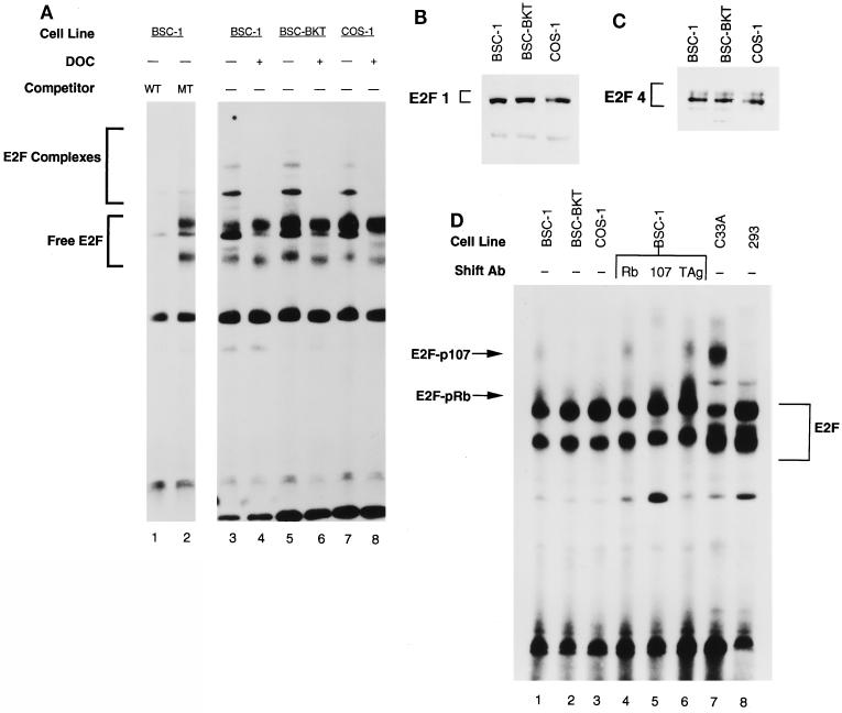
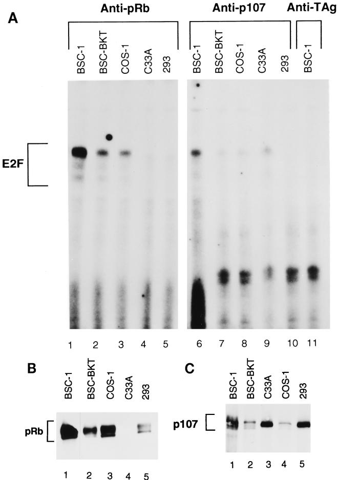
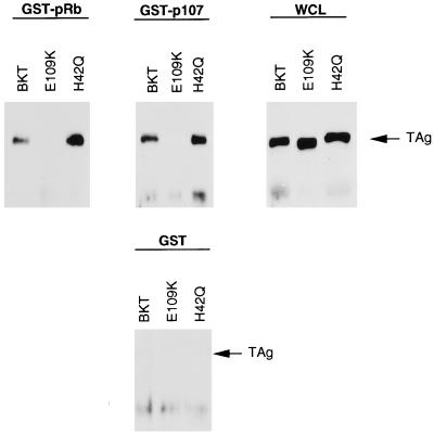
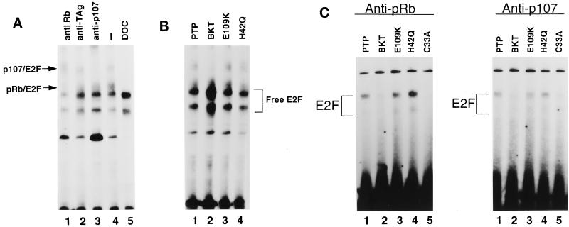
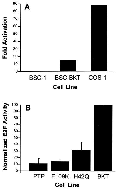
E2F activity is regulated in part by the retinoblastoma family of tumor suppressor proteins. Viral oncoproteins, such as simian virus 40 (SV40) large-T antigen (TAg), adenovirus E1A, and human papillomavirus E7, can disrupt the regulation of cellular proliferation by binding to pRb family members and dissociating E2F-pRb family protein complexes. BK virus (BKV), which infects a large percentage of the human population and has been associated with a variety of human tumors, encodes a TAg homologous to SV40 TAg. It has been shown that BKV TAg, when expressed at low levels, does not detectably bind to pRb family members, yet it induces a serum-independent phenotype and causes a decrease in the overall levels of pRb family proteins. The experiments presented in this report show that, despite the lack of TAg-pRb interactions, BKV TAg can induce transcriptionally active E2F and that this induction does in fact require an intact pRb-binding domain as well as an intact J domain. In addition, E2F-pRb family member complexes can be detected in both BKV and SV40 TAg-expressing cells. These results suggest the presence of alternate cellular mechanisms for the release of E2F in addition to the well-established model for TAg-pRb interactions. These results also emphasize a role for BKV TAg in the deregulation of cellular proliferation, which may ultimately contribute to neoplasia.


Figures

FIG. 1
FIG. 1
Increased free E2F in the presence of BKV TAg. (A) Whole-cell extracts were prepared from subconfluent BSC-1, BSC-BKT, and COS-1 cells. Extracts were incubated with a labeled E2F probe for 30 min at room temperature and resolved by native 5% PAGE. Competition reactions were performed in the presence of a 150-fold excess of cold wild-type (WT) competitor (lane 1) or mutant (MT) competitor (lane 2). For lanes 4, 6, and 8, extracts were preincubated with 0.8% deoxycholate (DOC) for 15 min on ice, followed by 1.2% NP-40 for 15 min on ice, and then used for DNA binding reactions. Free E2F, E2F bound to the DNA probe. E2F Complexes, E2F-pRb family member complexes bound to the DNA probe. (B and C) Whole-cell extracts (25 μg) were separated by SDS–10% PAGE, transferred to nitrocellulose, and probed with KH95 (anti-E2F1; B) or C-20 (anti-E2F4; C). (D) Whole-cell extracts were prepared, and equivalent amounts of protein from each cell line were used in a DNA binding reaction as described for panel A. For supershift experiments (lanes 4 to 6), extracts from BSC-1 cells were preincubated for 15 min at room temperature with C-15 (anti-pRb), a mixture of SD6, SD9, and SD15 (anti-p107), or PAb430 (anti-TAg) as a negative control before the addition of the DNA probe. Ab, antibody.
FIG. 2
FIG. 2
Presence of E2F-pRb and E2F-p107 complexes in TAg-expressing cells. (A) Total protein (50 μg) from whole-cell extracts was immunoprecipitated with C-15 (anti-pRb; lanes 1 to 5), C-18 (anti-p107; lanes 6 to 10), or PAb430 (anti-TAg; lane 11). Immunoprecipitates were treated with deoxycholate to disrupt protein-protein complexes, and 4 μl of supernatant was assayed for DNA binding activity under the same conditions as those described in the legend to Fig. 1. (B and C) Western blot analyses of immunoprecipitates used in panel A. Supernatant (12 μl) was separated by SDS–8% PAGE, transferred to nitrocellulose, and probed with C-15 (B) or C-18 (C). Note that the order of COS-1 and C33A cells is reversed in the two blots.
FIG. 3
FIG. 3
In vitro complex formation between mutant BKV TAgs and proteins pRb and p107. Whole cell-lysate (150 μg) from BSC-tet cells transiently transfected with pBKT-tet, pE109K-tet, and pH42Q-tet were incubated with equivalent amounts of each purified GST-Sepharose-bound fusion protein. Bound complexes were released and separated by SDS-PAGE, transferred to nitrocellulose, and probed with PAb416. For whole-cell lysate samples (WCL), 50 μg of lysate from each cell line was used.
FIG. 4
FIG. 4
E2F complexes in cell lines expressing mutant TAgs. (A) Supershift experiments were performed on PTP cells as described in the legend to Fig. 1D. For lane 5, PTP cell extract was preincubated with deoxycholate (DOC) and NP-40 as described in the legend to Fig. 1A. (B) Whole-cell extracts were prepared, and equivalent amounts of protein were used in DNA binding reactions as described in the legend to Fig. 1A. (C) Immunoprecipitations followed by deoxycholate reactions were performed as described in the legend to Fig. 2A, except that for anti-p107 immunoprecipitates, a cocktail of monoclonal antibodies SD6, SD9, and SD15 was used.
FIG. 5
FIG. 5
Increased transcriptionally active E2F in the presence of BKV TAg requires both the pRb-binding and the J domains. (A) Cells were transfected with 3 μg of pE2F-CAT or 3 μg of pΔE2F-CAT and 1 μg of pAdCMVβ. CAT assays were performed 48 h after transfection. Signals from the autoradiograms were quantitated with AMBIS Image Acquisition and Analysis software and normalized to the β-galactosidase activity for each extract. Fold activation, CAT activity from the pE2F-CAT construct divided by CAT activity from the pΔE2F-CAT construct. Data represent the results from three independent experiments. (B) CAT assays were performed as described in panel A. The fold activation of E2F in wild-type BKT cells after normalization to the background is 14.95. This level was set to 100%, and the activation from the mutant TAg-expressing cell lines is represented as a percentage of wild-type BKV TAg activity. Numbers represent averages from four separate experiments.
FIG. 6
FIG. 6
The J domain but not the pRb-binding domain of BKV TAg is required for the induction of serum-independent growth. For each cell line, 2 × 10⁴ cells were plated in medium containing 0.1% serum. Cells from duplicate wells were counted every other day. Values shown are averages from three independent experiments. Symbols: ○, BKT; ▪, E109K; •, PTP; ▴, H42Q.

References

    1. Arthur R R, Shah K V, Baust S J, Santos G W, Saral R. Association of BK viruria with hemorrhagic cystitis in recipients of bone marrow transplants. N Engl J Med. 1986;315:230–234. - PubMed
    1. Bagchi S, Weinmann R, Raychaudhuri P. The retinoblastoma protein copurifies with E2F-I, an E1A-regulated inhibitor of the transcription factor E2F. Cell. 1991;65:1063–1072. - PubMed
    1. Bandara L R, Adamczewski J P, Hunt T, La Thangue N B. Cyclin A and the retinoblastoma gene product complex with a common transcription factor. Nature. 1991;352:249–251. - PubMed
    1. Barbosa M S, Edmonds C, Fisher C, Schiller J T, Lowy D R, Vousden K H. The region of the HPV E7 oncoprotein homologous to adenovirus E1a and SV40 large T antigen contains separate domains for Rb binding and casein kinase II phosphorylation. EMBO J. 1990;9:153–160. - PMC - PubMed
    1. Beijersbergen R L, Carlee L, Kerkhoven R M, Bernards R. Regulation of the retinoblastoma protein-related p107 by G1 cyclin complexes. Genes Dev. 1995;9:1340–1353. - PubMed

Publication types

MeSH terms

LinkOut - more resources