. 1998 Feb;116(2):797-803.
doi: 10.1104/pp.116.2.797.

Plastid ontogeny during petal development in Arabidopsis

Affiliations

Plastid ontogeny during petal development in Arabidopsis

K A Pyke et al. Plant Physiol. 1998 Feb.

Abstract

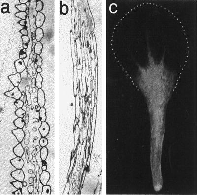
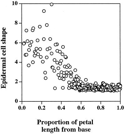
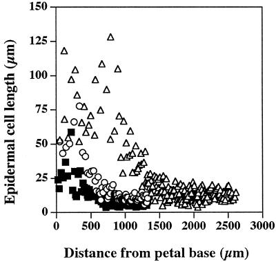
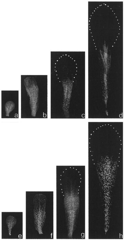
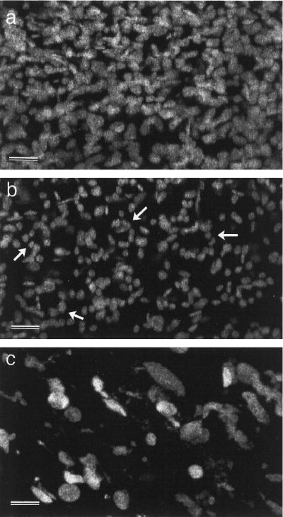
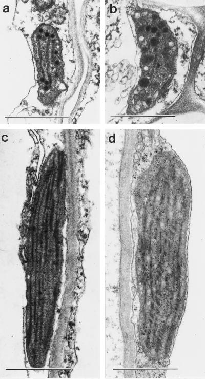
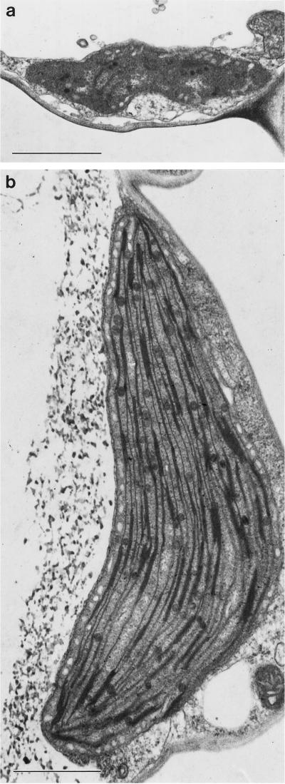
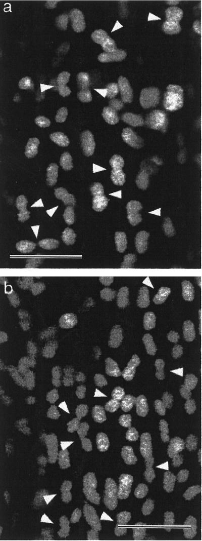
Imaging of chlorophyll autofluorescence by confocal microscopy in intact whole petals of Arabidopsis thaliana has been used to analyze chloroplast development and redifferentiation during petal development. Young petals dissected from unopened buds contained green chloroplasts throughout their structure, but as the upper part of the petal lamina developed and expanded, plastids lost their chlorophyll and redifferentiated into leukoplasts, resulting in a white petal blade. Normal green chloroplasts remained in the stalk of the mature petal. In epidermal cells the chloroplasts were normal and green, in stark contrast with leaf epidermal cell plastids. In addition, the majority of these chloroplasts had dumbbell shapes, typical of dividing chloroplasts, and we suggest that the rapid expansion of petal epidermal cells may be a trigger for the initiation of chloroplast division. In petals of the Arabidopsis plastid division mutant arc6, the conversion of chloroplasts into leukoplasts was unaffected in spite of the greatly enlarged size and reduced number of arc6 chloroplasts in cells in the petal base, resulting in few enlarged leukoplasts in cells from the white lamina of arc6 petals.


Figures

Figure 1
Figure 1
Structure of mature Arabidopsis petals. a, Transverse section through the upper white petal lamina. b, Transverse section through the stalk at the basal part of the petal. c, Entire intact petal viewed by conventional fluorescence microscopy showing chlorophyll autofluorescence in the petal stalk and the lack of fluorescence in the upper white petal lamina. The extent of the white petal lamina is shown by the dotted line.
Figure 2
Figure 2
The relationship between the shape of petal epidermal cells viewed in transverse section and the relative position of the epidermal cell along the length of mature Arabidopsis petals. Data were collected from three different petals and pooled, since variability between individual petals was very low. Values of cell shape were maximum. Feret diameter/minimum Feret diameter, see Methods.
Figure 3
Figure 3
The relationship between the length of epidermal cells viewed in transverse section and their distance from the petal base for petals at different developmental stages: ▪, stage 10; ○, stage 12; and ▵, mature.
Figure 4
Figure 4
Developing Arabidopsis petals of the wild type (a–d) and the arc6 mutant (e–h) viewed by confocal microscopy. Red chlorophyll autofluorescence is shown as gray. The outline of the white lamina lacking fluorescing plastids is outlined by dotted lines. a and e, Stage 9; b and f, stage 10; c and g, stage 12; and d and h, mature.
Figure 5
Figure 5
Confocal images of fluorescing chloroplasts from the basal part of mature Arabidopsis petals. a, Wild-type mesophyll parenchyma chloroplasts. b, Wild-type epidermal chloroplasts. c, arc6 chloroplasts from both epidermal and mesophyll cells. Arrows in b indicate circular arrays of chloroplasts. Bars = 5 μm.
Figure 6
Figure 6
Electron micrographs of leukoplasts from the upper petal lamina (a and b) and chloroplasts from the green stalk of mature wild-type Arabidopsis petals (c and d) in mesophyll parenchyma cells (a and c) and epidermal cells (b and d). Bars = 1 μm.
Figure 7
Figure 7
Electron micrographs of a leukoplast from the upper white petal lamina (a) and chloroplasts from the green stalk of a mature Arabidopsis petal (b) of the mutant arc6. Bars = 1 μm, the same magnification as in Figure 6.
Figure 8
Figure 8
Confocal images of fluorescent chloroplasts from the base of different mature wild-type petals (a and b) showing the large proportion of the chloroplast population with dumbbell phenotypes typical of chloroplasts in division (arrowheads). Bars = 5 μm.

Similar articles

Cited by

References

    1. Brett DW, Sommerard AP. Ultrastructural development of plastids in the epidermis and starch layer of glossy Ranunculus petals. Ann Bot. 1986;58:903–910.
    1. Dupree P, Pwee K-H, Gray JC. Expression of photosynthetic gene-promoter fusions in leaf epidermal cells of transgenic tobacco plants. Plant J. 1991;1:115–120.
    1. Falk H. Chromoplasts of Tropaeolum majus L.: structure and development. Planta. 1976;128:15–22. - PubMed
    1. Kirk JTO, Tilney-Bassett RAE (1978) The Plastids. Elsevier/North Holland Biomedical Press, Amsterdam, The Netherlands
    1. Lawrence SD, Cline K, Moore GA. Chromoplast development in ripening tomato fruit: identification of cDNAs for chromoplast-targeted proteins and characterisation of a cDNA encoding a plastid-localised low-molecular weight heat shock protein. Plant Mol Biol. 1997;33:483–492. - PubMed

Publication types