. 1998 Mar 9;140(5):991-1002.
doi: 10.1083/jcb.140.5.991.

A dominant mutant of inner centromere protein (INCENP), a chromosomal protein, disrupts prometaphase congression and cytokinesis

Affiliations

A dominant mutant of inner centromere protein (INCENP), a chromosomal protein, disrupts prometaphase congression and cytokinesis

A M Mackay et al. J Cell Biol. .

Abstract

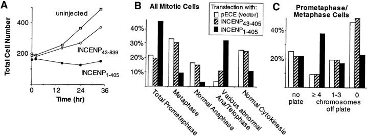
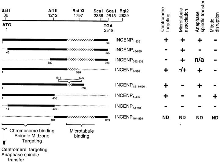
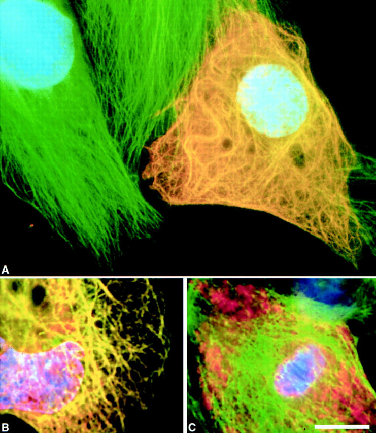
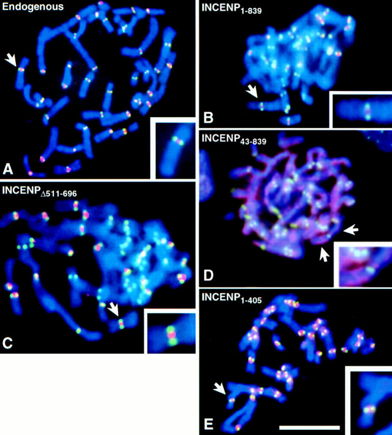
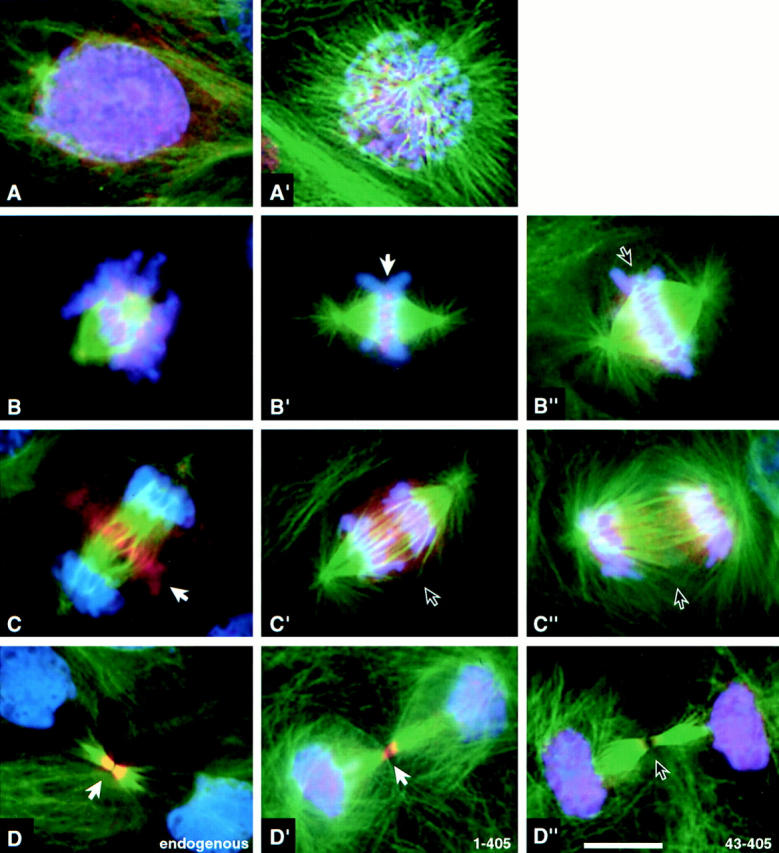
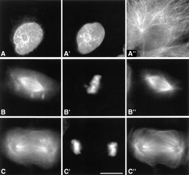
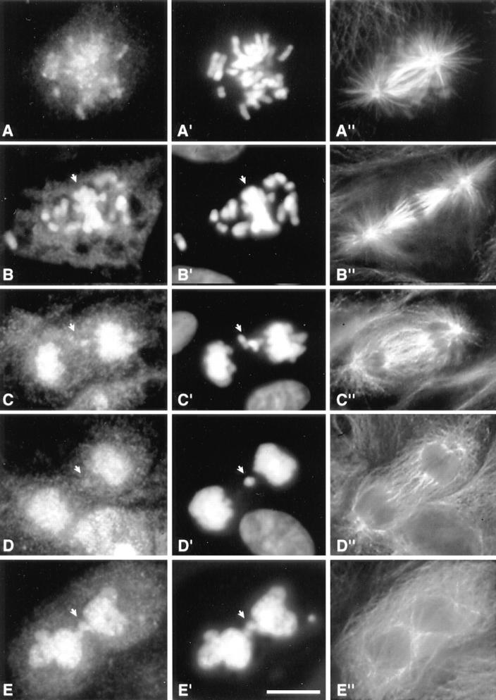
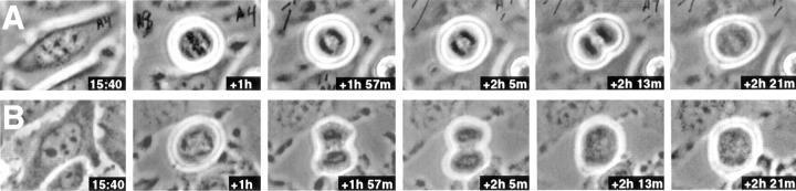
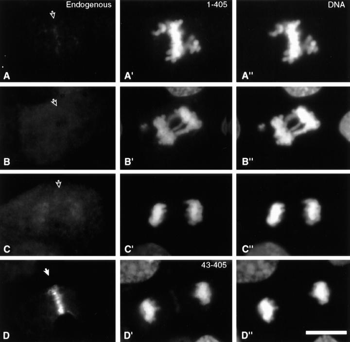
INCENP is a tightly bound chromosomal protein that transfers to the spindle midzone at the metaphase/anaphase transition. Here, we show that an INCENP truncation mutant (INCENP382-839) associates with microtubules but does not bind to chromosomes, and coats the entire spindle throughout mitosis. Furthermore, an INCENP truncation mutant (INCENP43-839) previously shown not to transfer to the spindle at anaphase (Mackay, A.M., D.M. Eckley, C. Chue, and W.C. Earnshaw. 1993. J. Cell Biol. 123:373-385), is shown here to bind chromosomes, but is unable to target to the centromere. Thus, association with the chromosomes, and specifically with centromeres, appears to be essential for INCENP targeting to the correct spindle subdomain at anaphase. An INCENP truncation mutant (INCENP1-405) that targets to centromeres but lacks the microtubule association region acquires strong dominant-negative characteristics. INCENP1-405 interferes with both prometaphase chromosome alignment and the completion of cytokinesis. INCENP1-405 apparently exerts its effect by displacing the endogenous protein from centromeres. These experiments provide evidence of an unexpected link between this chromosomal protein and cytokinesis, and suggest that one function of INCENP may be to integrate the chromosomal and cytoskeletal events of mitosis.


Figures

Figure 6
Figure 6
Phenotypic analysis of dominant-negative INCENP mutant in mammalian cells. (A) INCENP1–405 inhibits colony growth. Colonies of 16–32 HeLa cells were microinjected with plasmid constructs encoding either INCENP1–405 or INCENP43–839 at time t = 0 (abscissa). The aggregate number of cells in all injected colonies is summed on the ordinate. In this experiment, 52% of the cells in colonies injected with INCENP1–405 were binucleate. This contrasts with 8% binucleate cells in colonies injected with INCENP43–839 and 4% binucleate cells in uninjected colonies. (B) Expression of INCENP1–405 causes the accumulation of cells in prometaphase and in ana/ telophase. Slides were prepared from cultures transfected either with the pECE vector alone (158 cells scored), or the vector encoding either INCENP1–405 (129 cells scored) or INCENP43–405 (75 cells scored). For each transfected construct, mitotic cells were scored blind for their morphology in mitosis. For the latter two transfections, only cells expressing the transfected INCENP were included in the scoring. (C) For a more detailed analysis of prometaphase congression, a separate series of transfections was performed. Slides were prepared from cultures transfected either with the pECE vector alone (87 cells scored), or the pECE vector encoding either INCENP1–405 (100 cells scored) or INCENP43–405 (96 cells scored). As in B, transfected cells were scored blind for their morphology in mitosis.
Figure 1
Figure 1
INCENP open reading frame and deletion mutants used in this study. (Top) Map of the INCENP cDNA showing the position of important restriction sites. Below this are the various INCENP constructs used in this study. The region of putative coiled-coil (Berger et al., 1995) is stippled. (Bottom) Known functional regions required for chromosome binding and centromere targeting (this study) and for transfer to the spindle and microtubule binding (Mackay et al., 1993). The results of expression of each species in cultured cells are summarized on the right. Because our molecular cloning analysis (Mackay et al., 1993) revealed that the two INCENP polypeptides are likely to be the alternatively spliced products of a single gene, we now refer to this gene and both of its products as INCENP.
Figure 2
Figure 2
Deletion of the carboxy-terminal region of INCENP abolishes the ability of the protein to bind interphase microtubules. In these images, INCENP is red, microtubules are green, and DNA is blue. (A) Bundling of cytoplasmic microtubules in a jackpot cell expressing wild-type INCENP1–839. Neighboring nonexpressing cells have normal microtubule arrays. (B) INCENP43–839 associates with microtubules in a jackpot cell. (C) Failure of INCENP1–405 to associate with and bundle microtubules in a jackpot cell. Bar, 10 μm.
Figure 3
Figure 3
Deletion of residues 1–42 abolishes the ability of INCENP to concentrate in the inner centromere. LLCPK chromosomes were stained for INCENP (red), kinetochores (green), and DNA (blue). (A) Endogenous (porcine) INCENP is concentrated in the inner centromere. (B, C and E) Transfected full-length INCENP1–839, INCENPΔ511–696, and INCENP1–405 are all concentrated in the inner centromere. (D) Transfected INCENP43–839 fails to concentrate at centromeres, but instead paints the entire chromosome. Bar, 10 μm.
Figure 4
Figure 4
Distribution of endogenous INCENP, INCENP1–405 and INCENP43–405 in LLCPK cells undergoing normal mitosis. Control cells (A–D) or cells transfected with either INCENP1–405 (A′–D′) or INCENP43–405 (B′′–D′′) were stained for INCENP (red), microtubules (green) and DNA (blue). (A and A′) Interphase through prophase: endogenous INCENPs are nuclear. (B, B′, and B′′) Prometaphase through metaphase: both endogenous INCENPs and INCENP1–405 become highly concentrated at centromeres (B′ arrow). INCENP43–405 is distributed all along the chromosomes with no enrichment at centromeres (arrow). (C, C′, and C′′) Anaphase: endogenous INCENP transfers to the central spindle and cell cortex in the cleavage furrow. INCENP1–405 and INCENP43–405 remain on the separated sister chromatids. (D, D′, and D′′) Cytokinesis: endogenous INCENP labels the intercellular bridge intensely; nuclei are unlabeled. INCENP1–405 is found in both daughter nuclei and the intercellular bridge. INCENP43–405 is found in daughter nuclei and is absent from the intercellular bridge. Although quantitation of expression levels in transient transfections is problematic, it is our impression that cells expressing low levels of INCENP1–405 traverse mitosis normally, whereas cells expressing higher levels of the mutant protein frequently exhibit disruptions of mitosis. Bar, 10 μm.
Figure 5
Figure 5
Deletion of residues 1–381 abolishes the ability of INCENP to target to the spindle midzone during mitosis. LLCPK cells expressing INCENP382–839 (A–C) were stained for DNA (A′–C′) and tubulin (A′′–C′′). (A–A′′) INCENP382–839 is concentrated in nuclei during interphase. (B–B′′) INCENP382–839 fails to bind chromosomes during metaphase, instead uniformly coating the spindle microtubules. Several INCENP aggregates are also seen scattered in the cytoplasm. (C–C′′) INCENP382–839 coats the spindle microtubules at anaphase and does not target to the spindle midzone. Bar, 10 μm.
Figure 7
Figure 7
Phenotypes of cells undergoing aberrant mitosis induced by expression of INCENP1–405. (A–E) Transfected INCENP. (A′–E′) DNA stained with DAPI. (A′′–E′′) Microtubules. (A, A′, and A′′) Normal early prometaphase. (B, B′, and B′′) Prometaphase with many chromosomes that have failed to localize to the metaphase plate (arrow). (C, C′, and C′′) Anaphase with lagging chromosomes but a relatively normal central spindle. (D, D′, and D′′) Postmitotic cell with an unusual microtubule array that has characteristics of both anaphase and interphase. Prominent microtubule bundles are absent and the array appears to spread across the entire width of the cell. (E, E′, and E′′) Failed cytokinesis with an interphase microtubule array.
Figure 8
Figure 8
Examples of failures in cytokinesis in cells expressing INCENP1–405. (A and B) Examples of two HeLa cells injected with a plasmid encoding INCENP1–405 at time t = 0. Observation began 15 h, 40 min after microinjection. Both cells show indications of furrowing that ultimately reverse.
Figure 9
Figure 9
Expression of INCENP1–405, but not INCENP43–405, displaces the endogenous INCENP from its normal location. Endogenous INCENP detected using a polyclonal antibody specific for INCENP residues 404–839 is diffusely distributed in transfected cells expressing INCENP1–405 either during prometaphase (A, A′, and A′′), anaphase (B, B′, and B′′) or telophase (C, C′, and C′′). In contrast, the distribution of endogenous INCENP is normal in cells expressing INCENP43–405 (D, D′, and D′′). Staining for the endogenous protein is seen in A–D, with the expected location of INCENP indicated by an arrow in each panel. The expressed truncated INCENPs are seen in A′–D′. A′′–D′′ show the DAPI staining for DNA. Similar results were obtained with cells at all stages of the cell cycle. Bar, 10 μm.

Similar articles

Cited by

References

    1. Berger B, Wilson DB, Wolf E, Tonchev T, Milla M, Kim PS. Predicting coiled coils by use of pairwise residue correlations. Proc Natl Acad Sci USA. 1995;92:8259–8263. - PMC - PubMed
    1. Bernat RL, Borisy GG, Rothfield NF, Earnshaw WC. Injection of anticentromere antibodies in interphase disrupts events required for chromosome movement at mitosis. J Cell Biol. 1990;111:1519–1533. - PMC - PubMed
    1. Bernat RL, Delannoy MR, Rothfield NF, Earnshaw WC. Disruption of centromere assembly during interphase inhibits kinetochore morphogenesis and function in mitosis. Cell. 1991;66:1229–1238. - PubMed
    1. Buck RC, Tisdale JM. The fine structure of the mid-body of the rat erythroblast. J Cell Biol. 1962;13:109–115. - PMC - PubMed
    1. Caceres A, Binder LI, Payne MR, Bender P, Rebhun LI, Steward O. Differential subcellular localization of tubulin and the microtubule- associated protein MAP-2 in brain tissue as revealed by immunocytochemistry with monoclonal hybridoma antibodies. J Neurosci. 1983;4:394–410. - PMC - PubMed

Publication types

Substances