. 1998 Mar 9;140(5):1055-62.
doi: 10.1083/jcb.140.5.1055.

AP-2/Eps15 interaction is required for receptor-mediated endocytosis

Affiliations

AP-2/Eps15 interaction is required for receptor-mediated endocytosis

A Benmerah et al. J Cell Biol. .

Abstract

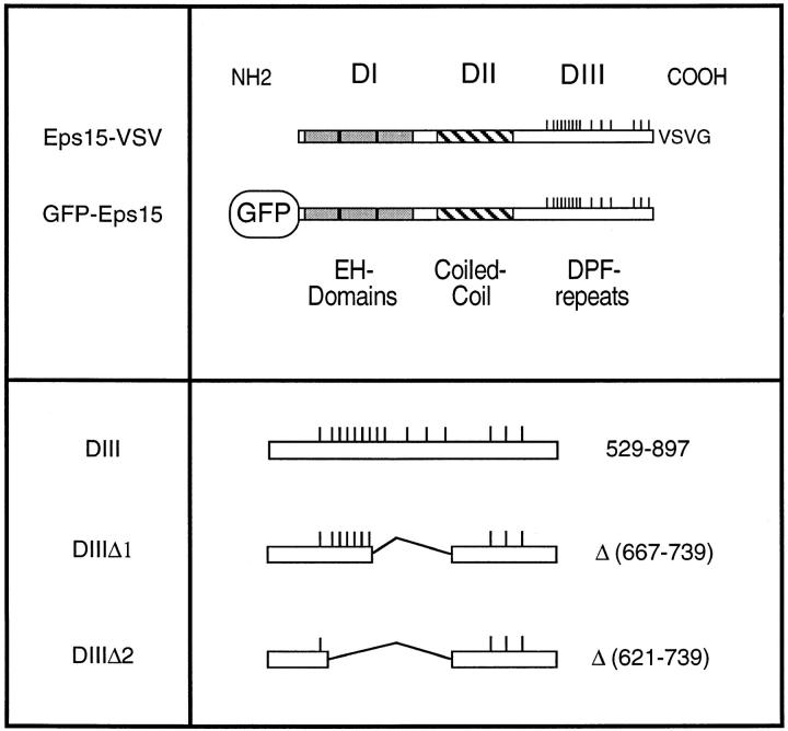
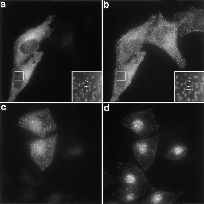
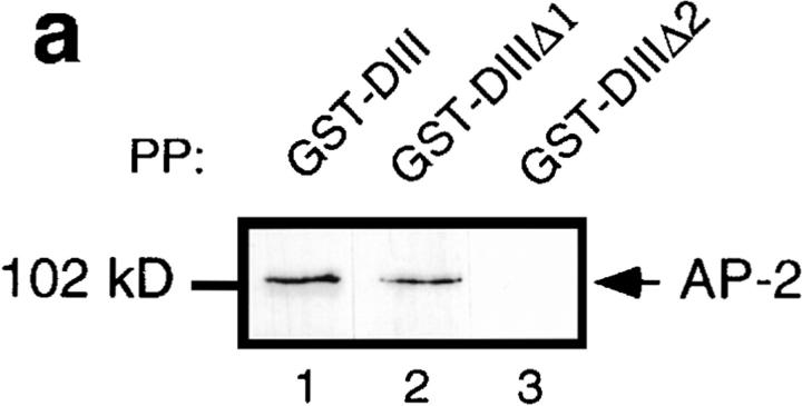
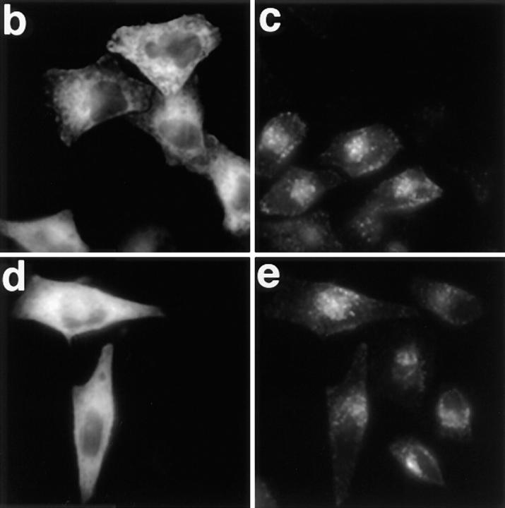
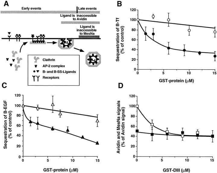
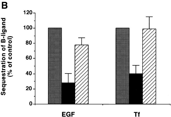
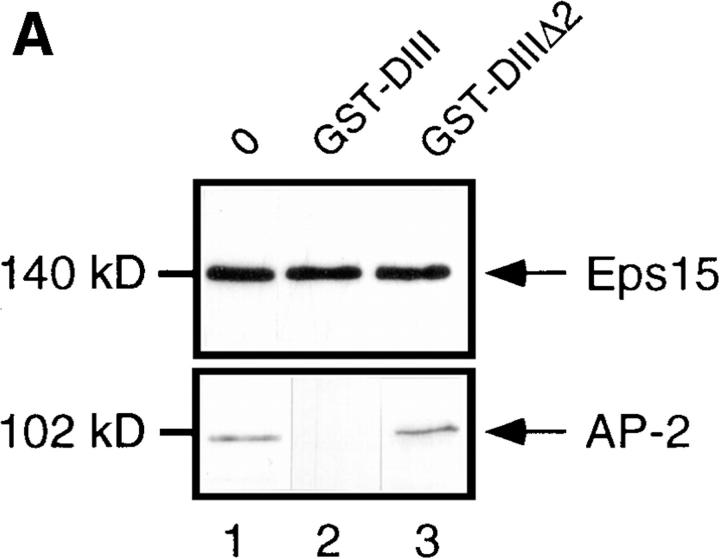
We have previously shown that the protein Eps15 is constitutively associated with the plasma membrane adaptor complex, AP-2, suggesting its possible role in endocytosis. To explore the role of Eps15 and the function of AP-2/Eps15 association in endocytosis, the Eps15 binding domain for AP-2 was precisely delineated. The entire COOH-terminal domain of Eps15 or a mutant form lacking all the AP-2-binding sites was fused to the green fluorescent protein (GFP), and these constructs were transiently transfected in HeLa cells. Overexpression of the fusion protein containing the entire COOH-terminal domain of Eps15 strongly inhibited endocytosis of transferrin, whereas the fusion protein in which the AP-2-binding sites had been deleted had no effect. These results were confirmed in a cell-free assay that uses perforated A431 cells to follow the first steps of coated vesicle formation at the plasma membrane. Addition of Eps15-derived glutathione-S-transferase fusion proteins containing the AP-2-binding site in this assay inhibited not only constitutive endocytosis of transferrin but also ligand-induced endocytosis of epidermal growth factor. This inhibition could be ascribed to a competition between the fusion protein and endogenous Eps15 for AP-2 binding. Altogether, these results show that interaction of Eps15 with AP-2 is required for efficient receptor-mediated endocytosis and thus provide the first evidence that Eps15 is involved in the function of plasma membrane-coated pits.


Figures

Figure 1
Figure 1
Structural organization of Eps15 and Eps15 constructs.
Figure 2
Figure 2
Subcellular distribution of transfected GFP-Eps15 and effect on Tf endocytosis. (a and b) HeLa cells grown on coverslips were transiently transfected with GFP-Eps15, fixed, and then stained for AP-2 using the AP-6 antibody. The GFP-Eps15 fusion protein was visualized directly by the green fluorescence emitted by the GFP (a). AP-6 antibody was detected with a secondary anti-IgG2b antibody labeled with Texas red (b). The inserts show a higher magnification of the same area. Arrows point to representative spots stained in both green and red (a and b). (c and d) HeLa cells transiently transfected with GFP-Eps15 were incubated in the presence of 100 nM Texas red–conjugated Tf for 15 min at 37°C, washed twice in cold PBS, and then fixed. (c) Green fluorescence emitted by the GFP construct and (d) Texas red–conjugated Tf. a and b, and c and d represent the same fields.
Figure 3
Figure 3
Overexpression of the Eps15-binding site for AP-2 inhibits Tf endocytosis in vivo. (a) Different GST fusion proteins encoding either the COOH-terminal domain of Eps15 or deleted forms of this domain (Fig. 1) were used to precipitate cell lysates (PP). The presence of AP-2 in the precipitates was revealed by Western blotting using the anti–α-adaptin antibody 100/2. HeLa cells were transiently transfected with the GFP-DIII construct (b and c) or with the GFP-DIIIΔ2 construct lacking all the AP-2–binding sites (d and e). Transfected cells were incubated in the presence of 100 nM Texas red–conjugated Tf for 15 min at 37°C, and washed twice with cold PBS. Left panels represent the green fluorescence emitted by the GFP constructs, and the right panels represent the staining observed for Texas red–conjugated Tf. b and c, and d and e represent the same fields.
Figure 3
Figure 3
Overexpression of the Eps15-binding site for AP-2 inhibits Tf endocytosis in vivo. (a) Different GST fusion proteins encoding either the COOH-terminal domain of Eps15 or deleted forms of this domain (Fig. 1) were used to precipitate cell lysates (PP). The presence of AP-2 in the precipitates was revealed by Western blotting using the anti–α-adaptin antibody 100/2. HeLa cells were transiently transfected with the GFP-DIII construct (b and c) or with the GFP-DIIIΔ2 construct lacking all the AP-2–binding sites (d and e). Transfected cells were incubated in the presence of 100 nM Texas red–conjugated Tf for 15 min at 37°C, and washed twice with cold PBS. Left panels represent the green fluorescence emitted by the GFP constructs, and the right panels represent the staining observed for Texas red–conjugated Tf. b and c, and d and e represent the same fields.
Figure 4
Figure 4
GST-DIII inhibits both EGF and Tf endocytosis in perforated A431 cells. (A) Schematic representation of the perforated cell assay and the different steps measured by the inaccessibility to avidin and to MesNa. (B and C) The sequestration of B-Tf (B) and B-EGF (C) was measured in perforated A431 cells under standard assay conditions (see Materials and Methods) in the presence of increasing concentrations of GST-DIII (closed symbols) or GST-DIIIΔ2 (open symbols). (D) The sequestration of B-SS-Tf into constricted coated pits and coated vesicles was detected by the inaccessibility to avidin (□), whereas the sequestration of B-SS-Tf into coated vesicles only is measured by the resistance to MesNa (▪). The experiments were performed as in B and C, except that the incubation at 37°C was shortened to 15 min. The data represent averages (±SD) of three experiments.
Figure 5
Figure 5
GST-DIII competes with endogenous Eps15 for AP-2 binding. (A) Cell lysates were preincubated for 30 min on ice with no GST fusion protein (lane 1), or with 10 μM GST-DIII (lane 2), or GST-DIIIΔ2 (lane 3), and then immunoprecipitated with the anti-Eps15 antibody 6G4, specific for an epitope located in the NH2-terminal domain of Eps15, which does not recognize the GST-DIII construct (Benmerah et al., 1996). The presence of Eps15 and AP-2 in the precipitates was analyzed by Western Blotting using the 6G4 (top panel) or the 100/2 (bottom panel) antibody. (B) Sequestration assays for B-Tf and B-EGF were performed as described in Fig. 4 with 12 μM GST-DIII in the absence (black bars) or presence (striped bars) of purified AP-2 (0.3 mg/ml). Controls (gray bars) were done in the absence or presence of purified AP-2, to match the experimental conditions, in perforated A431 cells incubated without GST-DIII.
Figure 5
Figure 5
GST-DIII competes with endogenous Eps15 for AP-2 binding. (A) Cell lysates were preincubated for 30 min on ice with no GST fusion protein (lane 1), or with 10 μM GST-DIII (lane 2), or GST-DIIIΔ2 (lane 3), and then immunoprecipitated with the anti-Eps15 antibody 6G4, specific for an epitope located in the NH2-terminal domain of Eps15, which does not recognize the GST-DIII construct (Benmerah et al., 1996). The presence of Eps15 and AP-2 in the precipitates was analyzed by Western Blotting using the 6G4 (top panel) or the 100/2 (bottom panel) antibody. (B) Sequestration assays for B-Tf and B-EGF were performed as described in Fig. 4 with 12 μM GST-DIII in the absence (black bars) or presence (striped bars) of purified AP-2 (0.3 mg/ml). Controls (gray bars) were done in the absence or presence of purified AP-2, to match the experimental conditions, in perforated A431 cells incubated without GST-DIII.

Similar articles

Cited by

References

    1. Ahle S, Mann A, Eichelsbacher U, Ungewickell E. Structural relationships between clathrin assembly proteins from the Golgi and the plasma membrane. EMBO (Eur Mol Biol Organ) J. 1988;7:919–929. - PMC - PubMed
    1. Beck KA, Keen JH. Interaction of phosphoinositide cycle intermediates with the plasma membrane-associated clathrin assembly protein AP-2. J Biol Chem. 1991;266:4442–4447. - PubMed
    1. Benedetti H, Raths S, Crausaz F, Riezman H. The END3 gene encodes a protein that is required for the internalization step of endocytosis and for actin cytoskeleton organization in yeast. Mol Biol Cell. 1994;5:1023–1037. - PMC - PubMed
    1. Benmerah A, Gagnon J, Bègue B, Mégarbané B, Dautry-Varsat A, Cerf-Bensussan N. The tyrosine kinase substrate Eps15 is constitutively associated with the plasma membrane adaptor AP-2. J Cell Biol. 1995;131:1831–1838. - PMC - PubMed
    1. Benmerah A, Bègue B, Dautry-Varsat A, Cerf-Bensussan N. The ear of α-adaptin interacts with the COOH-terminal domain of the Eps15 protein. J Biol Chem. 1996;271:12111–12116. - PubMed

Publication types

MeSH terms