. 1998 Feb 1;116(2):659-69.
doi: 10.1104/pp.116.2.659.

Oxidative Burst and Hypoosmotic Stress in Tobacco Cell Suspensions

Affiliations

Oxidative Burst and Hypoosmotic Stress in Tobacco Cell Suspensions

AC Cazalé et al. Plant Physiol. .

Abstract

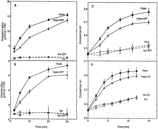
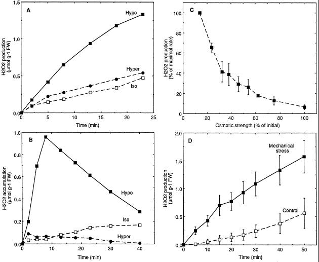
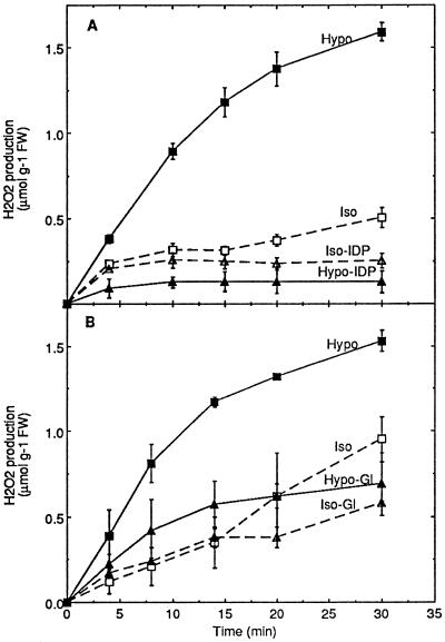
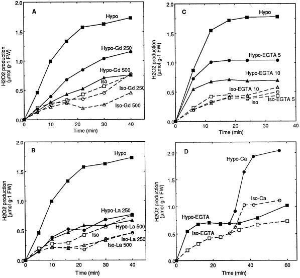
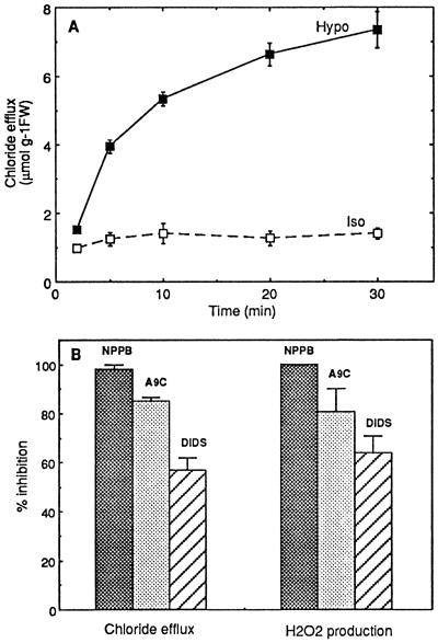
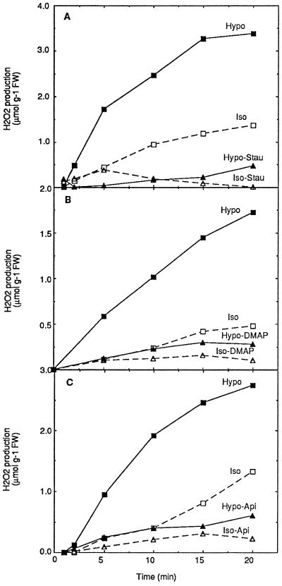
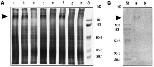
Oxidative burst constitutes an early response in plant defense reactions toward pathogens, but active oxygen production may also be induced by other stimuli. The oxidative response of suspension-cultured tobacco (Nicotiana tabacum cv Xanthi) cells to hypoosmotic and mechanical stresses was characterized. The oxidase involved in the hypoosmotic stress response showed similarities by its NADPH dependence and its inhibition by iodonium diphenyl with the neutrophil NADPH oxidase. Activation of the oxidative response by hypoosmotic stress needed protein phosphorylation and anion effluxes, as well as opening of Ca2+ channels. Inhibition of the oxidative response impaired Cl- efflux, K+ efflux, and extracellular alkalinization, suggesting that the oxidative burst may play a role in ionic flux regulation. Active oxygen species also induced the cross-linking of a cell wall protein, homologous to a soybean (Glycine max L.) extensin, that may act as part of cell volume and turgor regulation through modification of the physical properties of the cell wall.


Figures

Figure 6
Figure 6
Effect of inhibitors of H2O2 production on the ion fluxes induced by hypoosmotic stress. Aliquots of cell suspension were treated by IDP (A–C, Hypo-IDP and Iso-IDP) or glucosamine (D, Hypo-Gl and Iso-Gl) as described in the legend of Figure 2 for each of the inhibitors, except the replacement of 10 mm by 1 mm Mes-Tris, pH 5.2, for the pH measurement experiments. Means ± se of at least two independent experiments are reported in each case. FW, Fresh weight.
Figure 1
Figure 1
Oxidative burst induced by hypoosmotic (A–C) or mechanical (D) stress in suspension-cultured cells. H2O2 production (A) and accumulation in extracellular medium (B) by cells transferred at 0 time to hypoosmotic (Hypo, 40 mOsm), isoosmotic (Iso, 160 mOsm), or hyperosmotic (Hyper, 600 mOsm) medium buffered with 10 mm Mes-Tris, pH 5.2. One representative experiment of three independent experiments is illustrated in each case. C, Osmotic strength dependence of the oxidative burst triggered by transfer in hypoosmotic conditions. Aliquots of cells were transferred at 0 time in hypoosmotic media differing in osmotic strength (corresponding to differing Suc contents) and AOS production was followed. Maximal rate (100%) corresponds to H2O2 production rate for maximal osmotic shock. Means ± se of two independent experiments are reported. D, H2O2 production of cells subjected to a mechanical stress (see Methods). Means ± se of four independent experiments are reported. Aliquots of cell suspension were equilibrated for 3 h in isoosmotic (A–C) or in culture medium (D) buffered with 10 mm Mes-Tris, pH 5.2, before stress treatments. FW, Fresh weight.
Figure 2
Figure 2
Inhibition by IDP (A) and glucosamine (B) of the oxidative burst induced by hypoosmotic stress in tobacco cell suspensions. A, Aliquots of cell suspension were equilibrated for 3 h in isoosmotic medium before transfer at 0 time to hypoosmotic (Hypo), isoosmotic (Iso), hypoosmotic containing 20 μm IDP (Hypo-IDP), or isoosmotic containing 20 μm IDP (Iso-IDP) media. The inhibitor was also added during the last 15 min of the equilibration time at the concentration used after transfer. B, Aliquots of cell suspension were equilibrated for 2 h in buffered culture medium and for an additional 1 h in the same medium containing 10 mm glucosamine, before transfer in hypoosmotic medium containing glucosamine (Hypo-Gl) or isoosmotic medium containing glucosamine (Iso-Gl). Corresponding controls were treated similarly, except for the replacement of glucosamine by 30 mm Man during the last 1 h of equilibration and after transfer in hypoosmotic (Hypo) or isoosmotic (Iso) conditions. Means ± se of two independent experiments are reported in each case. FW, Fresh weight.
Figure 3
Figure 3
Inhibition of the hypoosmotically induced oxidative burst by the presence of Gd (A) or La (B) and by the lack of extracellular Ca2+ (C and D) in cell suspensions. A and B, Aliquots of cell suspension were equilibrated for 3 h in isoosmotic medium before transfer at time 0 to hypoosmotic (Hypo), isoosmotic (Iso), or hypoosmotic medium containing 250 or 500 μm La(NO3)3 or GdCl3 (Hypo-La and Hypo-Gd) or isoosmotic medium containing the same inhibitor concentrations (Iso-La and Iso-Gd). The inhibitor La(NO3)3 or GdCl3 was also added during the last 15 min of the equilibration time, at the concentration used after transfer. C and D, Aliquots of cell suspension were equilibrated for 3 h in isoosmotic medium deprived of Ca2+ before transfer at 0 time in hypoosmotic medium containing 5 or 10 mm EGTA (Hypo-EGTA) or without EGTA (Hypo) and in isoosmotic medium containing 5 or 10 mm EGTA (Iso-EGTA) or without EGTA (Iso). In D, 3 mm CaCl2 was added after about 30 min to the cells previously transferred in hypoosmotic (Hypo-Ca) or isoosmotic (Iso-Ca) media containing 10 mm EGTA. One representative experiment out of three independent experiments is illustrated in each part of the figure. FW, Fresh weight.
Figure 4
Figure 4
Cl efflux induced by hypoosmotic stress (A) and inhibition of hypoosmotically induced responses by anion channel blockers NPPB, A9C, and DIDS (B). A, Aliquots of cell suspension were equilibrated for 3 h in isoosmotic medium before transfer at 0 time to hypoosmotic (Hypo) or isoosmotic (Iso) medium. Means ± se of two independent experiments are reported. B, Aliquots of cell suspension were equilibrated for 3 h in isoosmotic medium before transfer at time 0 to hypoosmotic medium containing 100 μm NPPB, 100 μm A9C, or 100 μm DIDS. Inhibitions by each molecule of the Cl efflux (left part) and H2O2 production (right part) initial rates, in comparison with control cells deprived of inhibitor, are reported. Each inhibitor was also added during the last 15 min of the equilibration time at the concentration used after transfer. Means ± se of at least two independent experiments are reported in each case. FW, Fresh weight.
Figure 5
Figure 5
Involvement of protein phosphorylation in the activation of oxidative burst by hypoosmotic stress. Aliquots of cell suspension were equilibrated for 3 h in isoosmotic medium before transfer at 0 time to hypoosmotic (Hypo), isoosmotic (Iso), or hypoosmotic medium containing 0.5 μm staurosporine (A, Hypo-Stau), 500 μm DMAP (B, Hypo-DMAP), 500 μm apigenin (C, Hypo-Api), or isoosmotic medium containing the same inhibitor concentrations (A, Iso-Stau; B, Iso-DMAP; and C, Iso-Api). One representative experiment of three independent experiments is illustrated in each case. FW, Fresh weight.
Figure 7
Figure 7
Cross-linking of cell wall proteins by oxidative burst induced by hypoosmotic or mechanical stress in tobacco cells. A, SDS-PAGE and silver staining of cell wall proteins extracted from untreated cells (lane a), 5 min after cell transfer in isoosmotic medium (lane b), hypoosmotic medium (lane c), hypoosmotic medium containing 20 μm IDP (lane d), after 20 min of mechanical stress in culture medium (lane e) or culture medium containing 20 μm IDP (lane f), after 5 min of in vivo treatment of cells by 1 mm H2O2 (lane g) or 5 min of in vitro treatment of proteins by 2 mm H2O2 (lane h). B, Western blot of cell wall proteins corresponding to cells transferred in hypoosmotic medium containing 20 μm IDP (a) or hypoosmotic medium without IDP (b). St, Molecular mass standards (in kilodaltons). Arrowheads in A and B indicate the 115-kD bands.

References

    1. Baker CJ, Orlandi EW, Mock NM. Harpin, an elicitor of the hypersensitive response in tobacco caused by Erwinia amylovora, elicits active oxygen production in suspension cells. Plant Physiol. 1993;102:1341–1344. - PMC - PubMed
    1. Blum H, Beier H, Gross HJ. Improved silver staining of plant proteins, RNA and DNA in polyacrylamide gels. Electrophoresis. 1987;8:93–99.
    1. Bottin A, Véronési C, Pontier D, Esquerré-Tugayé MT, Blein JP, Rusterucci C, Ricci P. Differential responses of tobacco cells to elicitors from two Phytophtora species. Plant Physiol Biochem. 1994;32:373–378.
    1. Braam J, Davis RW. Rain-, wind-, and touch-induced expression of calmodulin and calmodulin-related genes in Arabidopsis. Cell. 1990;60:357–364. - PubMed
    1. Bradley DJ, Kjellbom P, Lamb CJ. Elicitor and wound-induced oxidative cross-linking of a proline-rich plant cell wall protein: a novel, rapid defense response. Cell. 1992;70:21–30. - PubMed

LinkOut - more resources