. 1998 Feb 1;116(2):765-76.
doi: 10.1104/pp.116.2.765.

Calcium-Dependent Protein Phosphorylation May Mediate the Gibberellic Acid Response in Barley Aleurone

Affiliations

Calcium-Dependent Protein Phosphorylation May Mediate the Gibberellic Acid Response in Barley Aleurone

S Ritchie et al. Plant Physiol. .

Abstract

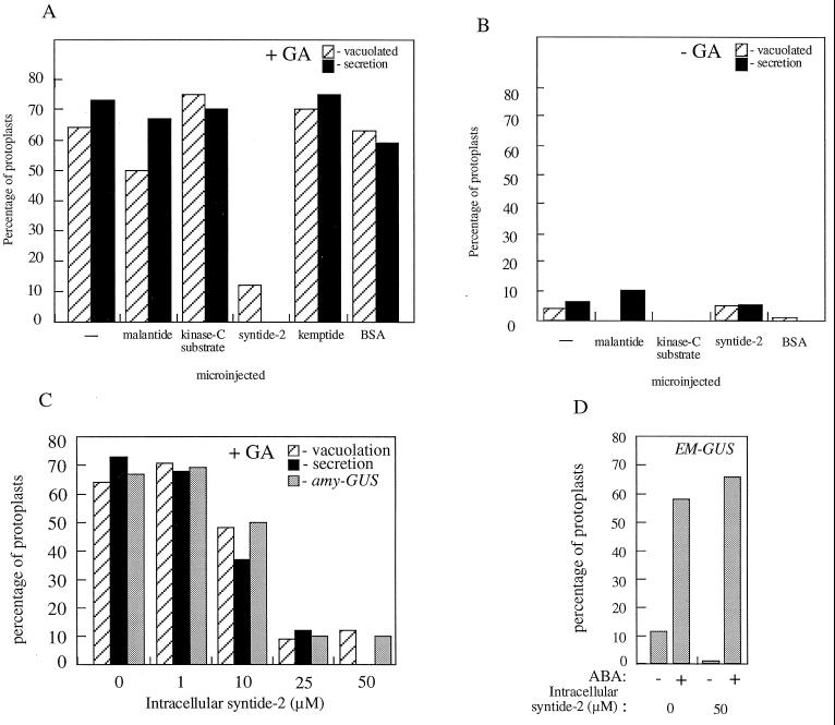
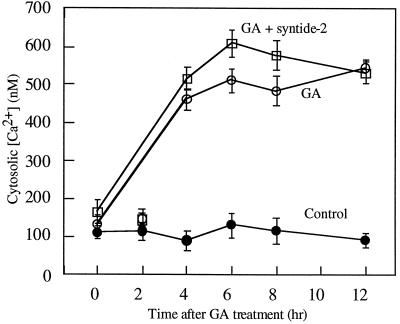
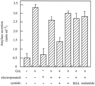
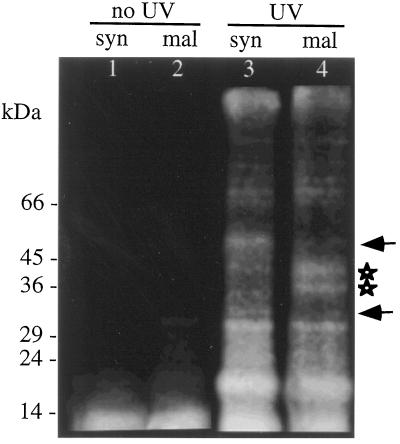
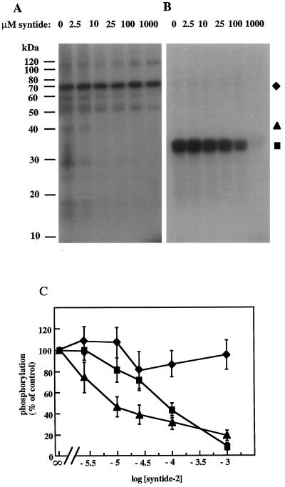
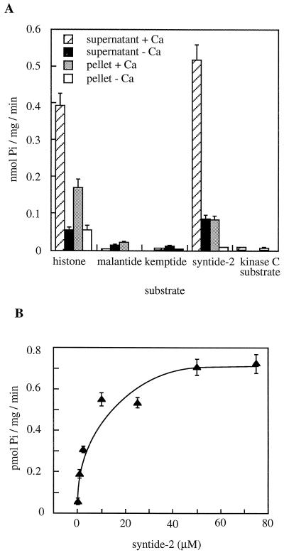
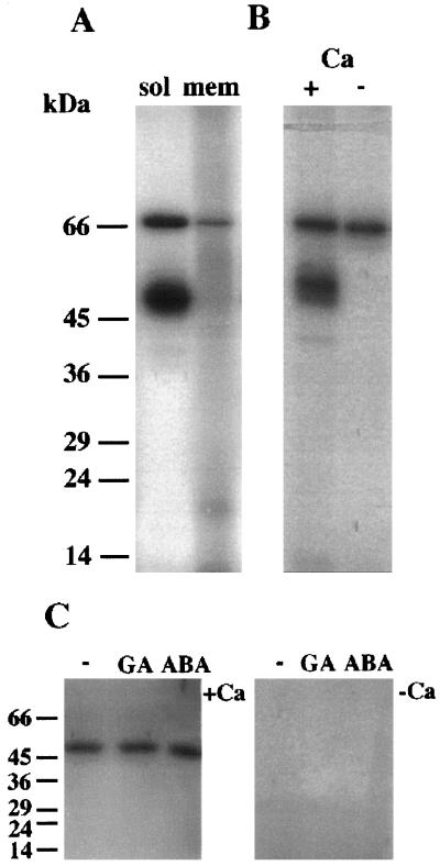
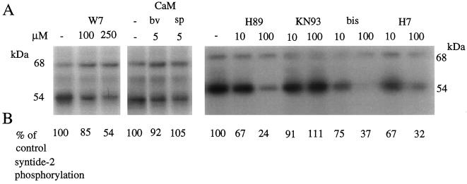
Peptide substrates of well-defined protein kinases were microinjected into aleurone protoplasts of barley (Hordeum vulgare L. cv Himalaya) to inhibit, and therefore identify, protein kinase-regulated events in the transduction of the gibberellin (GA) and abscisic acid signals. Syntide-2, a substrate designed for Ca2+- and calmodulin (CaM)-dependent kinases, selectively inhibited the GA response, leaving constitutive and abscisic acid-regulated events unaffected. Microinjection of syntide did not affect the GA-induced increase in cytosolic [Ca2+], suggesting that it inhibited GA action downstream of the Ca2+ signal. When photoaffinity-labeled syntide-2 was electroporated into protoplasts and cross-linked to interacting proteins in situ, it selectively labeled proteins of approximately 30 and 55 kD. A 54-kD, soluble syntide-2 phosphorylating protein kinase was detected in aleurone cells. This kinase was activated by Ca2+ and was CaM independent, but was inhibited by the CaM antagonist N-(6-aminohexyl)-5-chloro-1-naphthalene-sulfonamide (250 mum), suggesting that it was a CaM-domain protein kinase-like activity. These results suggest that syntide-2 inhibits the GA response of the aleurone via an interaction with this kinase, implicating the 54-kD kinase as a Ca2+-dependent regulator of the GA response in these cells.


Figures

Figure 1
Figure 1
The effect of microinjection of protein kinase peptide substrates on GA-induced amylase secretion, vacuolation, and amylase-GUS and EM-GUS expression. A and B, Freshly isolated protoplasts were microinjected with 50 μm of the indicated protein kinase substrate peptides or BSA and then treated for 24 h with (+GA) or without (−GA) 5 μm GA. C, Freshly isolated protoplasts were microinjected with a range of syntide-2 concentrations and then treated for 24 h with 5 μm GA. Secretion of amylase, development of GA-induced vacuolated morphology, and amylase-GUS expression were then assessed on a single-cell basis. D, Freshly isolated protoplasts were co-microinjected with 50 μm syntide-2 or BSA and the EM-GUS construct and treated with or without 10 μm ABA. Induction of EM-GUS expression was then assessed on a single-cell basis. Protoplasts were scored as showing induction of amylase-GUS or EM-GUS expression if they exhibited a GUS:LUC of more than 4000 (Gilroy, 1996). Protoplasts were scored as secreting amylase if they showed a cleared “halo” of >25 μm in the starch thin-film assay (Hillmer et al., 1992). Vacuolated protoplasts were those showing development to stage 3 or 4 as defined by Bush et al. (1986). The responses of at least nine microinjected protoplasts per treatment are shown.
Figure 2
Figure 2
The effect of microinjection of syntide-2 on GA-induced cytoplasmic Ca2+ elevation. Freshly isolated protoplasts were microinjected with dextran-conjugated indo-1, with or without 50 μm syntide-2. These protoplasts were then treated without hormone (control) or with 5 μm GA and their cytoplasmic [Ca2+] was monitored at the indicated times using confocal ratio imaging. The results represent the mean ± se of at least nine microinjected protoplasts per treatment.
Figure 3
Figure 3
Cytoplasmic distribution of fluorescently labeled syntide-2 loaded into protoplasts by microinjection. A, Fluorescence from a protoplast microinjected with FITC-dextran. B, Fluorescence from a protoplast microinjected with 1 μm cytoplasmic concentration of fluorescently labeled syntide-2. C, Membranous components of protoplasts visualized by incubation with 5 μm N-phenyl-1-naphthylamine. D, Nuclear fluorescence from a protoplast stained with 4′,6′-diamidino-2-phenylindole. E, Fluorescence from vacuoles labeled with 2′,7′-bis-(2-carboxyethyl)-5-(and 6)carboxyfluorescein, acetoxymethyl ester. Freshly isolated protoplasts were microinjected with FITC-labeled 10-kD dextran fluorescein (A) or fluorescein labeled syntide-2 (B), treated for 24 h with 5 μm GA, and then fluorescence visualized using the Zeiss LSM-410 confocal microscope. C to E, Protoplasts were treated for 24 h with 5 μm GA and then stained for the appropriate compartment. Results are representative of more than 10 replicates. v, Vacuole; n, nucleus. The scale bar represents 10 μm.
Figure 4
Figure 4
The effect of electroporation of protein kinase substrate peptides on GA-induced amylase secretion. Freshly isolated protoplasts were loaded with 25 μm syntide-2, malantide, or BSA by electroporation and treated for 48 h with or without 5 μm GA. Secreted amylase activity was then assessed. A set of nonelectroporated control protoplasts was treated identically. Results represent the mean ± se of three separate experiments.
Figure 5
Figure 5
Fluorograph of proteins labeled in vivo by photoaffinity syntide-2 or photoaffinity malantide. Protoplasts were electroporated with fluorescent, photoaffinity-labeled syntide-2 or malantide and allowed to recover for 1 h. The protoplasts were then frozen in liquid N2 to freeze molecular interactions, and total proteins were extracted in the dark (lanes 1 and 2). In parallel experiments the peptide-loaded protoplasts were UV irradiated to cross-link the photoaffinity peptides to closely associated proteins and then extracted (lanes 3 and 4). Proteins were then separated by SDS-PAGE. Peptide-labeled proteins were visualized using the fluorescein attached to the photoaffinity peptides. Note the 30- and 55-kD proteins selectively labeled by syntide-2 (arrows) and the 33- and 41-kD specific to malantide (stars). The positions of molecular mass markers are indicated in kilodaltons on the left.
Figure 6
Figure 6
The inhibition of histone and endogenous protein phosphorylation by syntide-2. Syntide-2 was added to in vitro substrate phosphorylation assays carried out without (A) or with (B) 25 μm histone. The samples were precipitated with 10% TCA and processed for SDS-PAGE and autoradiography. C, Phosphorylation levels from the 70- and 40-kD endogenous substrate bands and histone was assessed by cutting sections of the gel that corresponded to bands on the autoradiograph, and counting duplicate samples in a scintillation counter. Results are typical of two independent experiments. ▪, Histone; ♦, 70 kD; ▴, 40 kD.
Figure 7
Figure 7
Peptide phosphorylation activity in barley aleurone extract. A, Microsomal (pellet) and soluble (supernatant) fractions were prepared and their kinase activity assessed using 25 μm substrate peptides or histone. Assays were carried out in the presence of 50 μm CaCl2 (+Ca) or 1 mm EGTA (−Ca). B, A range of syntide-2 concentrations was used on the soluble fraction in assays containing 50 μm CaCl2, and the level of 32P incorporation into the peptide during the initial 2 min of reaction was calculated. The reaction rate was linear over this time period. The results are the mean ± se of two experiments, with two replicates per experiment.
Figure 8
Figure 8
Autophosphorylation characteristics of barley aleurone kinase activities. A, Autophosphorylating activities of kinases in soluble or microsomal fractions extracted from aleurone layers. Assays were carried out in the presence of 50 μm CaCl2 using 50 μg of protein per fraction. B, Autophosphorylation activities of kinases in a crude fraction were carried out in the presence of 50 μm CaCl2 (+) or 500 μm EGTA (−). C, Aleurone layers were treated for 24 h with no hormones (−), 5 μm GA, or 5 μm ABA. Crude extracts of proteins were then prepared, run on 12% SDS-PAGE, renatured, and assayed for autophosphorylating activity in gel. For these assays incubation with [32P]ATP at a concentration of 1 μm (50,000 dpm pmol−1) was carried out in the presence of 50 μm CaCl2 (+Ca) or 1 mm EGTA (−Ca). Note that only the 54-kD kinase is renatured under these conditions. The positions of molecular mass markers are indicated in kilodaltons on the left.
Figure 9
Figure 9
The effect of protein kinase inhibitors, CaM antagonist W7, and exogenous CaM on autophosphorylation and syntide-2 phosphorylation activities in the barley aleurone. Four protein kinase inhibitors, W7, or CaM (bv, bovine; sp, spinach [Sigma]) were added to autophosphorylation and syntide-2 phosphorylation assays at the indicated concentrations. Aleurone layers were extracted and assays were carried out in the presence of 50 μm CaCl2 as described in Methods. A, Autophosphorylation of the 68- and 54-kD proteins was visualized by autoradiography; B, the effect of protein kinase inhibitors on syntide-2 phosphorylation expressed as a percentage of control (no inhibitor) is shown below the appropriate inhibitor lane on the autoradiograph. The inhibitors used were obtained from Calbiochem: H89, specific for cAMP-dependent protein kinase; KN93, specific for Ca2+/CaM-dependent protein kinase II; bisindolylmaleimide (bis), specific for protein kinase C; and 1-(5-isoquinolinesulfonyl)-2-methylpiperazine (H7), a broad-range Ser/Thr kinase inhibitor. The experiments were repeated twice with two replicates each. The largest se for syntide-2 phosphorylation was 12%.

Similar articles

Cited by

References

    1. Anderberg RJ, Walker-Simmons MK. Isolation of a wheat cDNA clone for an abscisic acid-inducible transcript with homology to protein kinases. Proc Natl Acad Sci USA. 1992;89:10183–10187. - PMC - PubMed
    1. Armstrong F, Leung J, Grabov A, Brearley J, Blatt MR. Sensitivity to abscisic acid of guard cell K+ channels is suppressed by ABI1-1, a mutant Arabidopsis gene encoding a putative protein phosphatase. Proc Natl Acad Sci USA. 1995;92:9520–9524. - PMC - PubMed
    1. Aromatorio DK, Parker J, Brown WE. High-resolution analytical and preparative peptide mapping by a combination of ion-exchange chromatography and thin-layer chromatography. Methods Enzymol. 1983;91:384–391. - PubMed
    1. Ashizawa K, Wishart GJ, Hashimoto K, Tsuzuki Y. Dephosphorylation of a 30 kD protein kinase of fowl spermatozoa by the addition of myosin light chain substrate peptide inhibits the flagellar motility. Biochem Biophys Res Commun. 1995;215:706–712. - PubMed
    1. Banks PR, Paquette DM. Comparison of three common reactive fluorescent probes used for conjugation to biomolecules by capillary zone electrophoresis. Bioconjugate Chem. 1995;6:447–458. - PubMed

LinkOut - more resources