. 1998 Feb 15;507 ( Pt 1)(Pt 1):201-17.
doi: 10.1111/j.1469-7793.1998.201bu.x.

CA1 pyramidal to basket and bistratified cell EPSPs: dual intracellular recordings in rat hippocampal slices

Affiliations

CA1 pyramidal to basket and bistratified cell EPSPs: dual intracellular recordings in rat hippocampal slices

A B Ali et al. J Physiol. .

Abstract

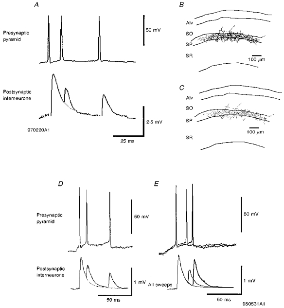
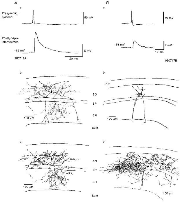
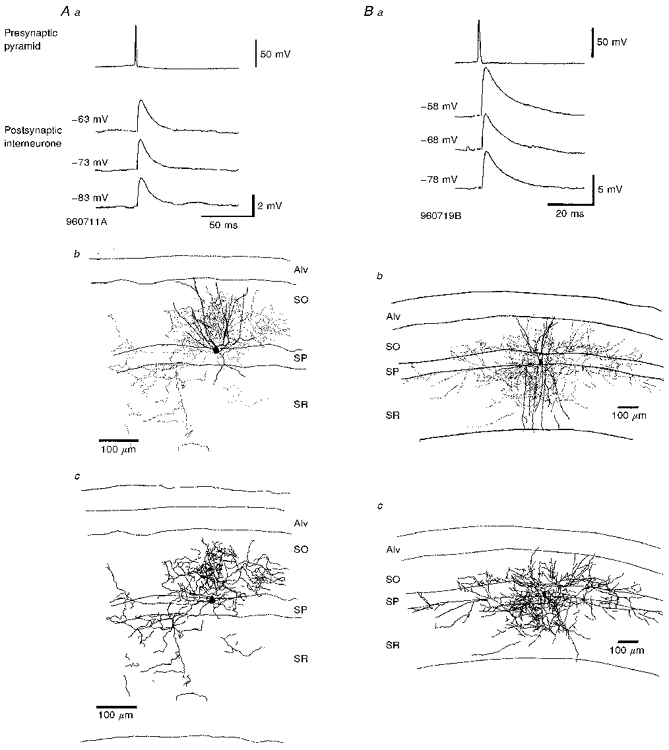
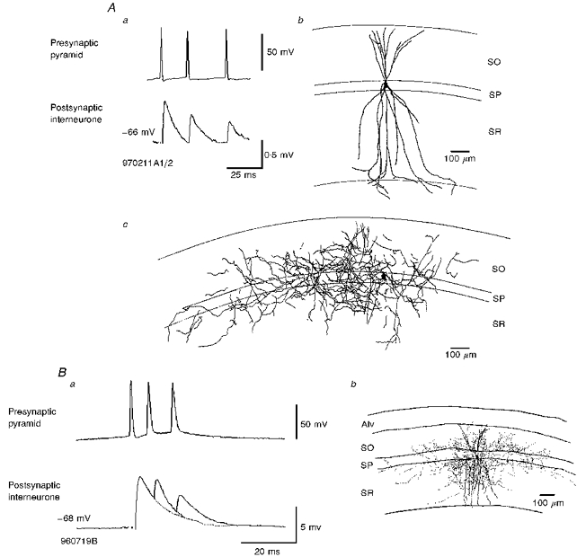
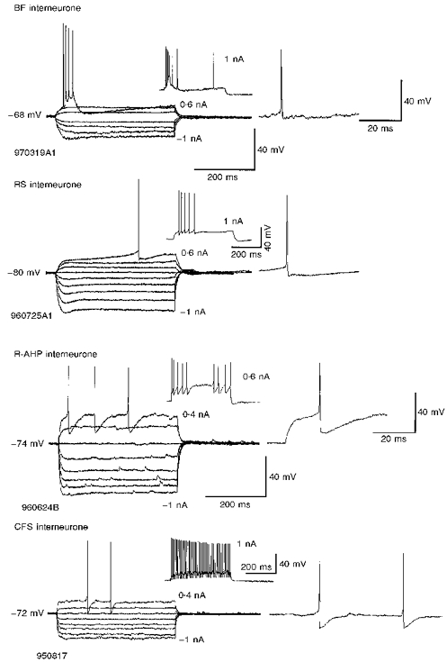
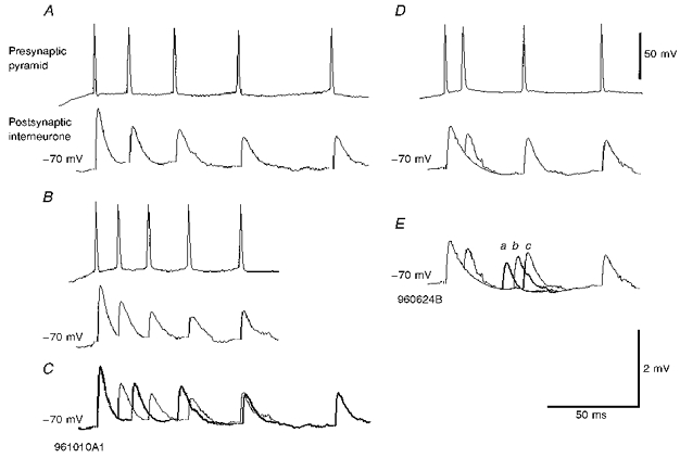
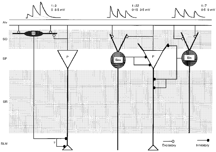
1. Dual intracellular recordings in the CA1 region of adult rat hippocampal slices and biocytin filling of synaptically connected cells were used to study the excitatory postsynaptic potentials (EPSPs) elicited in basket (n = 7) and bistratified interneurones (n = 7) by action potentials activated in simultaneously recorded pyramidal cells. 2. Interneurones could be subdivided according to their electrophysiological properties into classical fast spiking, burst firing, regular spiking and fast spiking cells with a rounded spike after-hyperpolarization. These physiological classes did not, however, correlate with morphological type. EPSPs were not recorded in regular spiking cells. 3. Average EPSP amplitudes were larger in bistratified cells (range, 0.5-9 mV) than in basket cells (range, 0. 15-3.6 mV) and the probability of obtaining a pyramidal cell-interneurone EPSP was also higher for the bistratified cells (1:7) than for the basket cells (1:22). EPSP 10-90 % rise times in bistratified cells (0.7-2 ms) and their widths at half-amplitude (3. 9-11.2 ms) were slightly longer than in basket cells (rise times, 0.4-1.6 ms; half-widths, 2.2-9.7 ms). 4. The majority of these EPSPs (6 of 8 tested) increased in amplitude and duration with postsynaptic depolarization, although in two (of 4) basket cells the voltage relation was conventional. 5. All EPSPs tested in both basket (n = 7) and bistratified cells (n = 5) decreased in amplitude with repetitive presynaptic firing. The average amplitudes of second EPSPs elicited within 15 ms of the first were between 34 and 94 % of the average amplitude of the first EPSP. Third and fourth EPSPs in brief trains were further depressed. This depression was associated with an increase in the incidence of apparent failures of transmission indicating a presynaptic locus.


Figures

Figure 4
Figure 4. EPSPs recorded in basket cells in response to APs in simultaneously recorded pyramids either increase, or decrease in amplitude with postsynaptic depolarization
Aa, averaged EPSPs recorded in basket cell at three different membrane potentials and in response to trains of three presynaptic spikes in a pyramidal neurone. As the postsynaptic cell was depolarized the EPSP increased in amplitude and duration. The averaged EPSPs following the second and third spikes were smaller than following the first. The postsynaptic basket cell soma and dendrites are indicated by the continuous lines in Ab and the boutons by the dots. Only the axon from one 60 μm section is included in this drawing but the distribution was similar in all other sections. The complete axon from that one section is illustrated in Ac. The axon and boutons ramified mostly within SP and proximal SO and SR, indicating that it was a basket cell (cell code 961010A1). Ba, averaged EPSPs recorded in response to APs in a presynaptic pyramid at three different membrane potentials. As the postsynaptic basket cell was depolarized the EPSP average amplitude decreased. The soma/dendrites (continuous lines) and boutons (dots) of this cell are illustrated in Bb. Only some dendrites could be drawn due to light labelling. The axon remained well labelled (2 of 4 sections reconstructed) and was mostly confined to stratum pyramidale, as can also be seen in Bc where the complete axonal arborization in these two sections is shown (code 950531A1).
Figure 6
Figure 6. Brief train depression in EPSPs recorded in basket cells in response to brief trains of APs in presynaptic pyramids
A, the averaged 2nd and 3rd EPSPs recorded in a basket cell in response to a brief train of three presynaptic pyramidal cell spikes were depressed when compared with the first EPSP. The axon of the cell was restricted to the cell body layer, i.e. this is a basket cell. The axonal distribution is shown in B and the boutons in C (cell code 970220A1). D, depression in a second postsynaptic basket cell in response to a brief train of APs in a presynaptic pyramid. See also Fig. 4Ba and the reconstruction of this cell in 4Bb and 4Bc (code 950531A1). E, paired pulse depression was dependent on interspike interval. Averaged responses to pairs of APs at two different interspike intervals are illustrated. Stronger depression was apparent at the briefer interval (normal trace) than at the longer interval (bold trace).
Figure 2
Figure 2. The largest (A) and one of the smaller (B) average pyramid-bistratified cell EPSPs recorded in this study with the reconstructions of the postsynaptic interneurones
In Aa the average EPSP was the largest recorded in this study and at -66 mV was 9.06 mV (cell code 960719A). The interneurone soma was located in stratum oriens (SO) and dendrites traversed SO and stratum pyramidale (SP) into stratum radiatum (SR), but did not enter stratum lacunosum-moleculare (SLM) (Ab). This cell was reconstructed using the Neurolucida system which allows the boutons to be shown separately from the rest of the axon (Ab). The dots in Ab illustrate the distribution of the boutons while the axonal arborization in full is shown in Ac with the position of the interneurone soma shown for comparison with Ab. The axon of this cell ramified mostly in SO and SR, with only a few branches in SP. Ba illustrates one of the smaller average EPSPs recorded, which had an average amplitude of 0.72 mV at -61 mV (cell code 960717B). The interneurone was reconstructed by hand using a drawing tube. Bb illustrates the soma and dendrites (Alv, alveus). The axon of this cell (Bc) ramified mostly in SO, but there was also a large amount in SP, with some in SR. As stated in the text this cell may be a radially oriented trilaminar cell. Here and in all figures, averaged EPSPs include between 50 and 300 sweeps.
Figure 3
Figure 3. Pyramid to bistratified cell EPSPs increase in amplitude and duration with postsynaptic depolarization
Aa and Ba, averaged EPSPs recorded in two different bistratified cells each at three different membrane potentials in response to single spikes in presynaptic pyramids. These EPSPs increased in average amplitude and duration with postsynaptic depolarization (cell codes 960711A and 960719B). Ab and Bb, in each case the reconstruction indicates the soma and dendrites of the postsynaptic bistratified cells and the dots represent the distribution of the terminal boutons as established at the light microscopic level. In Ab the axon terminated mostly in SO with some also evident in SR. The dendrites of this cell in SR are drawn truncated as their identification in one section was ambiguous. In Bb the boutons were located mostly in SO and SR, with only a few in SP. The dendrites of this cell extended throughout SR but did not enter SLM. Ac and Bc, the distribution of the complete axon of the same cells and only the soma of the interneurones is illustrated. The distribution of the axon was largely similar to the distribution of the boutons (Ab and Bb).
Figure 5
Figure 5. EPSPs recorded in bistratified cells in response to trains of spikes in single presynaptic pyramidal neurones exhibit brief train depression
Aa, averaged postsynaptic responses recorded in a bistratified cell to a brief train of three APs in a presynaptic pyramid. With each successive AP in the train, the EPSP decreased in average amplitude. The interneurone soma and dendrites are shown in the camera lucida reconstruction in Ab and the axon separately in Ac. The axon arborized mostly in SO and SR, but also, to a lesser extent, in SP. The dendrites extended throughout SO and SR and entered only the very proximal portion of SLM (cell code 970211A1/2). Ba, brief train depression of an EPSP in another bistratified cell (drawn in Bb and earlier in more detail in Fig. 2B c). The averaged EPSPs in this cell exhibited stronger brief train depression than in Aa, with the briefer interspike intervals recorded here (cell code 960719B).
Figure 1
Figure 1. Basket and bistratified cells displayed diverse electrophysiological characteristics
Interneurones of different morphological classes could display similar electrophysiological characteristics and those of the same class different electrophysiological features. They are classified here according to the shape and pattern of their discharge in response to depolarizing current injection into burst firing (BF), regular spiking (RS), classical fast spiking (CFS) or interneurones with a rounded spike after-hyperpolarization (R-AHP); see Table 1 for details of properties of each cell type. Burst firing interneurones responded to threshold depolarizations with a burst of high frequency action potentials (APs). Regular spiking interneurones were often difficult to distinguish from pyramidal neurones and were confirmed as interneurones only by morphological features, or by the IPSPs they elicited in other neurones. The example shown here resembles a neocortical late spiking cell (see Kawaguchi & Kubota, 1997). R-AHP interneurones were recognizable by the deep, rounded spike AHPs at threshold and non-accommodating fast spiking behaviour at more depolarized potentials (not illustrated). CFS interneurones had simple, fast spike AHPs and spike trains showed little accommodation or adaptation. Responses to subthreshold pulses are averages of between two and five sweeps.
Figure 7
Figure 7. Frequency-dependent depression in two pyramid to basket cell connections
In A and B averaged responses to two trains each of five presynaptic APs are shown. The first EPSP for both trains was the same. In A the interspike intervals were longer than in B and averaged 3rd, 4th and 5th EPSPs were less strongly depressed than in B. See comparison in C in which the bold trace repeats the averaged EPSPs in A superimposed on the averaged response shown in B (cell code 961010A1). D and E illustrate averaged EPSPs elicited by three types of trains, each of four presynaptic APs, in which the 3rd AP occurred at three different interspike intervals. Responses to the 1st, 2nd and 4th APs are the same for all three trains. One example is shown in D and in E, the three 3rd EPSPs are superimposed for comparison (a, b and c). Some recovery from the strong 3rd EPSP depression apparent at the briefest interval can be seen at the longer intervals (cell code 960624B).
Figure 8
Figure 8. Schematic of the excitatory connections made by CA1 pyramidal cells with three classes of interneurones
The EPSPs elicited in basket cells (Bas) and bistratified cells (Bis) exhibit frequency-dependent depression. In contrast, the EPSPs elicited in horizontally oriented oriens/alveus interneurones (H) display frequency-dependent facilitation (see Ali & Thomson, 1997, 1998). The probabilities of finding connections in randomly selected pyramid-interneurone pairs in these parallel studies are indicated and the ranges of average EPSP amplitudes recorded. For bistratified and basket cells, the largest EPSP amplitudes are for the first EPSP and for horizontally oriented O/A interneurones for the 4th or 5th EPSPs.

References

    1. Abbott LF, Varela JA, Sen K, Nelson SB. Synaptic depression and cortical gain control. Science. 1997;275:220–224. - PubMed
    1. Aika Y, Ren JQ, Kosaka K, Kosaka T. Quantitative analysis of GABA-like-immunoreactive and parvalbumin-containing neurons in the CA1 region of the rat hippocampus using a stereological method, the dissector. Experimental Brain Research. 1994;99:267–276. - PubMed
    1. Ali AB, Thomson AM. Brief train depression and facilitation at pyramid-interneurone connections in slices of rat hippocampus; paired recordings with biocytin filling. The Journal of Physiology. 1997;501.P:9. P.
    1. Ali AB, Thomson AM. Facilitating pyramid to horizontal oriens-alveus interneurone inputs: dual intracellular recordings in slices of rat hippocampus. The Journal of Physiology. 1998;507:185–199. - PMC - PubMed
    1. Betz WJ. Depression of transmitter release at the neuromuscular junction of the frog. The Journal of Physiology. 1970;206:620–644. - PMC - PubMed

Publication types

LinkOut - more resources