. 1997;6(6):333-47.

Identification of a second promoter in the human c-ets-2 proto-oncogene

Affiliations

Identification of a second promoter in the human c-ets-2 proto-oncogene

A Bègue et al. Gene Expr. 1997.

Abstract

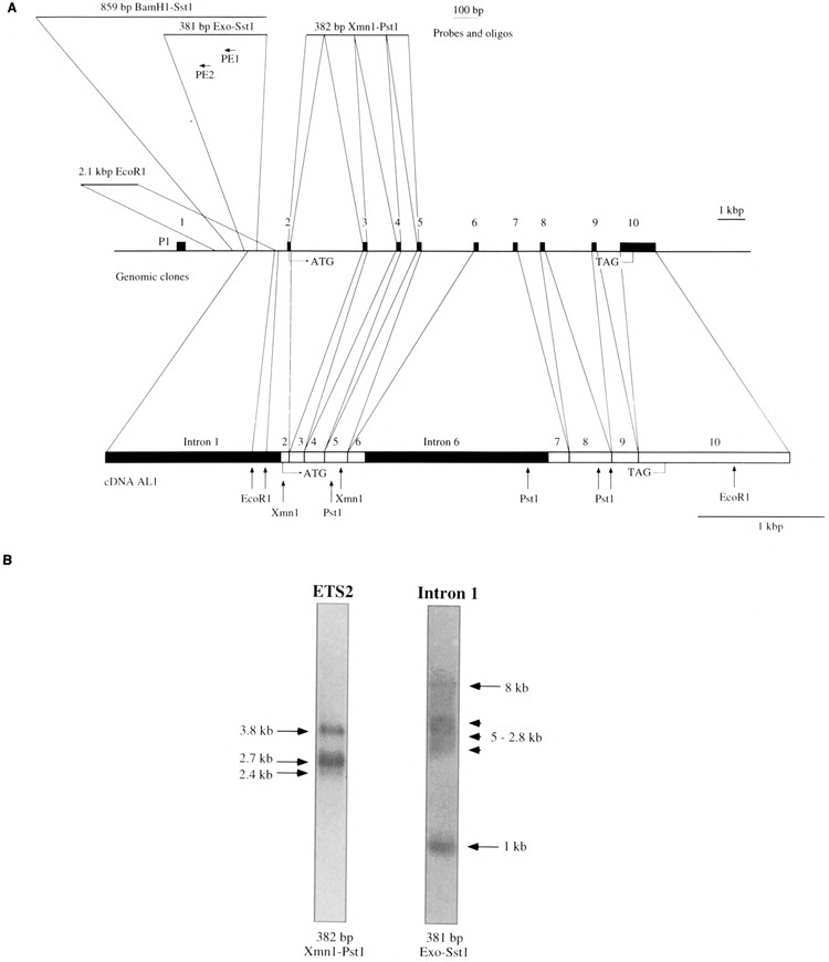
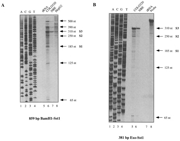
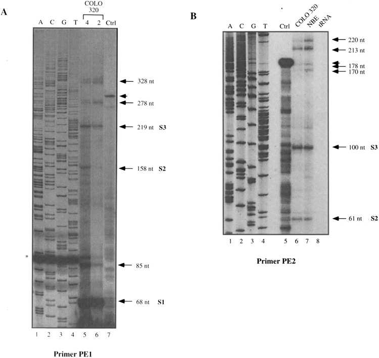
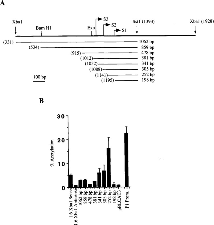
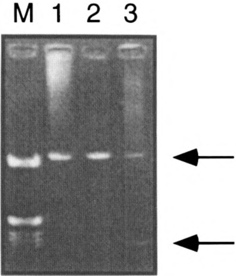
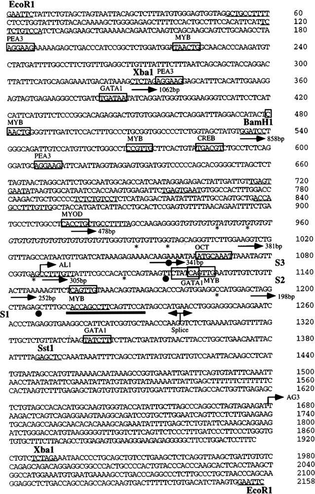
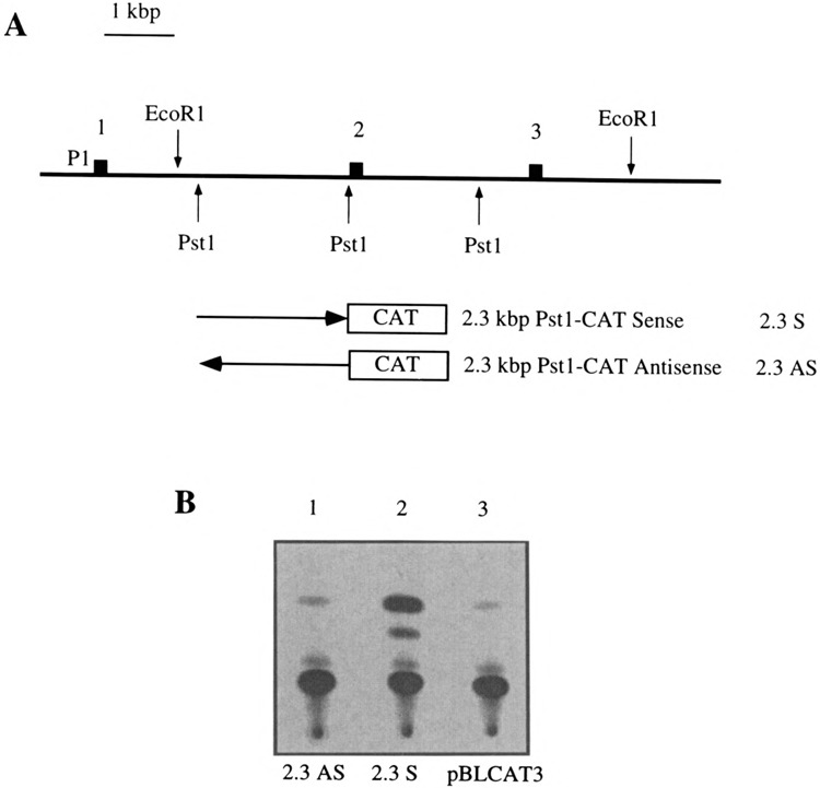
We localized and characterized a new regulatory element with promoter activity in the human c-ets-2 intron 1. This promoter governs the expression of 5' divergent c-ets-2 transcripts through multiple start sites dispersed within 300 bp. Among the multiple start sites detected, three are major transcriptional initiation points. We detected transcripts initiated from this new promoter in various cell lines such as COLO 320, NBE, or HepG2 cells. This promoter exhibits transcriptional activity when linked to the CAT gene, and deletion constructs reveal that it contains activating and repressing elements. The sequence of the promoter reveals putative binding sites for ETS, MYB, GATA, and Oct factors. In addition, we show that this promoter is functionally conserved in the chicken.


Figures

FIG. 1
FIG. 1
(A) Map of the human c-ets-2 gene. In the middle of the panel the genomic organization of c-ets-2 is depicted from exon 1 to exon 10 containing the stop codon. Exons are represented by black boxes. In the lower part of the panel the AL1 cDNA is represented at a different scale and limits between exons are indicated by vertical bars. Representative restriction sites are indicated as well as initiation and stop codons. The two intronic sequences contained in AL1 are indicated (intron 1 and intron 6) In the upper part of the panel the various probes used in this study are indicated together with their location in the c-ets-2 gene. The two primers used for primer extension experiments are also indicated. (B) Northern blot performed with 15 Mg of poly(A)+ RNAs extracted from COLO 320 cells and hybridized with a common c-ets-2 probe (ETS2: 382 bp XmnI-PstI) or an intron 1-specific probe (Intron 1:381 bp Exo-SstI). The exposure times of the two lanes are different.
FIG. 2
FIG. 2
RNAase protection experiments performed with the 859-bp BamHI-SstI probe (A) or the 381-bp Exo-SstI probe (B). Total RNAs (20 μg) from various cell lines indicated above each panel were hybridized with the corresponding probe. In each case a protection experiment was also performed with tRNAs as a negative control. The sizes of the protected fragments are indicated. The three major start sites S1, S2, and S3 are also depicted.
FIG. 3
FIG. 3
Primer extension experiments performed with the PE1 (A) or PE2 (B) oligonucleotides. For (A) two different amounts of COLO 320 (4 and 2 μg) total RNAs were used whereas for (B) COLO 320 and NBE total RNAs were used. Ctrl: Control experiment with in vitro synthetized cold RNA as explained in the text. The sizes of the extended fragments as the three major start sites S1, S2, and S3 are indicated. In (A) the star indicates an artefactual band in the sequencing reaction and the arrowhead indicates the extended product in the control lane.
FIG. 4
FIG. 4
(A) Map of the deletion constructs used to study the activity of the P2 human c-ets-2 promoter. The different constructs are named following the size of their respective inserted fragments. 5′ end of each construct is numbered according to its position in the sequence presented in Fig. 6. The size of the fragment is indicated at the right. The first 5′ base of each deletion is indicated with the numbering system used for Fig. 6. The three major start sites S1, S2, and S3 are also depicted together with representative restriction sites. (B) Activity of the c-ets-2 P2 human promoter and deletion constructs cloned in the pBLCAT3 vector and their transcriptional activity assayed in NBE cells. The averaged result of three experiments is presented with standard deviation. The activity mediated by the 1.6-kbp XbaI fragment cloned in sense or antisense orientation is shown together with the activity of the wild-type vector (pBLCAT3).
FIG. 5
FIG. 5
RT-PCR detection of transcripts initiated at the P2 promoter. RNAs from COLO 320 (lane 1), HepG2 (lane 2), and NBE (lane 3) cells were retrotranscribed using the oligo dT primer and amplified using RT1 and RT2 primers. (M) Molecular size marker. Arrows point to the two fragments discussed in the text.
FIG. 6
FIG. 6
Sequence of the c-ets-2 P2 human promoter. Restriction sites mentioned in the text are indicated. Putative sites for transcription factors are boxed and the name of the factor is indicated. The direct or inverted repeats observed in the sequence are underlined. The sequence of the GT repeat is italicized. The 5′ boundaries of the various deletion constructs used to measure the activity of the promoter are indicated with arrows. The three start sites S1, S2, and S3 are indicated by black dots below the sequence. Other additional minor start sites observed either in RNAase protection or in primer extension experiments are indicated by stars below the sequence.
FIG. 7
FIG. 7
(A) Map of the 5′ part of the chicken c-ets-2 gene. The first three exons are indicated by black boxes as well as representative restriction sites. The fragments used to measure the transcriptional activity mediated by intron 1 are indicated. The P1 promoter is indicated. These fragments were cloned in sense and antisense orientation in the pBLCAT3 vector. (B) Typical result of a CAT assay performed in the HD11 macrophagic cells. The 2.3AS and the 2.3S constructs were used together with the pBLCAT3 vector as a negative control.

References

    1. Bhat N. K.; Fisher R. J.; Fujiwara S.; Ascione R.; Papas T. S. Temporal and tissue-specific expression of mouse ets genes. Proc. Natl. Acad. Sci. USA 84:3161–3165; 1987. - PMC - PubMed
    1. Bhat N. K.; Komschlies K. L.; Fujiwara S.; Fisher R. J.; Mathieson B. J.; Gregorio T. A.; Young H. A.; Kasik J. W.; Ozato K.; Papas T. S. Expression of ets genes in mouse thymocyte subsets T cells. J. Immunol. 142:672–678; 1989. - PubMed
    1. Bhat N. K.; Thompson C. B.; Lindsten T.; June C. H.; Fujiwara S.; Koizum S.; Fisher R. J.; Papas T. S. Reciprocal expression of human ETS1 and ETS2 genes during T-cell activation: Regulatory role for the protooncogene ETS1. Proc. Natl. Acad. Sci. USA 87:3723–3727; 1990. - PMC - PubMed
    1. Boshart M.; Kluppel M.; Schmidt A.; Schutz C.; Luckow B. Reporter constructs with low background activity utilizing the cat gene. Gene 110:129–130; 1992. - PubMed
    1. Boulukos K. E.; Pognonec P.; Bègue A.; Galibert F.; Gesquière J. C.; Stéhelin D.; Ghysdael J. Identification in chickens of an evolutionary conserved cellular ets-2 gene (c-ets-2) encoding nuclear proteins related to the products of the c-ets proto-oncogene. EMBO J. 7:697–705; 1988. - PMC - PubMed

Publication types

MeSH terms

LinkOut - more resources