. 1998 Mar;180(5):1135-47.
doi: 10.1128/JB.180.5.1135-1147.1998.

An ABC transporter system of Yersinia pestis allows utilization of chelated iron by Escherichia coli SAB11

Affiliations

An ABC transporter system of Yersinia pestis allows utilization of chelated iron by Escherichia coli SAB11

S W Bearden et al. J Bacteriol. 1998 Mar.

Abstract

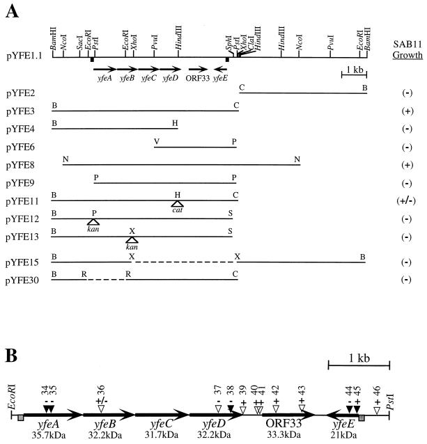
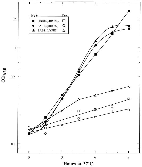
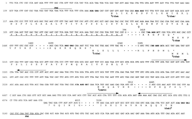
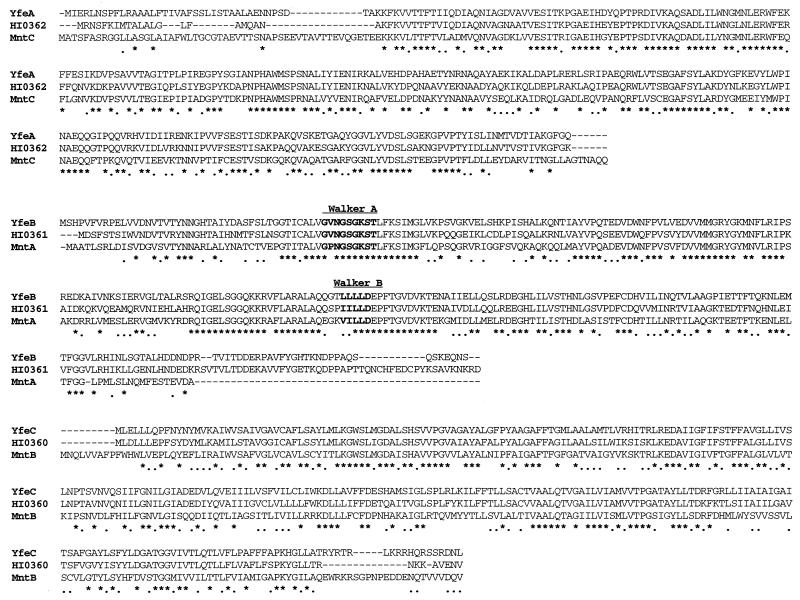
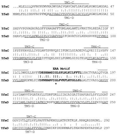
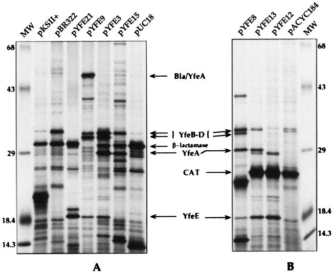
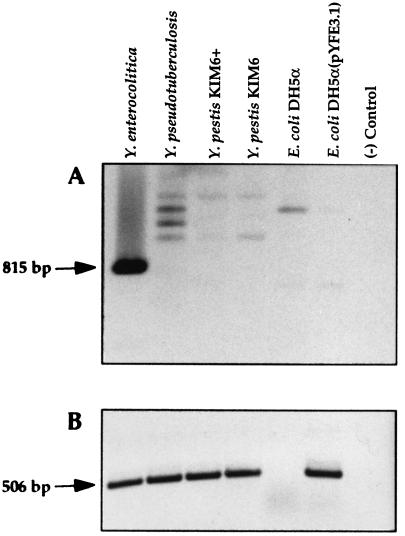
The acquisition of iron is an essential component in the pathogenesis of Yersinia pestis, the agent of bubonic and pneumonic plague. A cosmid library derived from the genomic DNA of Y. pestis KIM6+ was used for transduction of an Escherichia coli mutant (SAB11) defective in the biosynthesis of the siderophore enterobactin. Recombinant plasmids which had a common 13-kb BamHI fragment were isolated from SAB11 transductants in which growth but not enterobactin synthesis was restored on media containing the iron chelator EDDA [ethylenediamine-di(o-hydroxyphenyl acetic acid)]. Subcloning and transposon mutagenesis revealed a 5.6-kb region, designated yfe, essential for SAB11 growth stimulation. In vitro transcription-translation analysis identified polypeptides of 18, 29.5, 32, and 33 kDa encoded by the yfe locus. Sequence analysis shows this locus to be comprised of five genes in two separate operons which have potential Fur-binding sequences in both promoters. A putative polycistronic operon, yfeABCD, is Fur regulated and responds to iron and manganese. A functional Fur protein is required for the observed manganese repression of this operon. This operon encodes polypeptides which have strong similarity to the ATP-binding cassette (ABC) family of transporters and include a periplasmic binding protein (YfeA), an ATP-binding protein (YfeB), and two integral membrane proteins (YfeC and -D), which likely function in the acquisition of inorganic iron and possibly other ions. The approximately 21-kDa protein encoded by the separately transcribed yfeE gene may be located in the cell envelope, since a yfeE::TnphoA fusion is PhoA+. Mutations in this gene abrogate growth of SAB11 on iron-chelated media.


Figures

FIG. 1
FIG. 1
(A) Restriction map of pYFE1.1 and various subclones used to test for restoration of growth of E. coli SAB11. +, plasmids which promote SAB11 growth; −, no growth stimulation. Arrows indicate the directions of transcription of genes included in the yfe locus. Dashed lines and open triangles represent deletions and sites of antibiotic cassette insertion, respectively. (B) Map of relevant TnphoA insertions within the yfe locus. Filled triangles represent PhoA+ translational fusions. + and − signs above each insertion site indicate iron-deficient growth and no growth, respectively, of SAB11 carrying that insertion in pYFE1.1. Numbers above each triangle represent plasmids pYFE34 to -46 (pYFE1.1::TnphoA34 to -46), respectively (Table 1). Shaded boxes upstream of yfeA and yfeE represent potential Fur boxes.
FIG. 2
FIG. 2
Growth of E. coli HB101 or SAB11 transformed with either pBR322 or pYFE3. Filled symbols indicate strains grown in TG minimal medium supplemented with 10 μM FeCl3, while open symbols designate growth in TG without supplementation. OD620, optical density at 620 nm.
FIG. 3
FIG. 3
Partial DNA sequence representation of the yfeABCD and yfeE operons and ORF33, including single-letter amino acid translations of the 5′ and 3′ ends of each ORF. Only the coding strand of each gene is shown, and omitted nucleotide sequences are depicted as dots. The FBSs in the promoter regions of yfeA and yfeE are overlined and underlined, respectively. Nucleotides in boldface represent potential ribosomal binding sites. Nucleotides overlined with arrows, which follow the yfeD termination codon and the translational stop of yfeE, represent imperfect inverted repeat structures. The translational start sites of the Yfe polypeptides are indicated by arrows. YfeA′ indicates an alternate translational start site for the yfeA coding region. Underlined amino acids in YfeA depict the region of homology with N-terminal amino acid sequence of a 31-kDa iron-repressible periplasmic binding protein from H. influenzae (42). The vertical arrow marks a putative signal sequence cleavage site.
FIG. 4
FIG. 4
CLUSTALW amino acid sequence alignments of Y. pestis proteins YfeA, YfeB, and YfeC with components of a manganese transporter from Synechocystis sp. strain PCC 6803 (MntCAB) and with polypeptides from a putative ABC transporter from H. influenzae (HI0360, HI0361, and HI0362). Amino acids in boldface represent ATP-binding motifs (Walker A and Walker B). The consensus line displayed below the aligned sequences depicts identical amino acids as asterisks, with conserved residues shown as dots.
FIG. 5
FIG. 5
Amino acid sequence alignment of the putative cytoplasmic membrane proteins YfeC and YfeD. Identical amino acids are indicated by vertical lines, while conserved and semiconserved residues are shown by colons and dots, respectively. Overlined (YfeC) and underlined (YfeD) amino acids designate the predicted transmembrane (TM)-spanning domains of these polypeptides. Amino acids in boldface type represent the EAA motif, a conserved hydrophilic loop region common to the integral membrane component of bacterial binding protein-dependent transport systems (67). The invariant glycine residue is marked with an asterisk.
FIG. 6
FIG. 6
Autoradiograms of plasmid-encoded proteins labeled with 35S-amino acids by in vitro transcription-translation. Lanes MW, molecular mass markers. Other lane designations indicate the plasmids used. Maps of pYFE plasmids are shown in Fig. 1A. Relevant proteins are indicated by arrows. Bla/YfeA, β-lactamase–YfeA translational fusion; CAT, chloramphenicol acetyltransferase. Numbers on the left and right are molecular masses in kilodaltons.
FIG. 7
FIG. 7
PCR of DNAs derived from whole cells of Yersinia spp. or E. coli DH5α. (A) Oligonucleotide primers derived from a region of Y. enterocolitica yfuA were used to PCR amplify genomic DNAs from the indicated strains. The predicted product is indicated by the arrow. (B) Oligonucleotide primers were derived from a region of Y. pestis yfeA. The predicted amplicon is designated with an arrow. Reactions were performed with Taq DNA polymerase for 25 cycles of 94°C for 30 s, 50°C for 30 s, and 72°C for 30 s.

References

    1. Adhikari P, Berish S A, Nowalk A J, Veraldi K L, Morse S A, Mietzner T A. The fbpABC locus of Neisseria gonorrhoeae functions in the periplasm-to-cytosol transport of iron. J Bacteriol. 1996;178:2145–2149. - PMC - PubMed
    1. Adhikari P, Kirby S D, Nowalk A J, Veraldi K L, Schryvers A B, Mietzner T A. Biochemical characterization of a Haemophilus influenzae periplasmic iron transport operon. J Biol Chem. 1995;270:25142–25149. - PubMed
    1. Altschul S F, Gish W, Miller W, Myers E W, Lipman D J. Basic local alignment search tool. J Mol Biol. 1990;215:403–410. - PubMed
    1. Angerer A, Klupp B, Braun V. Iron transport systems of Serratia marcescens. J Bacteriol. 1992;174:1378–1387. - PMC - PubMed
    1. Ausubel F M, Brent R, Kingston R E, Moore D D, Seidman J G, Smith J A, Struhl K. Current protocols in molecular biology. New York, N.Y: John Wiley & Sons; 1987.

Publication types

MeSH terms

Associated data