. 1998 Mar;180(5):1261-9.
doi: 10.1128/JB.180.5.1261-1269.1998.

Insertional inactivation of genes encoding the crystalline inclusion proteins of Photorhabdus luminescens results in mutants with pleiotropic phenotypes

Affiliations

Insertional inactivation of genes encoding the crystalline inclusion proteins of Photorhabdus luminescens results in mutants with pleiotropic phenotypes

S B Bintrim et al. J Bacteriol. 1998 Mar.

Abstract

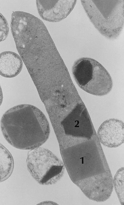
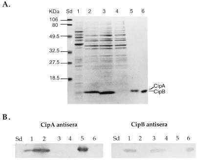
The entomopathogenic bacterium Photorhabdus luminescens exhibits phase variation when cultured in vitro. The variant forms of P. luminescens are pleiotropic and are designated phase I and phase II variants. One of the characteristic phenotypes of phase I cells is the production of two types of intracellular protein inclusions. The genes encoding the protein monomers that form these inclusions, designated cipA and cipB, were cloned and characterized. cipA and cipB encode hydrophobic proteins of 11,648 and 11,308 Da, respectively. The deduced amino acid sequences of CipA and CipB have no significant amino acid sequence similarity to any other known protein but have 25% identity and 49% similarity to each other. Insertional inactivation of cipA or cipB in phase I cells of P. luminescens produced mutants that differ from phase I cells in bioluminescence, the pattern and activities of extracellular products, biochemical traits, adsorption of dyes, and ability to support nematode growth and reproduction. In general, the cip mutants were phenotypically more similar to each other than to either phase I or phase II variants.


Figures

FIG. 1
FIG. 1
Micrograph of sectioned P. luminescens cells. The two different inclusion types (12), designated type 1 and type 2, are composed of CipB and CipA, respectively. Stationary-phase cells of P. luminescens NC1/1 were prepared according to standard methods and examined by transmission electron microscopy at the University of Wisconsin—Madison Electron Microscope Facility. Magnification, ×36,000.
FIG. 2
FIG. 2
Protein and immunoblot analyses of E. coli recombinants expressing CipA and CipB. (A) SDS-PAGE analysis of whole-cell lysates and purified CipA and CipB on a 12% acrylamide gel. Lanes: Sd, molecular weight standards; 1, P. luminescens Hm/1; 2, E. coli DH5α expressing CipA (EC109); 3, E. coli DH5α expressing CipB (EC211); 4, E. coli DH5α control (EC30); 5, purified CipA; 6, purified CipB. The positions of CipA and CipB are indicated. Lanes containing cell lysates and purified inclusion proteins contained 10 and 1 μg of protein, respectively. (B) Corresponding immunoblot analysis of the same samples, using either CipA or CipB antiserum. Lanes are the same as in panel A.
FIG. 3
FIG. 3
(A) Partial nucleotide sequence of the 1,404-bp EcoRI-XbaI fragment of pCA9 which encodes CipA. Amino acids deduced from the nucleotide sequence are specified by standard one-letter abbreviations. The N-terminal amino acid sequence previously determined for CipA is underlined. The positions of putative promoters (−35 and −10 regions) (38), putative ribosome-binding site (36), and the BclI restriction site used for construction of the cipA mutant allele are indicated. Underlined nucleotide sequence downstream of cipA corresponds to the ERIC sequence (23). The positions of a putative stem-loop structure is marked by dashed arrows. (B) Operator-like region identified upstream of cipA. The positions of a putative promoter and ribosome-binding site are indicated. The regions of twofold symmetry are boxed.
FIG. 4
FIG. 4
Partial nucleotide sequence of the 1,624-bp DraI-Sau3AI fragment that contains cipB. Amino acids deduced from the nucleotide sequence are indicated by standard one-letter abbreviations. The N-terminal amino acid sequence previously determined for CipB is underlined. The positions of a putative promoter (−35 and −10 regions) (38), putative ribosome-binding site (36), and the BglII site used for construction of a mutant cipB allele are indicated. The positions of potential stem-loop structures are marked by dashed arrows.
FIG. 5
FIG. 5
Comparison of the deduced amino acid sequences of CipA and CipB. Alignments were constructed using the program BESTFIT from the Genetics Computer Group software package. Identical residues are indicated with vertical lines, and similar residues are indicated with colons. Gaps are represented by dashes.
FIG. 6
FIG. 6
Protein and immunoblot analyses of cipA and cipB mutants of P. luminescens NC1. (A) SDS-PAGE analysis of cell lysates on a 12% acrylamide gel. Lanes: Sd, molecular weight standards; 1, P. luminescens phase I variant (NC1/1); 2, P. luminescens NC1 phase II variant (NC1/2); 3, cipA mutant (NP173); 4, cipB mutant (NP151). Twenty micrograms of each cell lysate was loaded per lane. The positions of CipA and CipB are indicated. (B) Corresponding immunoblot analysis of the same samples, using either CipA or CipB antiserum. Lanes are the same as in panel A.
FIG. 7
FIG. 7
SDS-PAGE analysis of culture filtrates on a 12% acrylamide gel. Lanes: Sd, molecular weight standards; 1, P. luminescens NC1 phase I variant (NC1/1); 2, P. luminescens NC1 phase II variant (NC1/2); 3, cipA mutant (NP173); 4, cipB mutant (NP151). Thirty micrograms of each culture filtrate was loaded per lane.

References

    1. Akhurst R J. Morphological and functional phase variation in Xenorhabdus spp.: bacteria symbiotically associated with the insect pathogenic nematodes Neoplectana and Heterorhabditis. J Gen Microbiol. 1980;121:303–309.
    1. Akhurst R J. Antibiotic activity of Xenorhabdus spp.: bacteria symbiotically associated with insect pathogenic nematodes of the families Heterorhabditidae and Steinernematidae. J Gen Microbiol. 1982;128:3061–3065. - PubMed
    1. Akhurst R J, Boemare N E. A numerical taxonomic study of the genus Xenorhabdus (Enterobacteriaceae) and proposed elevation of the subspecies of X. nematophilus to species. J Gen Microbiol. 1988;134:1835–1845. - PubMed
    1. Akhurst, R. J. Personal communication.
    1. Altschul S F, Gish W, Miller W, Myers E W, Lipman D J. Basic local alignment search tool. J Mol Biol. 1990;215:403–410. - PubMed

Publication types

Substances

Associated data

LinkOut - more resources