

Endotoxins modulate the autocrine function of organ cultured donor corneas and increase the incidence of endothelial cell death
- PMID: 9497472
- PMCID: PMC1722073
- DOI: 10.1136/bjo.81.12.1093
Endotoxins modulate the autocrine function of organ cultured donor corneas and increase the incidence of endothelial cell death
Abstract
Background/aims: Bacterial endotoxin is a potent inflammatory stimulator, the local and systemic responses thereby elicited being mediated via the release of cytokines from diverse cell types. Under physiological conditions, the corneal endothelium is protected from these toxins by the epithelial and vascular barriers, but in organ culture these safeguards are no longer operative, and such substances will therefore have ready access to this cell layer. The consequences of such exposure may take the form of overt damage to the endothelium and/or a more discreet influence on the cornea's immunological status, the effects of which may be realised only after transplantation, by its poor performance. The media bathing organ cultured donor corneas were monitored for the presence of various cytokine mediators of the inflammatory response before and after incubation with endotoxin, and these data compared with those pertaining to endothelial cell morphology and numerical density.
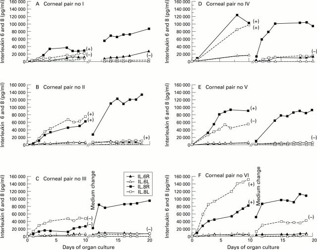
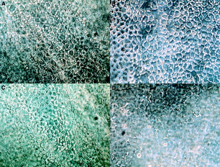
Methods: Six pairs of fellow donor corneas were cultured for an initial equilibration period of 10 days and then transferred to fresh medium; thereafter, one of each pair was incubated in the absence, and the other in the presence, of endotoxin (50 micrograms/ml = 25,000 units/ml), and culturing continued for a further 10 days. Samples of medium were withdrawn at regular intervals throughout the 20 days and screened for the cytokines IL-1, IL-2, IL-4, IL-5, IL-6, IL-8, IL-10, GM-CSF, AND TNF by ELISA; endothelial cell morphology and area density were assessed on days 0, 10, and 20.
Results: Spiking of organ culture media with endotoxin led to a substantial increase in the level of IL-8, and a smaller one in that of IL-6, but none of the other cytokines were detected. In five of the six stimulated corneas, these changes coincided with an increased incidence of endothelial cell loss, compared with that incurred by the fellow control, and the surviving population also evinced signs of degeneration not seen in the latter.
Conclusion: Endotoxin induced increases in the levels of IL-6 and IL-8 appear to be correlated with endothelial cell loss. Since no adverse effects of this toxin on long term cultured monolayers of human corneal endothelial cells have been previously observed, the damage incurred in corneal organ culture may well be attributable to the influence of cytokines produced by other corneal cells or a non-intrinsic (passenger) cell population, such as macrophages, Langerhans cells or lymphocytes present under these latter conditions.
Figures
References
Publication types
MeSH terms
Substances
LinkOut - more resources
Full Text Sources