. 1998 Mar;72(3):1711-24.
doi: 10.1128/JVI.72.3.1711-1724.1998.

African swine fever virus infection in the argasid host, Ornithodoros porcinus porcinus

Affiliations

African swine fever virus infection in the argasid host, Ornithodoros porcinus porcinus

S B Kleiboeker et al. J Virol. 1998 Mar.

Abstract

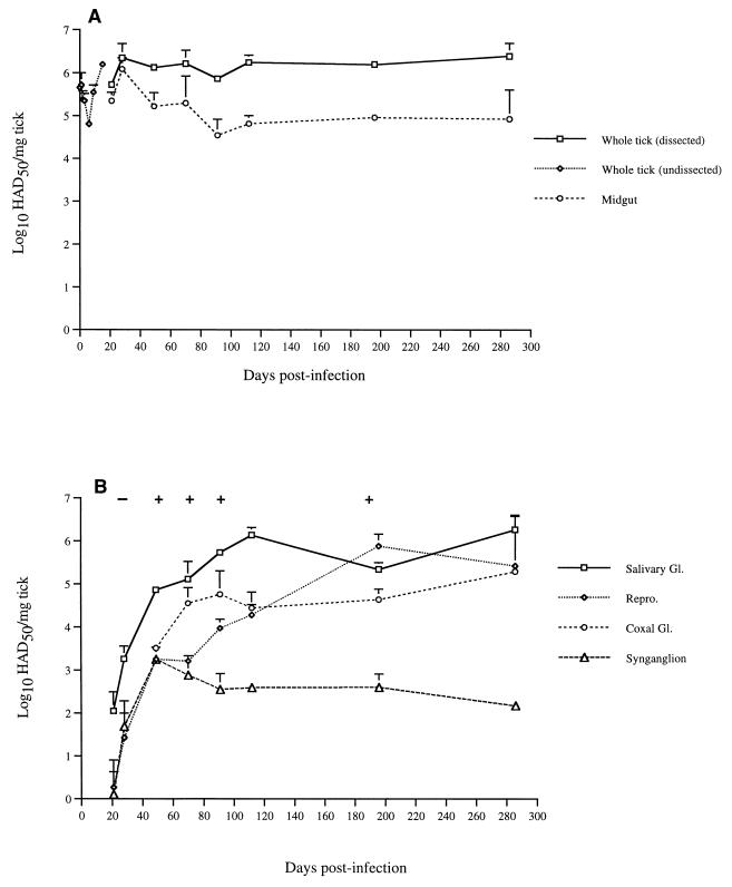
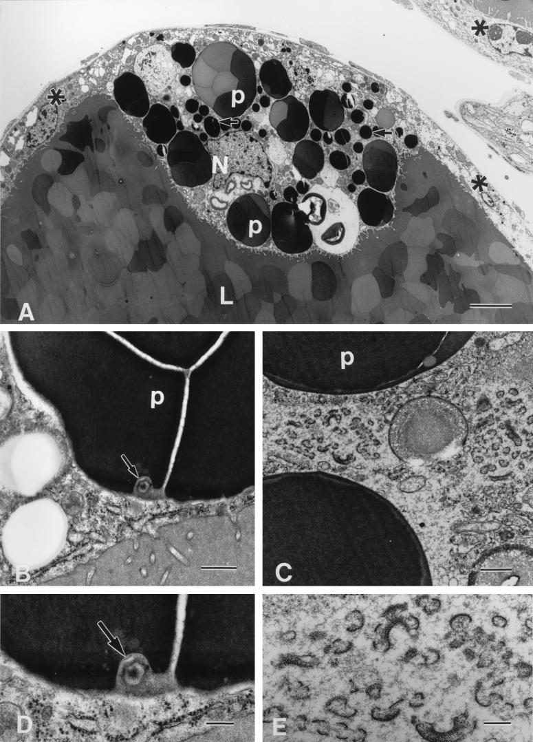
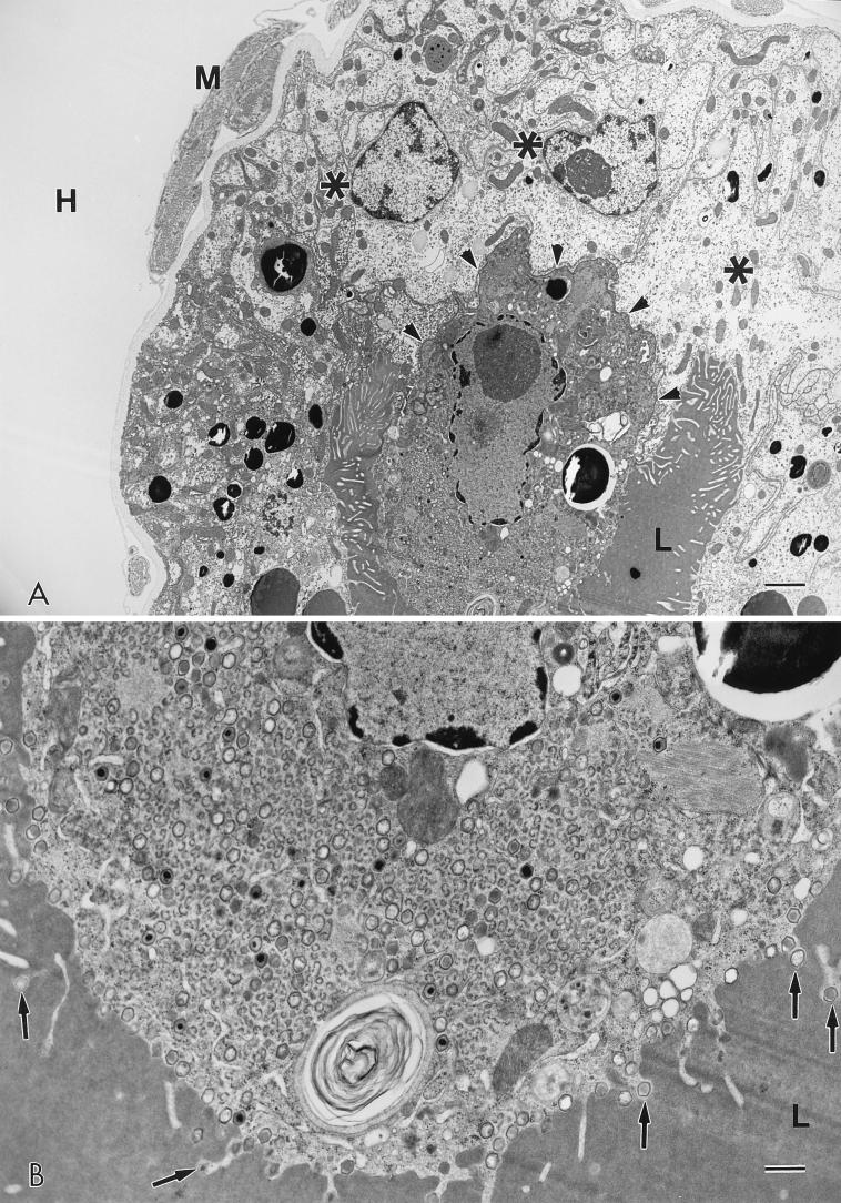
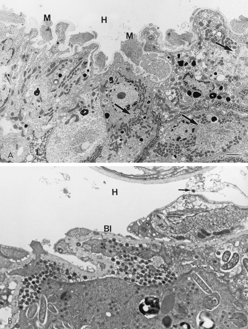
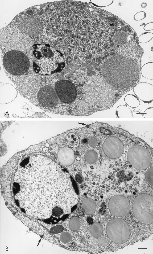
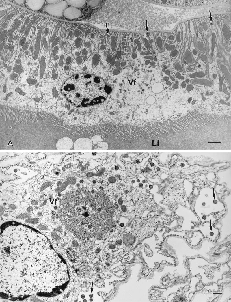
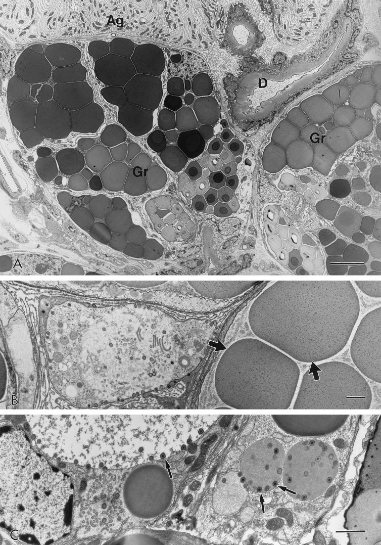
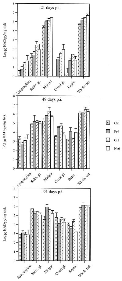
The pathogenesis of African swine fever virus (ASFV) infection in Ornithodoros porcinus porcinus was examined in nymphal ticks infected with the ASFV isolate Chiredzi/83/1. At times postinfection (p.i.) ranging from 6 h to 290 days, ticks or dissected tick tissues were titrated for virus and examined ultrastructurally for evidence of virus replication. The ASFV infection rate in ticks was 100% in these experiments, and virus infection was not associated with a significant increase in tick mortality. Initial ASFV replication occurred in phagocytic digestive cells of the midgut epithelium. Subsequent infection and replication of ASFV in undifferentiated midgut cells was observed at 15 days p.i. Generalization of virus infection from midgut to other tick tissues required 2 to 3 weeks and most likely involved virus movement across the basal lamina of the midgut into the hemocoel. Secondary sites of virus replication included hemocytes (type I and II), connective tissue, coxal gland, salivary gland, and reproductive tissue. Virus replication was not observed in the nervous tissue of the synganglion, Malpighian tubules, and muscle. Persistent infection, characterized by active virus replication, was observed for all involved tick tissues. After 91 days p.i., viral titers in salivary gland and reproductive tissue were consistently the highest detected. Successful tick-to-pig transmission of ASFV at 48 days p.i. correlated with high viral titers in salivary and coxal gland tissue and their secretions. A similar pattern of virus infection and persistence in O. porcinus porcinus was observed for three additional ASFV tick isolates in their associated ticks.


Figures

FIG. 1
FIG. 1
ASFV titers in Ch1-infected ticks. Whole ticks and dissected tick midguts (A) plus additional tissues from the same ticks (B) were titered at times p.i. Values are expressed as mean titers ± standard errors of the means. Unsuccessful (−) and successful (+) tick-to-pig transmission attempts are indicated.
FIG. 2
FIG. 2
ASFV uptake and early stages of replication in midgut digestive cells. (A) At 24 h postfeeding, intact erythrocytes have been taken into phagolysosomes (p) from the blood meal in the lumen (L). Hematin crystals (arrows) are scattered throughout the cytoplasm. Undifferentiated epithelial cells (asterisks) occur on either side of the digestive cell. N, nucleus of digestive cell. Bar, 5 μm. (B) Phagolysosome (p) of a digestive cell 72 h postfeeding. Ingested erythrocyte has an enveloped ASFV particle associated with it (arrow). Bar, 0.5 μm. (C) Nascent virus factory in digestive cell at 96 h postfeeding. Curvilinear forms of assembling virus particles occur in a more electron-lucent region of the cytoplasm. p, phagolysosomes; bar, 0.5 μm. (D) High-magnification view of ASFV particle (arrow) in panel B showing possible deterioration of virus structure. Bar, 0.2 μm. (E) High-magnification view of nascent virus factory in panel C. Bar, 0.2 μm.
FIG. 3
FIG. 3
Replication of ASFV in midgut digestive cells at 15 days postfeeding. (A) A digestive cell with an extensive virus factory projects into the midgut lumen (L) and is separated from undifferentiated epithelial cells (asterisks) via septate junctions (arrowheads). H, hemocoel; M, muscle; bar, 5 μm. (B) High-magnification view of the mature virus factory in panel A showing virus in various stages of assembly. Virions (arrows) are budding into the lumen (L). Bar, 0.5 μm.
FIG. 4
FIG. 4
Replication and generalization of ASFV in undifferentiated midgut cells at 21 days postfeeding. (A) Three undifferentiated epithelial cells with virus factories (large arrows). M, muscle; H, hemocoel; L, lumen; bar, 5 μm. (B) Accumulation of ASFV under the basal lamina (Bl). Arrow, virus free in the hemocoel (H); bar, 1 μm.
FIG. 5
FIG. 5
ASFV in hemocytes. (A) Type I hemocyte containing an extensive virus factory with many crystalline arrays at the periphery. Bar, 1 μm. (B) Type II hemocyte with virus factory and budding virus particles (arrows). Bar, 0.5 μm.
FIG. 6
FIG. 6
ASFV in coxal gland. (A) A small virus factory (Vf), mature particles (arrows), and a condensed-chromatin-containing nucleus in the proximal tubule of the coxal gland. Lt, lumen of tubule; bar, 1 μm. (B) Cell of the filtration membrane portion of coxal gland with a virus factory (Vf) and released virions (arrows). Bar, 0.5 μm.
FIG. 7
FIG. 7
ASFV in salivary gland. (A) Mock-infected salivary gland at 30 days postfeeding to show tissue structure. The salivary gland is composed of agranular (Ag) and granular (Gr) portions. Connective tissue cells occur at the edge and in the center of the gland. D, cuticle-lined ducts. (B) At 30 days p.i., replicating ASFV is found only in the connective tissue cells in the center of the gland. Secretory granules (arrows) do not contain virus. (C) At 42 days p.i., virus is observed budding into secretory granules (arrows). Bars: panel A, 5 μm; panels B and C, 1 μm.
FIG. 8
FIG. 8
Tissue titrations of ASFV-infected O. porcinus porcinus ticks. O. porcinus porcinus ticks (stages N2 to N4) collected from natural habitat were infected with ASFV isolates (Pr4, Cr1, and No6). At times p.i., ticks were dissected and tissue titers were determined. Values are expressed as mean titers ± standard errors of the means. Saliv., salivary; gl., gland.

References

    1. Booth T F, Davies C R, Jones L D, Staunton D, Nuttall P A. Anatomical basis of Thogoto virus infection in BHK cell culture and in the Ixodid tick vector, Rhipicephalus appendiculatus. J Gen Virol. 1989;70:1093–1104. - PubMed
    1. Booth T F, Steele G M, Marriott A C, Nuttall P A. Dissemination, replication, and trans-stadial persistence of Dugbe virus (Nairovirus, Bunyaviridae) in the tick vector Amblyomma variegatum. Am J Trop Med Hyg. 1991;45:146–157. - PubMed
    1. Borca, M. V., C. Carrillo, L. Zsak, W. W. Laegreid, G. F. Kutish, J. G. Neilan, and D. L. Rock. Deletion of a CD2-like gene from African swine fever virus affects viral pathogenesis in domestic swine. Submitted for publication. - PMC - PubMed
    1. Brown F. The classification and nomenclature of viruses: summary of results of meetings of the International Committee on Taxonomy of Viruses in Sendai, September 1984. Intervirology. 1986;25:141–143. - PubMed
    1. Chernesky M A, McLean D M. Localization of Powassan virus in Dermacentor andersoni ticks by immunofluorescence. Can J Microbiol. 1969;15:1399–1408. - PubMed

LinkOut - more resources