. 1998 Mar;72(3):1925-30.
doi: 10.1128/JVI.72.3.1925-1930.1998.

Mapping the interacting domains between the rabies virus polymerase and phosphoprotein

Affiliations

Mapping the interacting domains between the rabies virus polymerase and phosphoprotein

M Chenik et al. J Virol. 1998 Mar.

Abstract

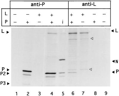
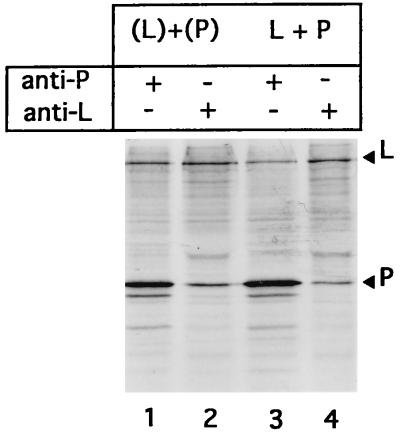
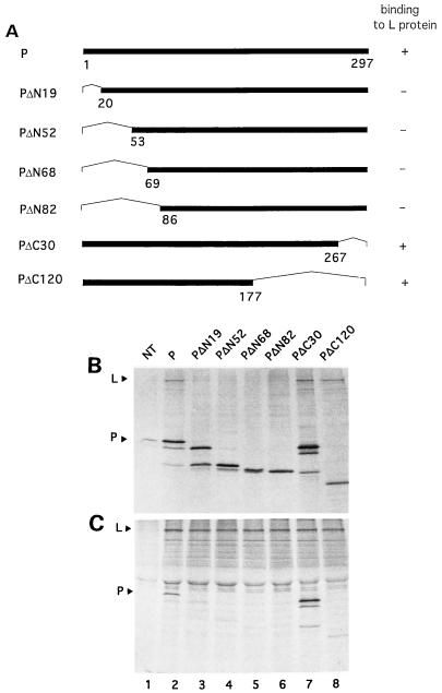
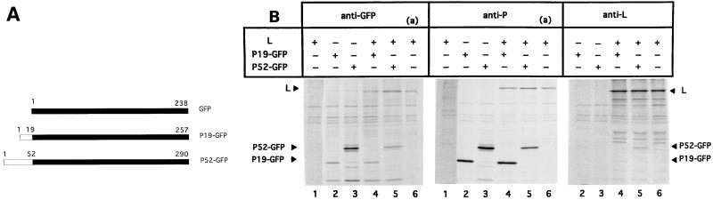
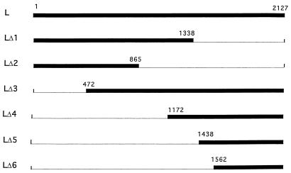
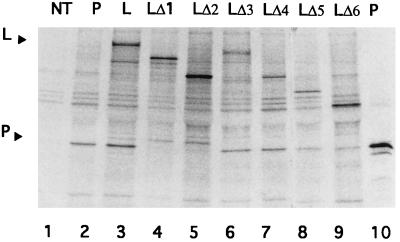
The RNA polymerase of rabies virus consists of two subunits, the large (L) protein and the phosphoprotein (P), with 2,127 and 297 amino acids, respectively. When these proteins were coexpressed via the vaccinia virus-T7 RNA polymerase recombinant in mammalian cells, they formed a complex as detected by coimmunoprecipitation. Analysis of P and L deletion mutants was performed to identify the regions of both proteins involved in complex formation. The interaction of P with L was not disrupted by large deletions removing the carboxy-terminal half of the P protein. On the contrary, P proteins containing a deletion in the amino terminus were defective in complex formation with L. Moreover, fusion proteins containing the 19 or the 52 first residues of P in frame with green fluorescent protein (GFP) still bound to L. These results indicate that the major L binding site resides within the 19 first residues of the P protein. We also mapped the region of L involved in the interaction with P. Mutant L proteins consisting of the carboxy-terminal 1,656, 956, 690, and 566 amino acids all bound to the P protein, whereas deletion of 789 residues within the terminal region eliminated binding to P protein. This result demonstrates that the carboxy-terminal domain of L is required for the interaction with P.


Figures

FIG. 1
FIG. 1
Detection of L-P complex in infected and transfected cells by immunoprecipitation. BSR cells were infected with rabies virus (i; lane 5) or with vTF7-3 (lanes 1 to 4 and 6 to 9). vTF7-3-infected cells were then either transfected with 5 μg of a plasmid encoding the P (lanes 2 and 8) or L (lanes 3 and 7) protein or cotransfected with both plasmids (lanes 4 and 6). At 24 h after infection or transfection, proteins were labeled with [35S]methionine for 3 h. Cell extracts were prepared and immunoprecipitated with A17 anti-P (lanes 1 to 5) and anti-L (lanes 6 to 9) antibodies. Immune complexes were collected with protein A-Sepharose and analyzed by SDS-PAGE (12% gel).
FIG. 2
FIG. 2
P-L complex formation in the absence of coexpression. A mixture of cytoplasmic extracts from cells separately transfected with P and L plasmids [(L) + (P)] or cell extracts from cotransfected cells (L+P) were immunoprecipitated with anti-P (lanes 1 and 3) and anti-L (lanes 2 and 4) antibodies. The immunoprecipitates were analyzed by SDS-PAGE (14% gel).
FIG. 3
FIG. 3
Mapping the L binding site on the rabies virus P protein. (A) Schematic representation of the truncated P proteins. Dark bars represent the protein product of each deleted P gene, with amino acid positions indicated. The thin angled lines indicate deleted regions. The plasmids encoding truncated P proteins have been described elsewhere (7, 8). P-L binding data are summarized at the right. (B and C) Analysis of interaction between the L protein and the truncated P proteins by immunoprecipitation. Cells were not transfected (NT; lane 2) or infected with vTF7-3 (lanes 1 to 8) and then cotransfected with 5 μg of plasmids encoding the complete L and P proteins (lane 2) or truncated P proteins PΔN19 (lane 3), PΔN52 (lane 4), PΔN68 (lane 5), PΔN82 (lane 6), PΔC30 (lane 7), and PΔ120 (lane 8). Cell extracts (35S labeled) were immunoprecipitated with A17 anti-P antibody (B) or anti-L antibody (C). The immunoprecipitates were analyzed by SDS-PAGE (14% gel).
FIG. 4
FIG. 4
The first 19 amino acids of P are sufficient for L-P interaction. (A) Schematic representation of the P-GFP fusion proteins. The 19 or 52 amino-terminal residues of P (white bars) are fused to GFP (dark bars); amino acids are indicated. (B) Cells were infected with vTF7-3 (lanes 1 to 6). vTF7-3-infected cells were then transfected with 5 μg of a plasmid encoding the complete L protein (lanes 1 and 6), P19-GFP (lanes 2), or P52-GFP (lanes 3). Cells were also cotransfected with plasmids encoding L and P19-GFP (lanes 4) or P52-GFP (lanes 5). Cell extracts (35S labeled) were immunoprecipitated with the indicated antibodies (anti-GFP, 25E6 anti-P, and anti-L). Cell extracts from transfected cells with a plasmid encoding L protein were also immunoprecipitated with the anti-L antibody to indicate the position of the L protein [(a)]. The precipitated proteins were analyzed by SDS-PAGE (14% gel). Longer exposures of the controls are included (lanes 1).
FIG. 5
FIG. 5
Schematic representation of the truncated L proteins. Dark bars represent the protein product of each deleted L gene, with amino acid positions indicated. The thin lines indicate deleted regions.
FIG. 6
FIG. 6
Mapping the L binding site on the rabies virus P protein. Cells were not transfected (NT; lane 1) or infected with vTF7-3 (lanes 1 to 10) and then cotransfected with 5 μg of plasmids expressing the complete P and L proteins (lane 3) or the complete P protein and truncated L proteins LΔ1 (lane 4), LΔ2 (lane 5), LΔ3 (lane 6), LΔ4 (lane 7), LΔ5 (lane 8), and LΔ6 (lane 9). Cell extracts (35S labeled) were immunoprecipitated with anti-L antibody (lanes 1 and 3 to 9). Extracts of cells transfected with plasmid encoding the complete P protein (lanes 2 and 10) were also immunoprecipitated with anti-P MAb 25E6 as described above (lane 2) and MAb A17 (lane 10). The immunoprecipitates were analyzed by SDS-PAGE (12% gel).

References

    1. Banerjee A K. Transcription and replication of rhabdoviruses. Microbiol Rev. 1987;51:66–87. - PMC - PubMed
    1. Barik S, Banerjee A K. Phosphorylation by cellular casein kinase II is essential for the transcriptional activity of vesicular stomatitis virus phosphoprotein P. Proc Natl Acad Sci USA. 1992;89:6570–6574. - PMC - PubMed
    1. Barik S, Banerjee A K. Sequential phosphorylation of the phosphoprotein of vesicular stomatitis virus by cellular and viral protein kinases is essential for transcription activation. J Virol. 1992;66:1109–1118. - PMC - PubMed
    1. Canter D M, Jackson R L, Perrault J. Faithful and efficient in vitro reconstitution of vesicular stomatitis virus transcription using plasmid-encoded L and P proteins. J Virol. 1993;70:4538–4548. - PubMed
    1. Canter D M, Perrault J. Stabilization of vesicular stomatitis virus L polymerase protein by P protein binding: a small deletion in the C-terminal domain of L abrogates binding. Virology. 1996;219:376–386. - PubMed

Publication types

MeSH terms

LinkOut - more resources