. 1998 Mar;72(3):1974-82.
doi: 10.1128/JVI.72.3.1974-1982.1998.

The genome of salmonid herpesvirus 1

Affiliations

The genome of salmonid herpesvirus 1

A J Davison. J Virol. 1998 Mar.

Abstract

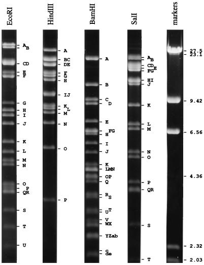
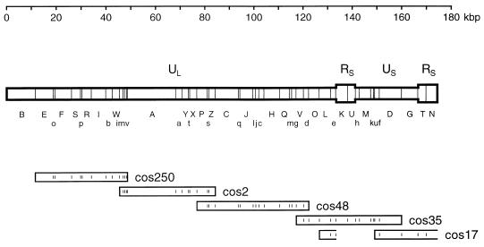
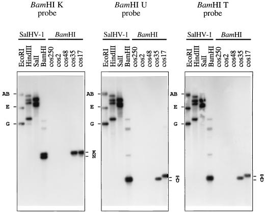
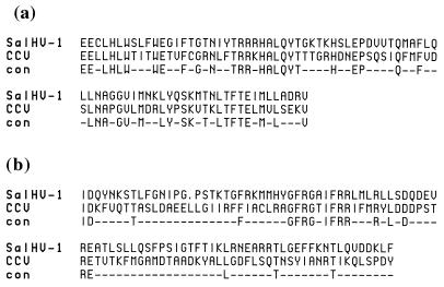
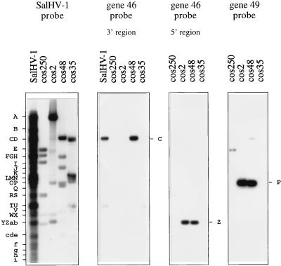
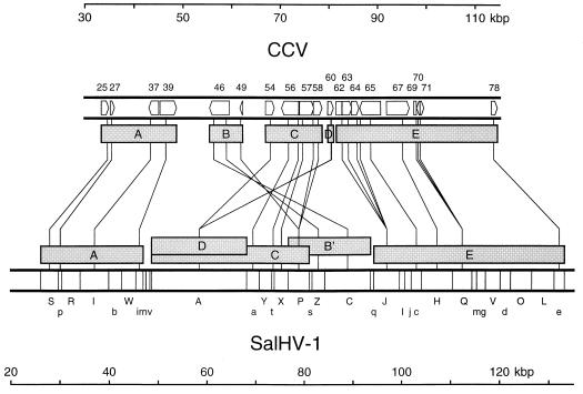
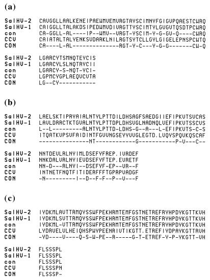
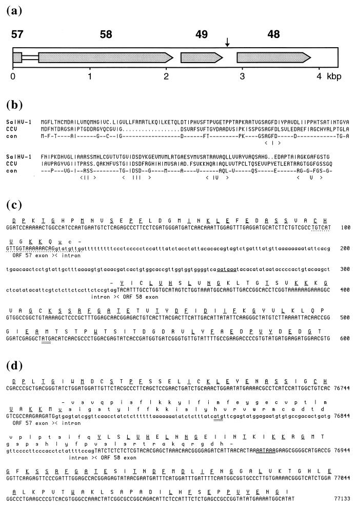
Salmonid herpesvirus 1 (SalHV-1) is a pathogen of the rainbow trout (Oncorhynchus mykiss). Restriction endonuclease mapping, cosmid cloning, DNA hybridization, and targeted DNA sequencing experiments showed that the genome is 174.4 kbp in size, consisting of a long unique region (U(L); 133.4 kbp) linked to a short unique region (U(S); 25.6 kbp) which is flanked by an inverted repeat (R(S); 7.7 kbp). U(S) is present in virion DNA in either orientation, but U(L) is present in a single orientation. This structure is characteristic of the Varicellovirus genus of the subfamily Alphaherpesvirinae but has evidently evolved independently, since an analysis of randomly sampled DNA sequence data showed that SalHV-1 shares at least 18 genes with channel catfish virus (CCV), a fish herpesvirus whose complete sequence is known and which is unrelated to mammalian herpesviruses. The use of oligonucleotide probes demonstrated that in comparison with CCV, the conserved SalHV-1 genes are located in U(L) in at least five rearranged blocks. Large-scale gene rearrangements of this type are also characteristic of the three mammalian herpesvirus subfamilies. The junction between two SalHV-1 gene blocks was confirmed by sequencing a 4,245-bp region which contains the dUTPase gene, part of a putative spliced DNA polymerase gene, and one other complete gene. The implications of these findings in herpesvirus taxonomy are discussed.


Figures

FIG. 1
FIG. 1
Restriction fragments of SalHV-1 DNA visualized by short-wavelength UV irradiation on a 0.5% agarose gel stained with ethidium bromide. Sizes are indicated in kilobase pairs.
FIG. 2
FIG. 2
Genome structure of SalHV-1. One of the two genome isomers is shown and is defined as the prototype. BamHI sites are marked, and fragment nomenclature is shown below in two ranks (A to Z and a to v). The inverted repeat (RS) is shown in a wider format than nonreiterated regions (UL and US). The locations of five cosmids are shown as rectangles below the fragment nomenclature, and BamHI sites are indicated. cos17 arose from a genome molecule in which US was inverted and thus appears to proceed from the right end of UL, through RS and into the right end of US, as indicated by the open ends of the rectangles.
FIG. 3
FIG. 3
Examples of hybridization data for an inverting region (US) flanked by an inverted repeat (RS) in the SalHV-1 genome. Restriction fragments of SalHV-1 DNA or cosmids were transferred from a 0.6% agarose gel and probed with radiolabeled BamHI K, U, or T at 75°C. Hybridizing BamHI fragments are indicated to the right of each panel, and EcoRI fragments are indicated to the left.
FIG. 4
FIG. 4
Amino acid sequence alignments of random SalHV-1 DNA sequences conceptually translated in appropriate reading frames with the CCV ORF 37 protein (residues 534 to 615) (a) and the CCV ORF 54 protein (residues 306 to 399) (b). Identical residues are indicated in the “con” line.
FIG. 5
FIG. 5
Examples of oligonucleotides hybridizing to SalHV-1 DNA. BamHI fragments of SalHV-1 or cosmids were transferred from a 0.8% agarose gel and probed with radiolabeled SalHV-1 DNA at 65°C or oligonucleotides at 42°C. Fragments to which oligonucleotides hybridized are shown to the right of each panel, and SalHV-1 BamHI fragments are shown on the left.
FIG. 6
FIG. 6
Scale representation of relative gene order in SalHV-1 and CCV. The locations and orientations of the 18 genes in the 30- to 115-kbp region of the 134-kbp CCV genome are shown at the top; ORFs 62, 69, and 71 are exons of a single gene. BamHI fragments in the 20- to 135-kbp region of the SalHV-1 genome are shown at the bottom. Regions in the CCV genome corresponding to SalHV-1 oligonucleotide probes are connected by lines to the centers of the SalHV-1 fragments to which they hybridized. Five gene blocks (A to E) are shown as shaded rectangles below the CCV genome, and corresponding blocks are shown above the SalHV-1 genome (B′ indicating an inversion).
FIG. 7
FIG. 7
Amino acid sequence alignments of conceptual translation products of random SalHV-1 DNA sequences with their counterparts in the SalHV-2 genome and the CCV gene 46 protein at residues 180 to 246 (a) and residues 759 to 837 (b) and the CCV ORF 62 protein at residues 345 to 401 (c). Residues conserved between SalHV-1 and SalHV-2 are indicated in the “con” line; those conserved between both viruses and CCV are indicated in the “CON” line.
FIG. 8
FIG. 8
(a) Summary of gene order in SalHV-1 BamHI P displayed in the same orientation as the genome layout depicted in Fig. 2. Predicted protein coding regions are shaded, and the proposed intron linking ORFs 57 and 58 is shown as a white rectangle. An AATAAA element potentially involved in polyadenylation is shown as a vertical arrow. (b) Alignment of the putative amino acid sequences of SalHV-1 and CCV dUTPases. Conserved residues are shown in the “con” line. Five recognized dUTPase motifs (I to V) are indicated (12). The CCV protein is shown as commencing at the second ATG codon in the relevant reading frame. (c and d) Locations of potential splice sites linking ORFs 57 and 58 in SalHV-1 (c) and CCV (d). The first 600 bp of SalHV-1 BamHI P and the corresponding region in the CCV genome are shown, proceeding from the 3′ end of ORF 57 through the 5′ end of ORF 58. Potential exons and introns are marked. Translated sequences are bracketed by stop codons which define the 3′ and 5′ limits of ORFs 57 and 58, respectively; the two ORFs overlap in CCV. The first ATG codon in ORF 58 is doubly underlined in each sequence. Conserved amino acid residues are singly underlined. Nucleic acid and amino acid residues outside the putative exons are shown in lowercase. AATAAA elements that could signal polyadenylation of transcripts from ORF 57 are underlined. The sequence corresponding to the oligonucleotide used to locate the 3′ end of SalHV-1 ORF 57 is underlined by dots.

References

    1. Benton M J. Vertebrate palaeontology. London, England: Harper Collins Academic; 1990. pp. 123–144.
    1. Bernard J, Mercier A. Sequence of two Eco RI fragments from salmonis herpesvirus 2 and comparison with ictalurid herpesvirus 1. Arch Virol. 1993;132:437–442. - PubMed
    1. Booy F P, Trus B L, Davison A J, Steven A C. The capsid architecture of channel catfish virus, an evolutionary distant herpesvirus, is largely conserved in the absence of discernible sequence homology with herpes simplex virus. Virology. 1996;215:134–141. - PubMed
    1. Chee M S, Bankier A T, Beck S, Bohni R, Brown C M, Cerny R, Horsnell T, Hutchison C A, III, Kouzarides T, Martignetti J A, Preddie E, Satchwell S C, Tomlinson P, Weston K M, Barrell B G. Analysis of the protein-coding content of the sequence of human cytomegalovirus strain AD169. Curr Top Microbiol Immunol. 1990;154:125–169. - PubMed
    1. Cunningham C, Davison A J. A cosmid-based system for constructing mutants of herpes simplex virus type 1. Virology. 1993;197:116–124. - PubMed

Publication types

Associated data

LinkOut - more resources