. 1998 Mar;72(3):1983-93.
doi: 10.1128/JVI.72.3.1983-1993.1998.

Nucleocapsid and matrix protein contributions to selective human immunodeficiency virus type 1 genomic RNA packaging

Affiliations

Nucleocapsid and matrix protein contributions to selective human immunodeficiency virus type 1 genomic RNA packaging

D T Poon et al. J Virol. 1998 Mar.

Abstract

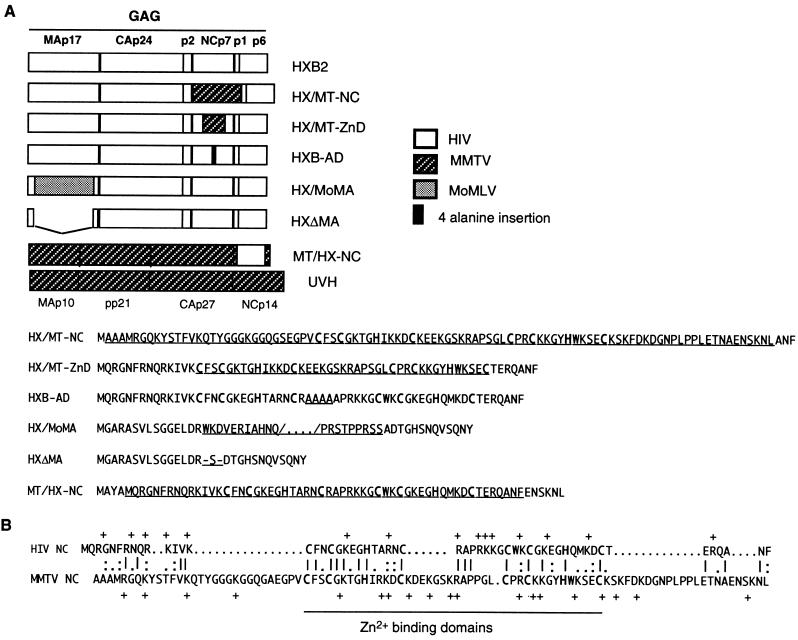
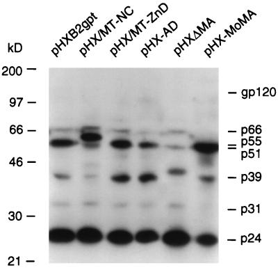
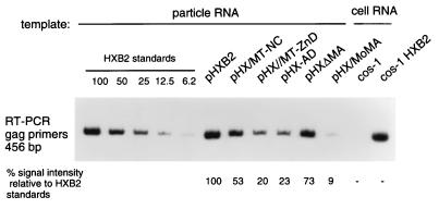
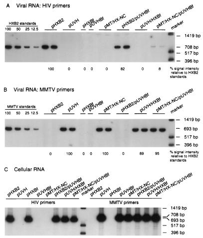
The nucleocapsid protein (NC) of retroviruses plays a major role in genomic RNA packaging, and some evidence has implicated the matrix protein (MA) of certain retroviruses in viral RNA binding. To further investigate the role of NC in the selective recognition of genomic viral RNA and to address the potential contribution of MA in this process, we constructed chimeric and deletion human immunodeficiency virus type 1 (HIV-1) mutants that alter the NC or MA protein. Both HIV and mouse mammary tumor virus (MMTV) NC proteins have two zinc-binding domains and similar basic amino acid compositions but differ substantially in total length, amino acid sequence, and spacing of the zinc-binding motifs. When the entire NC coding sequence of HIV was replaced with the MMTV NC coding sequence, we found that the HIV genome was incorporated into virions at 50% of wild-type levels. Viruses produced from chimeric HIV genomes with complete NC replacements, or with the two NC zinc-binding domains replaced with MMTV sequences, preferentially incorporated HIV genomes when both HIV and MMTV genomes were simultaneously present in the cell. Viruses produced from chimeric MMTV genomes in which the MMTV NC had been replaced with HIV NC preferentially incorporated MMTV genomes when both HIV and MMTV genomes were simultaneously present in the cell. In contrast, viruses produced from chimeric HIV genomes containing the Moloney NC, which contains a single zinc-binding motif, were previously shown to preferentially incorporate Moloney genomic RNA. Taken together, these results indicate that an NC protein with two zinc-binding motifs is required for specific HIV RNA packaging and that the amino acid context of these motifs, while contributing to the process, is less crucial for specificity. The data also suggest that HIV NC may not be the exclusive determinant of RNA selectivity. Analysis of an HIV MA mutant revealed that specific RNA packaging does not require MA protein.


Figures

FIG. 1
FIG. 1
(A) Schematic representation of Gag mutants and alignment of NC amino acids. The amino acid sequences of HIV-1 and MMTV NC chimeras and the relevant portions of MA protein mutants are indicated for each construct. (B) Sequence alignment for HIV and MMTV NC proteins.
FIG. 2
FIG. 2
Analysis of the protein content of HIV-1 mutant particles by Western blotting. Viral protein lysates, normalized to contain comparable amounts of p24, were subjected to SDS-PAGE (10% gel), transferred to nitrocellulose, and probed with HIV-1-positive human serum. The construct used to generate each lysate is indicated above the relevant lane. Viral proteins were visualized after 12 h of autoradiography.
FIG. 3
FIG. 3
Electron micrographs of transfected cos-1 cells. (A) Particles derived from construct pHXB2gpt; (B) particles derived from construct pHX/MT-NC; (C) particles derived from construct pHX/MT-ZnD; (D) particles derived from construct pMT/HX-NC. Pictures were taken at a magnification of ×18,000 for all panels.
FIG. 4
FIG. 4
Relative nucleic acid content of viral particles determined by RT-PCR. RNA incorporation was measured for all mutant viruses in duplicate in three independent experiments using this assay, and the average values are summarized in Table 3; the results of one representative experiment are shown. RNA samples from pelleted virions containing equal amounts of p24 were reverse transcribed by using primer HIVc1686 and subjected to PCR amplification in the presence of [32P]dCTP, using the HIVc1686 primer paired with primer HIV1230. The negative control included a sample from cos-1 cellular RNA. As a positive control, a sample from an RT-PCR with RNA extracted from cos-1 cells transfected with a wild-type HIV-1 plasmid was included. Equal volumes of RT-PCR samples were subjected to PAGE and autoradiography. The intensity of a band corresponding to the amplification of genomic RNA from mutated viruses was compared to the intensity of bands produced by amplifying serially diluted RNA from the wild-type parental virus HXB2gpt (standards).
FIG. 5
FIG. 5
RNase protection assay on cellular and viral RNAs. (A) Schematic representation of the radiolabeled antisense riboprobe obtained from plasmid pGAC linearized with AvaI. It extends from the ClaI site through the first splice donor (SD) site and beyond the viral transcription initiation site to the AvaI site. The predicted protected fragment sizes are also indicated. LTR, long terminal repeat. (B) RNase protection assay on cellular and viral RNAs to simultaneously detect spliced and unspliced messengers. Ten-microgram aliquots of the total cellular RNA sample isolated from each transfection and of a viral RNA sample equivalent to 100 ng of p24, isolated from virions present in each supernatant, were annealed to 1 ng (4.6 × 10⁴ cpm/ng) of the riboprobe. The fastest-migrating band appearing in the panel is an artifact of the assay. The intensity of each band was quantitated as described in Materials and Methods.
FIG. 6
FIG. 6
Relative homologous and heterologous RNA content of HIV particles determined by RT-PCR. RNA samples corresponding to equal amounts of p24 were reverse transcribed by using primer HIVc1686 (A) or primer MMTVc2234 (B). Equivalent aliquots of the RT reaction were subjected to PCR amplification in the presence of [32P]dCTP. Equal volumes of RT-PCR samples were subjected to PAGE and autoradiography. Two experiments were performed on RNA samples derived from two independent transfections, and the results shown are from one set of RNAs. Each sample was tested in duplicate. (A) Detection of genomic HIV-1 RNAs in a subset of mutant viruses. Primer HIVc1686 was paired with primer HIV979 to amplify a fragment of 708 bp. The intensity of bands was quantitated as described for Fig. 4. (B) Detection of genomic MMTV RNAs in a subset of mutant viruses. Primer MMTVc2234 was paired with primer MMTV1542 to amplify a band of 693 bp. (C) Amplification of cell-derived HIV-1 and MMTV genomic RNAs upon transfection. Primer HIVc1686 was paired with primer HIV979 to amplify a fragment of 708 bp. Primer MMTVc2234 was paired with primer MMTV1542 to amplify a band of 693 bp.
FIG. 7
FIG. 7
Amplification of HIV and MMTV fragments selected for quantitative analysis of HIV and MMTV RNA incorporation. (A) Representative PCR amplification using equimolar amounts of HIV and MMTV plasmid DNA templates. The intensity of bands after 14, 16, 18, and 20 cycles is shown. (B) Representative RT-PCR amplification using equimolar amounts of HIV and MMTV in vitro-transcribed RNA templates. The intensity of bands after 14, 16, 18, and 20 cycles is shown. Each sample was tested in duplicate, and the PhosphorImager values were for HIV 569,824 (14 cycles), 1,525,234 (16 cycles), 2,937,182 (18 cycles), and 3,932,910 (20 cycles) and for MMTV 546,690 (14 cycles), 1,664,639 (16 cycles), 3,373,286 (18 cycles), and 4,224,588 (20 cycles). (C) Estimates of the intensity of the bands representing amplified fragments after various amplification cycles. The level of signal detected in each fragment after 20 cycles was defined as 100%. Values detected at 18, 16, and 14 cycles are expressed as the percentage of the value at 20 cycles. The percentages and their standard errors are the average of four independent experiments. Percentage, instead of scanned values, was used to avoid variability in numbers due to the use of isotopes with different specific activities in different experiments. (D) Curves representing the percentage of incorporated radioactivity at 14, 16, 18, and 20 cycles of RT-PCR (the thin line represents the HIV curve; the thick line represents the MMTV curve).
FIG. 8
FIG. 8
Analysis of the protein content of MMTV-1 particles by Western blotting. Viral protein lysates, normalized to contain comparable amounts of MMTV particles, were subjected to SDS-PAGE (10% gel), transferred to nitrocellulose, and probed with an MMTV-positive polyclonal rabbit serum. The construct used to generate each lysate is indicated above the relevant lane. Viral proteins were visualized after 24 h of autoradiography. The intensity of the bands representing gp78, gp70, gp50, and p27 was quantitated by using a Molecular Dynamics PhosphorImager with ImageQuant software (Molecular Dynamics).
FIG. 9
FIG. 9
Relative homologous and heterologous RNA content of MMTV particles determined by RT-PCR. RNA samples corresponding to equal amounts of p24 (in the case of HIV particles) or equal amounts of protein lysates (in the case of MMTV particles) were reverse transcribed by using primer HIVc1686 (A) or primer MMTVc2234 (B). Equivalent aliquots of the RT reaction were subjected to PCR amplification in the presence of [32P]dCTP. Equal volumes of RT-PCR samples were subjected to PAGE and autoradiography. Two experiments were performed on RNA samples derived from two independent transfections, and the results shown are from one set of RNAs. Each sample was tested in duplicate. (A) Detection of genomic HIV-1 RNAs in a subset of mutant viruses. Primer HIVc1686 was paired with primer HIV979 to amplify a fragment of 708 bp. The intensity of bands was quantitated as described for Fig. 4. (B) Detection of genomic MMTV RNAs in a subset of mutant viruses. Primer MMTVc2234 was paired with primer MMTV1542 to amplify a band of 693 bp. (C) Amplification of cell-derived HIV-1 and MMTV genomic RNAs upon transfection. Primer HIVc1686 was paired with primer HIV979 to amplify a fragment of 708 bp. Primer MMTVc2234 was paired with primer MMTV1542 to amplify a band of 693 bp.

Similar articles

Cited by

References

    1. Aldovini A, Young R A. Mutations of RNA and protein sequences involved in human immunodeficiency virus type 1 packaging result in production of noninfectious virus. J Virol. 1990;64:1920–1926. - PMC - PubMed
    1. Aldovini A, Feinberg M B. Transfection of molecularly cloned HIV genomes. In: Aldovini A, Walker B D, editors. Techniques in HIV research. New York, N.Y: Stockton Press, Inc.; 1990. pp. 147–175.
    1. Aldovini A, Young R A. Construction and analysis of HIV and SIV mutants. Methods Mol Genet. 1994;4A:3–17.
    1. Ausubel F M, Brent R, Kingston R E, Moore D D, Seidman J G, Smith J A, Struhl K, editors. Current protocols in molecular biology. New York, N.Y: John Wiley and Sons, Inc.; 1987.
    1. Bennet R P, Nelle T D, Willis J W. Functional chimeras of the Rous sarcoma virus and human immunodeficiency virus Gag proteins. J Virol. 1993;67:6487–6498. - PMC - PubMed

Publication types

MeSH terms

Substances

LinkOut - more resources