. 1998 Mar;72(3):1994-2001.
doi: 10.1128/JVI.72.3.1994-2001.1998.

Association of the human papillomavirus type 11 E1 protein with histone H1

Affiliations

Association of the human papillomavirus type 11 E1 protein with histone H1

C S Swindle et al. J Virol. 1998 Mar.

Abstract

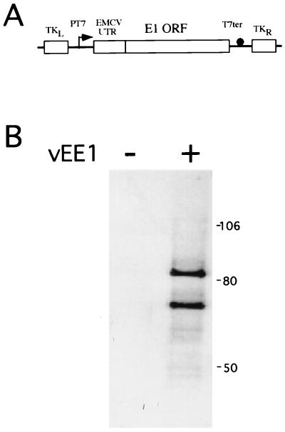
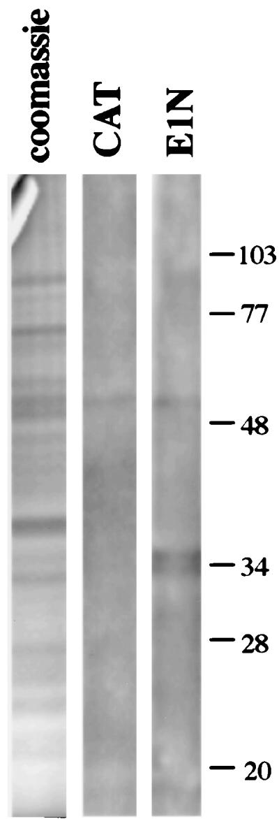
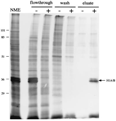
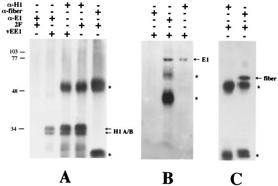
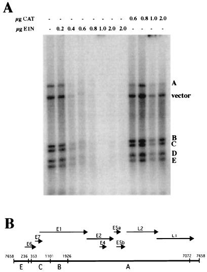
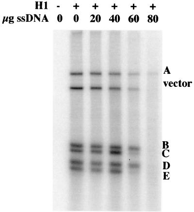
The E1 and E2 proteins are the only virus-encoded factors required for human papillomavirus (HPV) DNA replication. The E1 protein is a DNA helicase responsible for initiation of DNA replication at the viral origin. Its recruitment to the origin is facilitated by binding to E2, for which specific recognition elements are located at the origin. The remaining replication functions for the virus, provided by the host cell's replication machinery, may be mediated by further interactions with E1 and E2. Histone H1 was identified as an HPV type 11 (HPV-11) E1-binding protein by far-Western blotting and by microsequence analyses of a 34-kDa protein purified by E1 affinity chromatography. E1 also bound in vitro to H1 isolated under native conditions in association with intact nucleosomes. In addition, E1 and H1 were coimmunoprecipitated by an E1 antiserum from a nuclear extract prepared from cells expressing recombinant E1. Bound H1 was displaced from HPV-11 DNA by the addition of E1, suggesting that E1 can promote replication initiation and elongation by alteration of viral chromatin structure and disruption of nucleosomes at the replication fork. Furthermore, a region of the HPV-11 genome containing the origin of replication was identified which had weaker affinity for H1 than that of the remaining genome. This result suggests that the presence of a DNA structure at or near the HPV origin facilitates initiation of DNA replication by exclusion of H1. These results are similar to those of studies of simian virus 40 DNA replication, in which a large T antigen-H1 interaction and an H1-resistant region at the origin of DNA replication have also been demonstrated.


Figures

FIG. 1
FIG. 1
Expression of recombinant E1 from vEE1. (A) mRNA expression was under the control of the bacteriophage T7 RNA polymerase promoter (PT7) and terminator (T7ter). Cap-independent translation was directed by the EMCV UTR, which was fused to the 5′ end of the E1 ORF. The resulting operon was recombined into the TK gene (TKL and TKR) of vaccinia virus. The arrow indicates the direction of transcription, and the closed circle represents the T7 terminator of transcription. (B) Equal volumes of whole-cell lysates from HeLa cells infected with vTF-7.3 and vEE1 (+) or with vTF-7.3 alone (−) were resolved by SDS-PAGE on a 10% polyacrylamide gel and assayed for recombinant E1 protein by Western blot analysis with the E1 antiserum RL-070, biotinylated secondary antibody, and 35S-streptavidin. The image was generated by PhosphorImager analysis (Molecular Dynamics). The positions of molecular mass markers (in kilodaltons) are shown on the right.
FIG. 2
FIG. 2
Far-Western blot analysis of HeLa cell nuclear proteins with the E1N protein. A 0.5-mg quantity of nuclear protein was resolved by SDS-PAGE on a 12.5% polyacrylamide slab gel, transblotted to nitrocellulose, and renatured. Strips cut from the blot were probed with the CAT or E1N protein. E1N-reactive proteins were detected by reaction with His-probe antiserum and enhanced chemiluminescence. The left strip is total nuclear matrix protein, stained with Coomassie blue, derived from the same gel as the far-Western blots. The positions of molecular mass markers (in kilodaltons) are shown on the right.
FIG. 3
FIG. 3
Purification of E1-bound proteins by E1 affinity chromatography. Protein from a dialyzed HeLa cell NME was loaded onto an E1N (+) or a CAT (−) affinity column. Bound proteins were washed with Ni2+ wash buffer and were eluted with lysis buffer containing 0.5% SDS. Equal volumes from the flowthrough, wash, and eluate fractions were resolved by SDS-PAGE and detected by silver staining. Crude NME was loaded in the far left lane. The positions of molecular mass markers (in kilodaltons) are shown on the left. The arrow indicates the position of the protein doublet.
FIG. 4
FIG. 4
Association of histone H1, isolated under native conditions, with the E1 affinity column. Protein from a nucleosome extract (lanes 3 and 4) or nucleosome extract buffer alone (lanes 5 and 6) was loaded onto an E1N (lanes 4 and 6) or a CAT (lanes 3 and 5) affinity column, washed with Ni2+ wash buffer supplemented with 8 M urea, and eluted with pH 6.0 lysis buffer. Equal volumes of protein from the columns’ eluates were resolved by SDS-PAGE on a 15% polyacrylamide gel. Resolved proteins were detected by Coomassie brilliant blue staining. Proteins from crude NME and the flowthrough from the NME-loaded E1 column were loaded in lanes 1 and 2, respectively. Protein molecular mass standards (in kilodaltons) were loaded in the far-left lane.
FIG. 5
FIG. 5
Coimmunoprecipitation of histone H1 and HPV-11 E1. Nuclear extracts derived from HeLa cells expressing the HPV-11 E1 (vEE1) or adenovirus fiber (2F) protein, each from vaccinia virus, were incubated with (+) or without (−) E1 antiserum (α-E1), anti-H1 antibody (α-H1), and antifiber antibody (α-fiber). Immunoprecipitates were assayed for H1 (A), E1 (B), or fiber (C) by Western blot analysis and enhanced chemiluminescence. The asterisks denote the heavy and light chains of immunoglobulins used in the immunoprecipitations. The band in panel B denoted by the uppermost asterisk is derived from the E1 antiserum and is likely incompletely denatured immunoglobulin. Positions of molecular mass markers (in kilodaltons) are shown on the left.
FIG. 6
FIG. 6
Displacement of H1 from HPV-11 DNA by E1. Two hundred nanograms of 32P-labeled HPV-11 DNA restriction fragments were bound to 200 ng of H1; this was followed by incubation with (+) or without (−) the indicated amounts (in micrograms) of E1N or CAT and, finally, digestion with DNase I. H1-bound restriction fragments were protected from digestion by DNase I; displacement of H1 by E1N is indicated by removal of labeled DNAs by DNase I digestion. The image was generated by PhosphorImager analysis (Molecular Dynamics). (B) Linearized map of HPV-11 DNA restriction fragments A to E. Arrows denote viral protein ORFs. The noncoding region between the E6 and L1 ORFs is the URR and contains the HPV-11 origin of DNA replication.
FIG. 7
FIG. 7
Differential affinities of HPV-11 restriction fragments for binding to histone H1. 32P-labeled HPV-11 DNA restriction fragments were bound to 200 ng of H1 in the presence (+) or absence (−) of the indicated amounts of salmon sperm DNA (ssDNA) followed by digestion with DNase I. H1-bound restriction fragments were protected from digestion by DNase I; competition for H1 binding by salmon sperm DNA resulted in removal of labeled DNAs by DNase I digestion. Differential affinity for H1 among the restriction fragments is indicated by the differing amount of competition at 60 μg of salmon sperm DNA. HPV DNA restriction fragments are labeled according to the map in Fig. 6B. The image was generated by PhosphorImager analysis (Molecular Dynamics).

Similar articles

Cited by

References

    1. Adams C C, Workman J L. Nucleosome displacement in transcription. Cell. 1993;72:305–308. - PubMed
    1. Altschul S F, Gish W, Miller W, Myers E W, Lipman D J. Basic local alignment search tool. J Mol Biol. 1990;215:403–410. - PubMed
    1. Ambrose C, Blasquez V, Bina M. A block in initiation of simian virus 40 assembly results in the accumulation of minichromosomes containing an exposed regulatory region. Proc Natl Acad Sci USA. 1986;83:3287–3291. - PMC - PubMed
    1. Angeletti P C, Engler J A. Tyrosine kinase-dependent release of an adenovirus preterminal protein complex from the nuclear matrix. J Virol. 1996;70:3060–3067. - PMC - PubMed
    1. Berezney R. The nuclear matrix: a heuristic model for investigating genomic organization and function in the cell nucleus. J Cell Biochem. 1991;47:109–123. - PubMed

Publication types

MeSH terms

LinkOut - more resources