. 1998 Mar;72(3):2002-9.
doi: 10.1128/JVI.72.3.2002-2009.1998.

The cell tropism of human immunodeficiency virus type 1 determines the kinetics of plasma viremia in SCID mice reconstituted with human peripheral blood leukocytes

Affiliations

The cell tropism of human immunodeficiency virus type 1 determines the kinetics of plasma viremia in SCID mice reconstituted with human peripheral blood leukocytes

G R Picchio et al. J Virol. 1998 Mar.

Erratum in

  • J Virol 1998 Sep;72(9):7707

Abstract

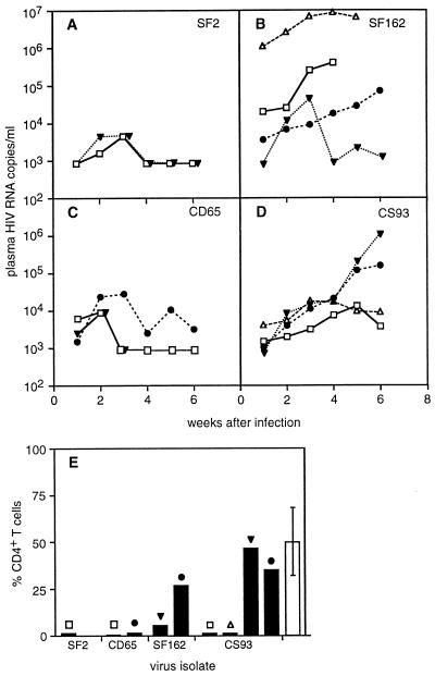
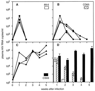
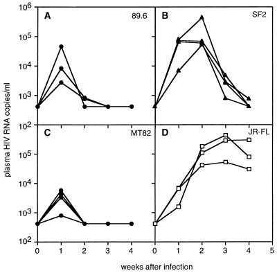
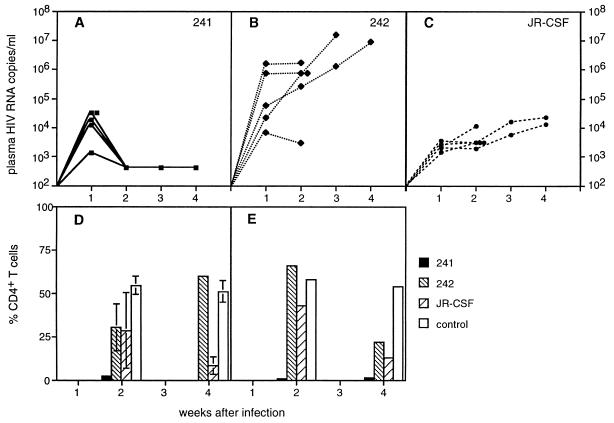
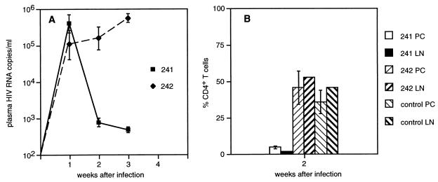
Most individuals infected with human immunodeficiency virus type 1 (HIV-1) initially harbor macrophage-tropic, non-syncytium-inducing (M-tropic, NSI) viruses that may evolve into T-cell-tropic, syncytium-inducing viruses (T-tropic, SI) after several years. The reasons for the more efficient transmission of M-tropic, NSI viruses and the slow evolution ofT-tropic, SI viruses remain unclear, although they may be linked to expression of appropriate chemokine coreceptors for virus entry. We have examined plasma viral RNA levels and the extent of CD4+ T-cell depletion in SCID mice reconstituted with human peripheral blood leukocytes following infection with M-tropic, dual-tropic, or T-tropic HIV-1 isolates. The cell tropism was found to determine the course of viremia, with M-tropic viruses producing sustained high viral RNA levels and sparing some CD4+ T cells, dual-tropic viruses producing a transient and lower viral RNA spike and extremely rapid depletion of CD4+ T cells, and T-tropic viruses causing similarly lower viral RNA levels and rapid-intermediate rates of CD4+ T-cell depletion. A single amino acid change in the V3 region of gp120 was sufficient to cause one isolate to switch from M-tropic to dual-tropic and acquire the ability to rapidly deplete all CD4+ T cells.


Figures

FIG. 1
FIG. 1
Plasma HIV RNA copy number in hu-PBL-SCID mice infected with the T-tropic HIV-1 isolate SF2 (A), the M-tropic isolate SF162 (B), the primary T-tropic patient isolate CD65 (C), or the primary M-tropic isolate CS93 (D). Each line represents serial measurements on an individual hu-PBL-SCID mouse from 1 to 6 weeks after infection. (E) Percentage of remaining CD4+ T cells in the peritoneal cavity (compared to total human CD3+ T cells) at 7 to 8 weeks after infection. This number was determined for individual mice (symbol above each column matches symbol for each animal in panels A through D) and compared to the mean (± standard error [SE]) for four uninfected hu-PBL-SCID mice (open column with error bar). All hu-PBL-SCID mice were derived from a single human donor who was Epstein-Barr virus seronegative and homozygous wild type at the CCR5 locus. The detection limit of RNA copy number was 800 in this experiment, so samples with undetectable viral RNA were assigned a value of 800. Serial samples from individual mice were saved and compared in the same Roche HIV Monitor Amplicor assay plate, and the available volume of mouse plasma determined the cut-off value for HIV RNA detection.
FIG. 2
FIG. 2
Plasma HIV RNA copy number in hu-PBL-SCID mice reconstituted with cells from another human donor (also EBV negative, CCR5 wild-type homozygous) and infected with the dual-tropic 89.6 HIV-1 isolate (A), the primary T-tropic isolate CD65 (B), or the primary M-tropic isolate CS93 (C). Panels A through C show the viral RNA levels in individual hu-PBL-SCID mice, and panel D shows the mean ± SE of each group of mice over the 5-week duration of the experiment. The limit of detection in this experiment was 200 copies/ml, and samples with no detectable viral RNA were assigned this value.
FIG. 3
FIG. 3
Plasma HIV RNA copy number in hu-PBL-SCID mice reconstituted with cells from a third human donor (also EBV negative, CCR5 wild-type homozygous) and infected with the dual-tropic 89.6 isolate (A), the T-tropic SF2 isolate (B), the T-tropic primary patient isolate MT82 (C), or the M-tropic JR-FL isolate (D). The limit of detection in this experiment was 400 copies/ml, and samples with no detectable viral RNA were assigned this value.
FIG. 4
FIG. 4
Plasma HIV RNA copy number and percentage of CD4+ T cells in hu-PBL-SCID mice generated from a single human donor. Five hu-PBL-SCID mice were infected either with HIV-1241, a dual-tropic, SI virus (A), or HIV-1242, an M-tropic, NSI virus that differs only by a glutamine-to-glutamic acid change in position 25 of the V3 loop (14) (B). An additional group of five mice was infected with the M-tropic JR-CSF isolate (C). Three mice in each group were used for determination of CD4+ T-cell levels at 2 weeks after infection, and the remaining two mice were examined after 4 weeks of infection. (D) Percentages ± SE of CD4+ T cells (of total human CD3+ T cells) recovered by peritoneal lavage of the animals compared to mean values for four uninfected control mice. Only one hu-PBL-SCID mouse infected with HIV-1242 was available at week 4, because the mouse with >107 HIV RNA copies/ml at week 3 after infection died. CD4+ T cells were also enumerated in regional lymph nodes (panel E) pooled from three mice (2 weeks after infection) or two mice (4 weeks after infection). Since these samples were pooled, no SE is shown.
FIG. 5
FIG. 5
Plasma HIV RNA copy number and recovery of CD4+ T cells in hu-PBL-SCID mice generated from a single donor who is heterozygous for the CCR5 Δ32 mutation. Five hu-PBL-SCID mice were infected with either HIV-1241 or HIV-1242 (as in the experiment shown in Fig. 4). The geometric mean RNA copy number (± relative SE) for each group of mice is shown for 1 to 3 weeks after infection (A). Three mice from each group were used to determine the extent of CD4+ T-cell depletion at 2 weeks after infection (B). Samples of peritoneal lavage cells (PC) were assayed from individual mice, and the mean ± SE is shown. Samples from lymph nodes (LN) were pooled prior to analysis, so no SE is displayed.
FIG. 6
FIG. 6
(A) Recovery of human CD4+ T cells from peritoneal lavage cells (filled bars) or local lymph nodes (hatched bars) of hu-PBL-SCID mice infected 2 weeks earlier with either HIV-1SF2 or HIV-1SF162. The numbers represent the mean ± SE of individual determinations on 4 to 5 mice per group and are expressed as a percentage of recovered total human T cells (CD3+). CD3+ T cells represented >90% of recovered human cells. Human cells represented 79 to 88% of all cells recovered from the peritoneal lavage of uninfected hu-PBL-SCID mice and from 37 to 89% of cells recovered from local lymph nodes. The numbers of recovered human cells declined in parallel with the loss of CD4+ T cells after HIV-1 infection in both sites. (B) Phenotype of human cells recovered from the peritoneal cavity of control, uninfected hu-PBL-SCID mice. Two-color immunofluorescence staining of cells was performed to identify memory/activated CD4 T cells, which express CD45RO, CD25 (IL-2R alpha chain), and/or CD69. (C) Phenotype of human cells recovered from the local lymph nodes draining the peritoneal cavity of hu-PBL-SCID mice and stained as in panel B.

Similar articles

Cited by

References

    1. Aldrovandi G, Feuer G, Gao L, Jamieson B, Kristeva M, Chen I, Zack J. The SCID-hu mouse as a model for HIV-1 infection. Nature. 1993;363:732–736. - PubMed
    1. Alkhatib G, Combadiere C, Broder C, Feng Y, Kennedy P, Murphy P, Berger E. CC CKR5: a RANTES, MIP-1α, MIP-1β receptor as a fusion cofactor for macrophage-tropic HIV-1. Science. 1996;272:1955–1958. - PubMed
    1. Autran B, Carcelain G, Li T, Blanc C, Mathez D, Tubiana R, Katlama C, Debre P, Leibowitch J. Positive effects of combined antiretroviral therapy on CD4+ T cell homeostasis and function in advanced HIV disease. Science. 1997;277:112–116. - PubMed
    1. Berger E. HIV entry and tropism: the chemokine receptor connection. AIDS. 1997;11(A):S3–S16. - PubMed
    1. Berson J F, Long D, Doranz B J, Rucker J, Jirik F R, Doms R W. A seven-transmembrane domain receptor involved in fusion and entry of T-cell-tropic human immunodeficiency virus type 1 strains. J Virol. 1996;70:6288–6295. - PMC - PubMed

Publication types