. 1998 Mar;72(3):2022-32.
doi: 10.1128/JVI.72.3.2022-2032.1998.

In vitro and in vivo biology of recombinant adenovirus vectors with E1, E1/E2A, or E1/E4 deleted

Affiliations

In vitro and in vivo biology of recombinant adenovirus vectors with E1, E1/E2A, or E1/E4 deleted

M Lusky et al. J Virol. 1998 Mar.

Abstract

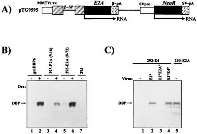
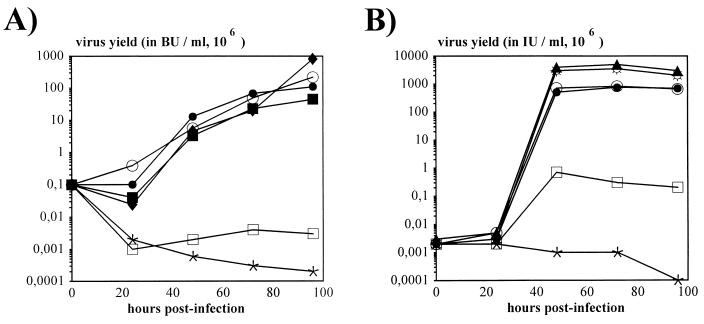
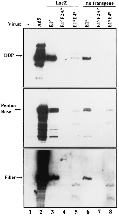
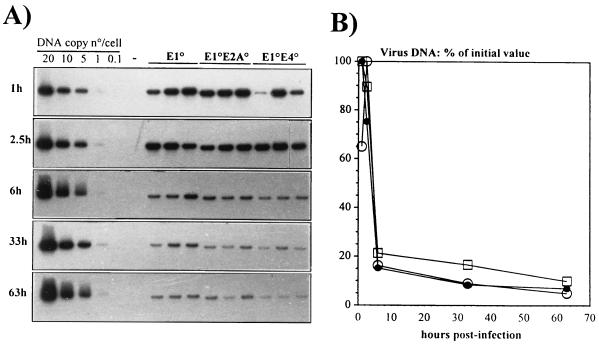
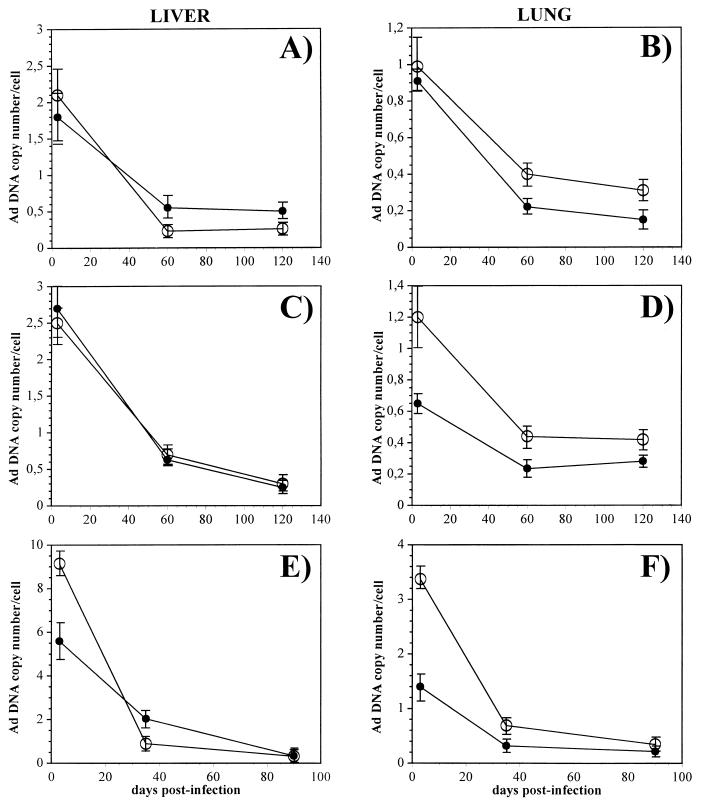
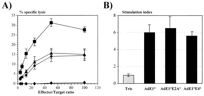
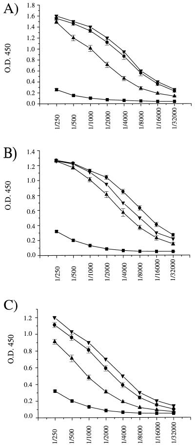
Isogenic, E3-deleted adenovirus vectors defective in E1, E1 and E2A, or E1 and E4 were generated in complementation cell lines expressing E1, E1 and E2A, or E1 and E4 and characterized in vitro and in vivo. In the absence of complementation, deletion of both E1 and E2A completely abolished expression of early and late viral genes, while deletion of E1 and E4 impaired expression of viral genes, although at a lower level than the E1/E2A deletion. The in vivo persistence of these three types of vectors was monitored in selected strains of mice with viral genomes devoid of transgenes to exclude any interference by immunogenic transgene-encoded products. Our studies showed no significant differences among the vectors in the short-term maintenance and long-term (4-month) persistence of viral DNA in liver and lung cells of immunocompetent and immunodeficient mice. Furthermore, all vectors induced similar antibody responses and comparable levels of adenovirus-specific cytotoxic T lymphocytes. These results suggest that in the absence of transgenes, the progressive deletion of the adenovirus genome does not extend the in vivo persistence of the transduced cells and does not reduce the antivirus immune response. In addition, our data confirm that, in the absence of transgene expression, mouse cellular immunity to viral antigens plays a minor role in the progressive elimination of the virus genome.


Figures

FIG. 1
FIG. 1
Structure of the E2A expression plasmid and steady-state levels of DBP protein in stable cell lines and after viral infection. (A) Schematic representation of the DBP expression plasmid pTG9595. Ad5 E2A sequences (nt 22440 to 24334) were inserted into an MMTV promoter-driven expression cassette (22) containing the rabbit β-globin splicing (β-SP) and polyadenylation (β-pA) signals. Expression of the neomycin resistance gene (NeoR) is regulated by the Simian virus 40 early promoter (SVpro) and late polyadenylation signal (SV-pA). LTR, long terminal repeat. (B) Western blot analysis of DBP protein in stable E1/E2A complementation cell lines. 293-E2A clones (9-16 and 9-72), established with pTG9595, and the control E2A complementation cell line gmDBP-6 (9) were compared for DBP expression in the presence (+) or absence (−) of dexamethasone (Dex). Total protein was extracted at 24 h postinduction, polypeptides (10 μg of protein) were separated on a 12% polyacrylamide–SDS gel, and the DBP protein was detected with the B6α72K anti-DBP monoclonal antibody (41) combined with enhanced chemiluminescence. (C) Comparison of DBP expression in 293-E2A cells (clone 9-72) and 293-E4 cells (clone 5-19; see Fig. 3) infected at an MOI of 6 IU/cell with AdE1°, AdE1°E2A°, and AdE1°E4° vectors. Total protein was extracted at 16 h p.i. (lanes 1 to 4) and 24 h after induction with dexamethasone (lane 5). DBP analysis was as described above.
FIG. 2
FIG. 2
Kinetics of viral growth. (A) Kinetics of AdE1° and AdE1°E2A° virus propagation in 293 and 293-E2A cells. Cells were infected at an MOI of 0.2 BU/cell. Titration of infectious viral progeny (in BU) at the indicated times p.i. was performed on 293-E2A cells. AdE1° (AdTG4656) (⧫, ○, and •) was used to infect 293 cells (⧫) and 293-E2A-9-72 cells in the presence (•) and absence (○) of dexamethasone induction. Similarly, AdE1°E2A° (AdTG9542) (▪, □, and ∗) was used to infect 293 cells (∗) and 293-E2A-9-72 cells in the presence (▪) and absence (□) of dexamethasone induction. (B) Kinetics of AdE1° and AdE1°E4° virus propagation in 293, 293-E4 (clone 293/1653il), and 293-E4ORF6+7 (clone 293/5-19) cells. Cells were infected at an MOI of 0.05 IU/cell. Titration of infectious viral progeny (in IU) at the indicated times p.i. was performed on 293 cells. AdE1° (AdTG4656) (⧫ and ☼) was used to infect 293 cells (⧫) and 239-E4ORF6+7 cells (☼); similarly, AdE1°E4° (AdTG8595 [•, □, and ∗] or AdTG5643 [○]) was used to infect 239-E4ORF6+7 (• and ○), 293-E4 (□), and 293 (∗) cells.
FIG. 3
FIG. 3
Structure of E4 expression plasmids and analysis of late viral proteins in 293-E2A and 293-E4 complementation cells. (A) Schematic representation of the E4 expression plasmid, pTG1653, containing the entire E4 region (Ad5 nt 32800 to 35826), including the E4 promoter (pro) and polyadenylation signal (pA). Expression of the puromycin resistance gene (PuroR) is regulated by the Simian virus 40 (SV) early promoter. (B) Schematic representation of the E4ORF6+7 expression plasmid, pTG5606. The tTA gene (26) as well as the E4ORF6+7 (Ad5 nt 32800 to 34219) genes are under the control of the minimal CMV immediate-early promoter fused to a heptameric tet operator (26), while the puromycin resistance gene is regulated by the simian virus 40 early promoter. (C) Analysis of late viral proteins in infected 293-E2A (clone 293/9-72; lanes 5 to 7 and 12 to 14) and 293-E4 (clone 293/5-19; lanes 1 to 4 and 8 to 11) complementation cells. Cells were infected with the indicated viruses at an MOI of 5 IU/cell (E1°, AdTG4656; E1°E4°, AdTG8595; E4°, AdTG9572) or 5 BU/cell (E1°E2°, AdTG9542). 293-E2A cells were induced with dexamethasone. All viruses contained the lacZ gene in place of the E1 region (Table 1), except for the vector AdTG9572, which contained an intact E1 region. Total protein extraction, electrophoresis, and Western blot analysis were performed as described in the legend to Fig. 1. Late viral proteins were detected with polyclonal antisera directed against the knob domain of fiber (serum E642; obtained from R. Gerard, Leuven, Belgium [31]) or against the penton base (serum SE262; obtained from P. Boulanger, Montpellier, France).
FIG. 4
FIG. 4
Expression of early and late viral proteins in noncomplementing human A549 cells. A549 cells were infected with the indicated vectors at an MOI of 500 BU/cell (lanes 3, 4, and 5) or 500 IU/cell (lanes 6, 7, and 8). Wild-type Ad5 was infected with an MOI of 0.5 IU/cell. At 72 h p.i. total protein was extracted and processed as described in the legend to Fig. 1.
FIG. 5
FIG. 5
Short-term stability of the vector DNAs in the livers of CBA/J mice. (A) Southern blot analysis of liver DNAs of CBA/J mice following intravenous administration of 4 × 10¹⁰ total viral particles of the indicated vectors. The experiment involved three mice per vector per time point. Total genomic liver DNA was extracted at the indicated times, digested with the restriction endonuclease BamHI, and analyzed by using a 32P-labeled restriction fragment from the E3-E4 region as probe. Control lanes contain 20, 10, 5, 1, and 0.1 viral genome copies, each mixed with 10 μg of mouse liver DNA (1 viral genome copy is equivalent to 30 pg of viral DNA). (B) Quantitative analysis of Ad vector DNA from the autoradiogram shown in panel A. The Southern blot was evaluated by densitometry scanning. The data are expressed as the percentage of viral DNA with respect to the initial value at 1 h p.i. □, E1°; •, E1°E2°; ○, E1°E4°.
FIG. 6
FIG. 6
Long-term persistence of vector DNA in the livers and lungs of CBA/J and SCID mice. Totals of 2 × 10⁹ IU of AdE1° (A and B) and AdE1°E4 (C and D) and 109 PFU of AdE1°E2A° (E and F) were injected intravenously into SCID (•) and CBA/J (○) mice. DNA from livers (A, C, and E) and lungs (B, D, and F) was prepared at the indicated times and analyzed by Southern blotting and densitometry scanning. Symbols represent the means ± SE for 8 (A to D) and 10 (E and F) CBA/J animals and for 6 (A and B), 5 (C and D), and 10 (E and F) SCID mice.
FIG. 7
FIG. 7
Induction of cellular immune response in CBA/J mice. (A) Mice were injected intraperitoneally with Tris (⧫) or with 109 IU of AdE1° (▴) and AdE1°E4° (•) and 109 PFU of AdE1°E2A° (▪) vectors. Splenocytes of the treated mice were tested for CTL activity against Ad-infected syngeneic cells. CTL activity was measured in a 4-h 51Cr assay. (B) Splenocytes from mock-infected (Tris) or Ad-infected mice were analyzed for their T-cell proliferative response to Ad particles applied to the culture plates. The stimulation index is the ratio between the values of [3H]thymidine incorporation by the stimulated cells and the unstimulated cells. Results are expressed as the means ± SE for four animals per group.
FIG. 8
FIG. 8
Anti-Ad antibodies in CBA/J mice. Mice were injected intravenously with 109 IU of AdE1° (A) and AdE1°E4° (C) and 109 PFU of AdE1°E2A° (B) vectors. Sera were collected at days 4 (▪), 35 (▴), and 90 (•) postinjection and analyzed by ELISA as previously described (37). Data are expressed as the means of the optical density at 450 nm (O.D. 450) ± SE determined for successive serial dilutions of the sera recovered from 5 to 10 mice per experimental group. Each plate contained the same positive control serum (▾).

Similar articles

Cited by

References

    1. Armentano D, Zabner J, Sacks C, Sookdeo C C, Smith M P, St. George J A, Wadsworth S C, Smith A E, Gregory R J. Effect of the E4 region on the persistence of transgene expression from adenovirus vectors. J Virol. 1997;71:2408–2416. - PMC - PubMed
    1. Barr D, Tubb J, Fergusson D, Scaria A, Lieber A, Wilson C, Perkins J, Kay M A. Strain related variations in adenovirally mediated transgene expression from mouse hepatocytes in vivo: comparisons between immunocompetent and immunodeficient inbred strains. Gene Ther. 1995;2:151–155. - PubMed
    1. Berkner K L. Development of adenovirus vectors for the expression of heterologous genes. BioTechniques. 1988;6:616–629. - PubMed
    1. Bondesson M, Ohman K, Mannervik M, Fan S, Aküsjarvi G. Adenovirus E4 open reading frame 4 protein autoregulates E4 transcription by inhibiting E1A transactivation of the E4 promoter. J Virol. 1996;70:3844–3851. - PMC - PubMed
    1. Bosma G C, Custer M C, Bosma M J. A severe combined immunodeficiency mutation in the mouse. Nature (London) 1983;301:527–530. - PubMed

Publication types

MeSH terms

LinkOut - more resources