. 1998 Mar;72(3):2079-88.
doi: 10.1128/JVI.72.3.2079-2088.1998.

Bovine papillomavirus type 1 genomes and the E2 transactivator protein are closely associated with mitotic chromatin

Affiliations

Bovine papillomavirus type 1 genomes and the E2 transactivator protein are closely associated with mitotic chromatin

M H Skiadopoulos et al. J Virol. 1998 Mar.

Abstract

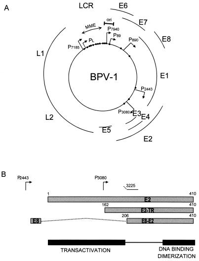
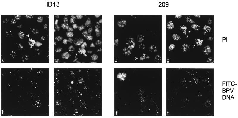
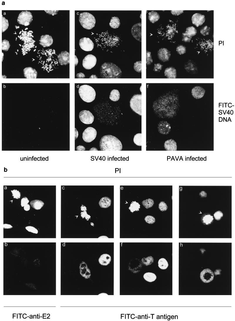
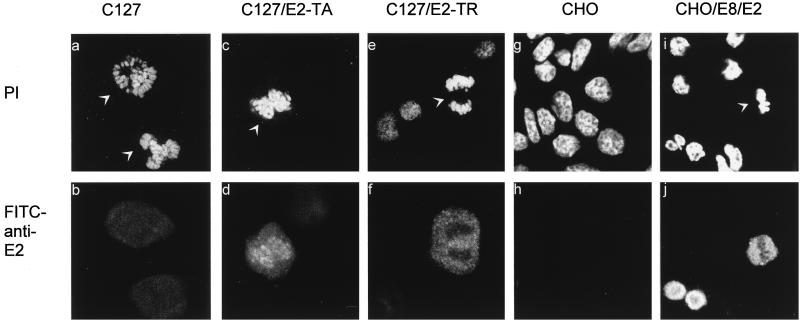
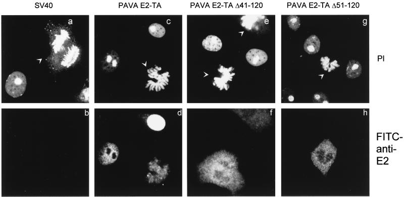
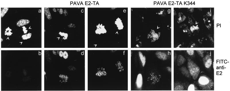
The bovine papillomavirus type 1 E2 transactivator protein is required for viral transcriptional regulation and DNA replication and may be important for long-term episomal maintenance of viral genomes within replicating cells (M. Piirsoo, E. Ustav, T. Mandel, A. Stenlund, and M. Ustav, EMBO J. 15:1-11, 1996). We have evidence that, in contrast to most other transcriptional transactivators, the E2 transactivator protein is associated with mitotic chromosomes in dividing cells. The shorter E2-TR and E8/E2 repressor proteins do not bind to mitotic chromatin, and the N-terminal transactivation domain of the E2 protein is necessary for the association. However, the DNA binding function of E2 is not required. We have found that bovine papillomavirus type 1 genomes are also associated with mitotic chromosomes, and we propose a model in which E2-bound viral genomes are transiently associated with cellular chromosomes during mitosis to ensure that viral genomes are segregated to daughter cells in approximately equal numbers.


Figures

FIG. 1
FIG. 1
(A) Diagram of the BPV-1 genome. The open reading frames E1 to E8 and L1 and L2 are shown. Promoters are represented by arrows, and E2-specific DNA binding sites are represented by small black circles. The LCR origin of replication (ori), and MME are also indicated. (B) Map of the E2 transactivator and repressor proteins. The full-length E2 protein is a transcriptional transactivator that can be expressed from the P2443 promoter. The E2-TR repressor protein is expressed from the P3080 promoter and initiated at an internal initiation codon. The E8/E2 repressor protein is encoded by a spliced message that links 11 amino acids of the E8 open reading frame to the C-terminal half of the E2 open reading frame.
FIG. 2
FIG. 2
BPV DNA was detected by FISH in C127 cells (a and b) and 137 cells (c to h). In panels a, c, e, and g, cellular DNA was detected by the propidium iodide (PI) signal. In panels b, d, f, and h, the same fields of cells are shown with the FITC-labeled BPV DNA signal. In panels a to f, cells were grown on slides and treated with colchicine for 30 min before fixation. In panels g and h, the cells were treated with colchicine for 30 min and the chromosomes were spread on slides as described in Materials and Methods. Mitotic cells are indicated with white arrowheads in the propidium iodide-stained images.
FIG. 3
FIG. 3
E2 proteins were detected in C127 cells (a, b, g, and h) and 137 cells (c to f, i, and j) by indirect immunofluorescence with the E2-specific B201 antibody. In panels a, c, e, g, and i, cellular DNA was detected by the propidium iodide (PI) signal. In panels b, d, f, h, and j, E2 protein was detected with an FITC-labeled secondary antibody in the same field of cells. Mitotic cells are indicated with white arrowheads in the propidium iodide-stained images.
FIG. 4
FIG. 4
BPV DNA was detected by FISH in ID13 cells (a to d) and 209 cells (e to h). In panels a, c, e, and g, cellular DNA was detected by propidium iodide (PI) staining. In panels b, d, f, and h, FITC-labeled BPV DNA was detected in the same fields of cells. Cells were grown on slides and treated with colchicine for 30 min before fixation. Mitotic cells are indicated with white arrowheads in the propidium iodide-stained images.
FIG. 5
FIG. 5
(a) SV40 and pPAVAkzE2-TA DNA was detected in COS-7 cells by FISH. Subpanels a and b show uninfected cells. Cells in subpanels c and d were infected with SV40, and those in subpanels e and f were infected with pPAVAkzE2-TA recombinant virus. In subpanels a, c, and e, cellular DNA was detected by propidium iodide (PI) staining. In subpanels b, d, and f, FITC-labeled BPV DNA was detected in the same fields of cells. Cells were treated with colchicine for 30 min, and the chromosomes were spread on slides as described in Materials and Methods. Mitotic cells are indicated with white arrowheads in the propidium iodide-stained images. (b) SV40 T antigen was detected in uninfected COS-7 cells by indirect immunofluorescence. In subpanels a, c, e, and g, cellular DNA was detected by propidium iodide (PI) staining. In subpanels d, f, and h, FITC-labeled secondary antibody was used to detect T antigen in the same fields of cells. As a negative control, cells in subpanel b were stained with an anti-E2 antibody. Cells were grown on slides and treated with colchicine for 30 min before fixation. Mitotic cells are indicated with white arrowheads in the propidium iodide-stained images.
FIG. 6
FIG. 6
E2 proteins were detected in C127 and CHO-derived cell lines expressing the E2-TA, E2-TR, and E8/E2 proteins by indirect immunofluorescence with the B201 (a to f) and B202 (g to j) E2-specific antibodies. The cell lines shown in each panel are as follows: C127 (a and b), C127/E2-TA (c and d), C127/E2-TR (e and f), CHO (g and h), and CHO/E8/E2 (i and j). In panels a, c, e, g, and i, cellular DNA was detected by propidium iodide (PI) staining. In panels b, d, f, h, and j, the E2 proteins were detected by the FITC signal in the same fields of cells. Mitotic cells are indicated with white arrowheads in the propidium iodide-stained images.
FIG. 7
FIG. 7
E2 proteins were detected in COS-7 cells infected with pPAVAkzE2-TA viruses by indirect immunofluorescence with the B201 E2-specific antibody. (a and b) SV40-infected COS-7 cells used as a control; (c and d) cells infected with pPAVAkzE2-TA; (e and f) cells infected with pPAVAkzE2-TAΔ41–120; (g and h) cells infected with pPAVAkzE2-TAΔ51–120. In panels a, c, e, and g, cellular DNA was detected by propidium iodide (PI) staining. In panels b, d, f, and h, FITC-labeled E2 protein is detected in the same fields of cells. Mitotic cells are indicated with white arrowheads in the propidium iodide-stained images.
FIG. 8
FIG. 8
E2 K344 is defective in DNA binding. The DNA binding domains (E2 residues 290 to 410) of wild-type and K344 E2 proteins were synthesized in vitro and tested for DNA binding in an electrophoretic mobility shift assay. The amount of oligonucleotide probe bound to the E2 proteins is plotted against the concentration of probe in the reaction mixtures. Background amounts of probe bound by the control lysate have been subtracted from the values shown. Values for wild-type E2 are represented by circles, and those for E2 K344 are represented by squares.
FIG. 9
FIG. 9
E2 proteins were detected in CV-1 cells infected with pPAVAkzE2-TA and pPAVAkzE2-TA K344 by immunofluorescence with the B201 E2-specific antibody. (a and b) Uninfected CV-1 cells; (c to f) pPAVAkzE2-TA-infected CV-1 cells; (g to j) pPAVAkzE2-TA K344-infected cells. In panels a, c, e, g, and i, cellular DNA was detected by propidium iodide (PI) staining. In panels b, d, f, h, and j, FITC-labeled E2 protein was detected in the same fields of cells. Mitotic cells are indicated with white arrowheads in the propidium iodide-stained images.

Similar articles

Cited by

References

    1. Adom J N, Gouilleux F, Richard-Foy H. Interaction with the nuclear matrix of a chimeric construct containing a replication origin and a transcription unit. Biochim Biophys Acta. 1992;1171:187–197. - PubMed
    1. Adom J N, Richard-Foy H. A region immediately adjacent to the origin of replication of bovine papilloma virus type 1 interacts in vitro with the nuclear matrix. Biochem Biophys Res Commun. 1991;176:479–485. - PubMed
    1. Bedell M A, Hudson J B, Golub T R, Turyk M E, Hosken M, Wilbanks G D, Laimins L A. Amplification of human papillomavirus genomes in vitro is dependent on epithelial differentiation. J Virol. 1991;65:2254–2260. - PMC - PubMed
    1. Blitz I L, Laimins L A. The 68-kilodalton E1 protein of bovine papillomavirus is a DNA binding phosphoprotein which associates with the E2 transcriptional activator in vitro. J Virol. 1991;65:649–656. - PMC - PubMed
    1. Bochkarev A, Barwell J A, Pfuetzner R A, Furey W J, Edwards A M, Frappier L. Crystal structure of the DNA-binding domain of the Epstein-Barr virus origin-binding protein EBNA 1. Cell. 1995;83:39–46. - PubMed

MeSH terms

LinkOut - more resources