. 1998 Mar;72(3):2150-9.
doi: 10.1128/JVI.72.3.2150-2159.1998.

Chimeric measles viruses with a foreign envelope

Affiliations

Chimeric measles viruses with a foreign envelope

P Spielhofer et al. J Virol. 1998 Mar.

Abstract

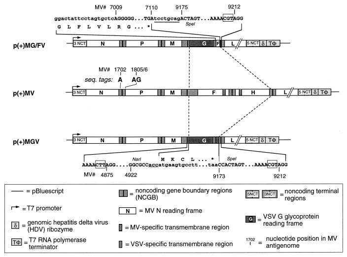
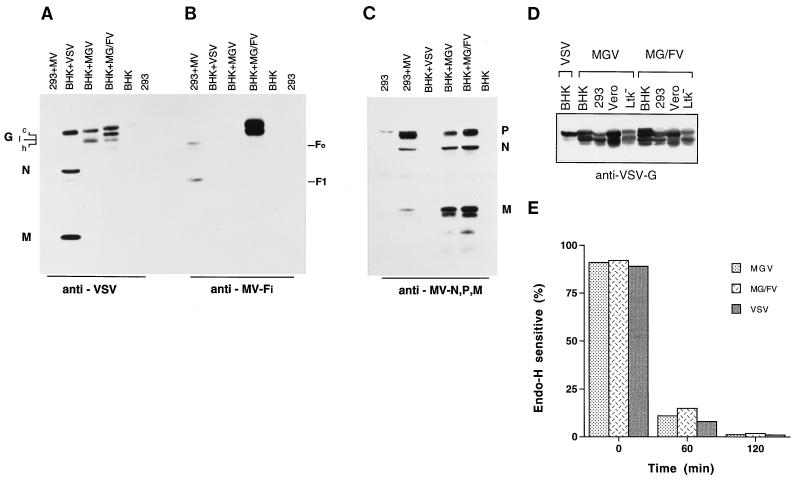
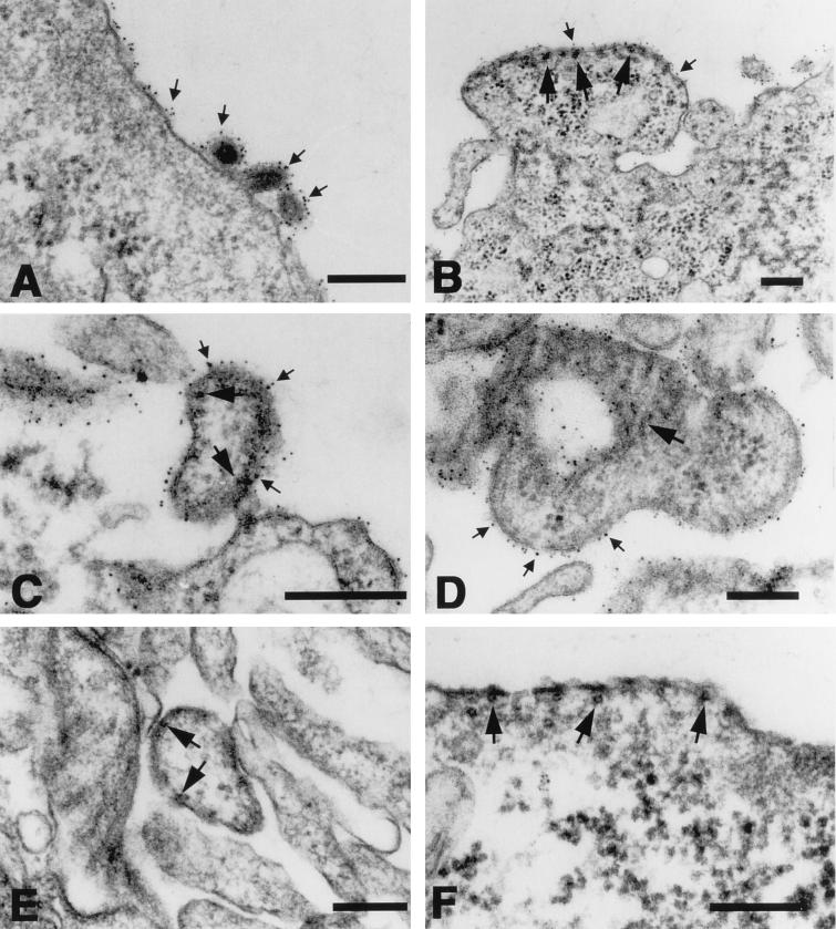
Measles virus (MV) and vesicular stomatitis virus (VSV) are both members of the Mononegavirales but are only distantly related. We generated two genetically stable chimeric viruses. In MGV, the reading frames of the MV envelope glycoproteins H and F were substituted by a single reading frame encoding the VSV G glycoprotein; MG/FV is similar but encodes a G/F hybrid in which the VSV G cytoplasmic tail was replaced by that of MV F. In contrast to MG/FV, MGV virions do not contain the MV matrix (M) protein. This demonstrates that virus assembly is possible in the absence of M; conversely, the cytoplasmic domain of F allows incorporation of M and enhances assembly. The formation of chimeric viruses was substantially delayed and the titers obtained were reduced about 50-fold in comparison to standard MV. In the novel chimeras, transcription and replication are mediated by the MV ribonucleoproteins but the envelope glycoproteins dictate the host range. Mice immunized with the chimeric viruses were protected against lethal doses of wild-type VSV. These findings suggest that it is feasible to construct MV variants bearing a variety of different envelopes for use as vaccines or for gene therapeutic purposes.


Figures

FIG. 1
FIG. 1
Features of plasmids which allow the rescue of the chimeric viruses MGV and MG/FV. The entire MV F and H open reading frame and parts of the untranslated region were substituted by the open reading frame of VSV G. The nucleotides in lowercase are derived from the VSV G gene, and those in uppercase represent MV sequences. The numbering of the nucleotides refers to the MV antigenomic sequence as given by Radecke and Billeter (27). The underlined nucleotides were added during the cloning procedure to comply with the rule of six (4) for p(+)MG/FV and p(+)MGV. The antigenomic sense of the specified RNAs is indicated by (+). Relevant regions of the nucleotide sequences near the ends of the VSV inserts and of the corresponding encoded amino acids are detailed. The sequence tags (MV 1702 and 1805/6) are present in all plasmids. Boxes around nucleotides indicate the nontranscribed gene boundary trinucleotides between genes M and F and between genes H and L, respectively. The MV cDNA sequence is available from the EMBL gene bank under accession no. Z66517.
FIG. 2
FIG. 2
Expression and transport of MGV, MG/FV, VSV, and MV proteins in BHK, 293, Vero, and Ltk cells. (A to C) Western blot analysis demonstrating the expression of G and G/F proteins as well as MV N, P, and M by the four viruses. Cells were infected with MGV and MG/FV for 48 h. For comparison, cells were infected with VSV and MV for 20 h. They were then lysed, and one-sixth of each lysates was subjected to SDS-PAGE (10% polyacrylamide) and blotted. The membranes were treated with the specified antibodies, polyclonal anti-VSV (A), monoclonal anti-Fi antibody (B), and polyclonal anti-MV N, P, and M antibodies (C). The protein positions are indicated. The various posttranslational forms of the proteins are indicated as c, I, and h. (D) expression of G and G/F in various cells. (E) Cellular transport of envelope glycoproteins encoded by MGV, MG/FV, and VSV. At 12 h after VSV infection and 48 h after chimera infection, cells were pulse-labeled, chased as indicated, and either treated or not treated with endo H. The percentage of glycoprotein sensitivity to endo H was quantitated with a phosphorimager.
FIG. 3
FIG. 3
Cytopathogenicity of MV, VSV, MGV, and MG/FV. Cells infected with the indicated virus were observed under a light microscope. The progress of the infection was monitored until clear CPE were visible; pictures were taken at different times after infection, as indicated. (A) Noninfected Vero cells; (B) MV infection at 36 h p.i.; (C) VSV infection at 30 h p.i.; (D) MGV infection at 156 h p.i.; (E) MG/FV infection at 144 h p.i. The arrow in panel D shows a fusion patch. Monolayers were photographed with a 40× objective for panels A, C, D, and E and a 20× objective for panel B.
FIG. 4
FIG. 4
Kinetics of infectious-particle formation. Vero cells plated equally in 36-mm well plates were infected with either MGV, MG/FV, VSV, or MV Edmonston B at a MOI of 0.1. At various time points, titers of cell-free and cell-associated virus were determined by immunofluorescence for MGV, MG/FV, and VSV and by plaque assay for MV. The titers of cell-free and cell-associated virus are compared in the same experiment. Data are means of two separate experiments with a deviation of less than 5 to 7%.
FIG. 5
FIG. 5
Purification and characterization of MGV and MG/FV particles in comparison to MV and VSV. (A) Virus particles were first pelleted on 20% (1.0 ml) and 60% (1.0 ml) sucrose cushions. The interphases were taken and diluted to 20% sucrose, applied to 20 to 60% sucrose gradients, and centrifuged to equilibrium. Fractions were analyzed by SDS-PAGE (12.5% polyacrylamide), immunoblotted, and probed with the antibodies indicated on the right. (B) The blots were reprobed with anti-MV M antibody in addition to anti-VSV. The sucrose density was determined by weighing 100 μl of each fraction. (C) Representative profile of the gradient.
FIG. 6
FIG. 6
Immunoelectron microscopy of Vero cells Infected with VSV, MGV, MG/FV, and MV. The infected cells were reacted, prior to their embedding for thin sectioning, with antibodies directed against the G protein of VSV followed by protein A coated with 6-nm colloidal gold. The VSV G protein (small arrows) is detectable on VSV virions (A) as well as on the surfaces of cells infected with MGV (B and C) and MG/FV (D). Dense alignment of gold granules is most prominent in association with segments of the plasma membrane which are associated with dense MV nucleoprotein (large arrows) (B to D), similar to that in MV-infected cells (E and F). Bar, 200 nm.

References

    1. Billeter M A, Cattaneo R, Schmid A, Eschle D, Kaelin K, Rebmann G, Udem S A, Sheppard R D, Baczko K, Liebert U G, Schneider-Schaulies S, Brinckmann U, ter Meulen V. Host and viral features in persistent measles virus infections of the brain. In: Kolakofsky D, Mahy B W J, editors. Genetics and pathogenicity of negative strand viruses. Amsterdam, The Netherlands: Elsevier Science Publishers BV; 1989. pp. 356–366.
    1. Blondel D, Harmison G G, Schubert M. Role of matrix protein in cytopathogenesis of vesicular stomatitis virus. J Virol. 1990;64:1716–1725. - PMC - PubMed
    1. Burge B W, Pfefferkorn E R. Phenotypic mixing between group A arboviruses. Nature. 1966;210:1397–1399. - PubMed
    1. Calain P, Roux L. The rule of six, a basic feature for efficient replication of Sendai virus defective interfering RNA. J Virol. 1993;67:4822–4830. - PMC - PubMed
    1. Cathomen T, Naim H Y, Cattaneo R. Measles viruses with altered envelope protein cytoplasmic tails gain cell fusion competence. J Virol. 1998;72:1224–1234. - PMC - PubMed

Publication types

MeSH terms

Substances

Associated data

LinkOut - more resources