. 1998 Mar;72(3):2259-64.
doi: 10.1128/JVI.72.3.2259-2264.1998.

The gene product of human cytomegalovirus open reading frame UL56 binds the pac motif and has specific nuclease activity

Affiliations

The gene product of human cytomegalovirus open reading frame UL56 binds the pac motif and has specific nuclease activity

E Bogner et al. J Virol. 1998 Mar.

Abstract

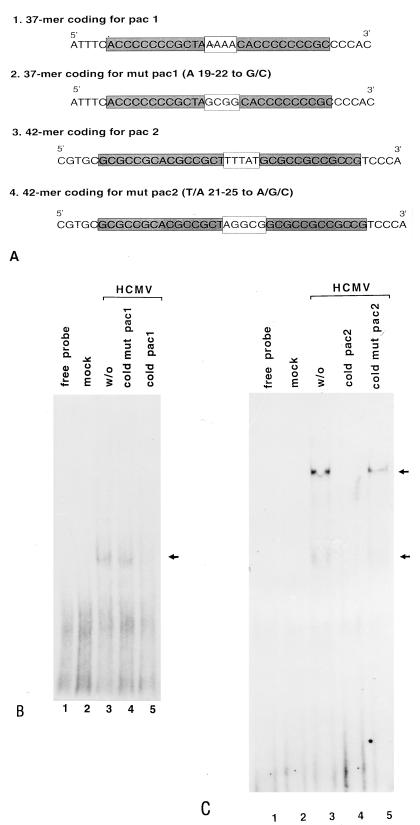
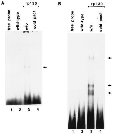
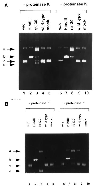
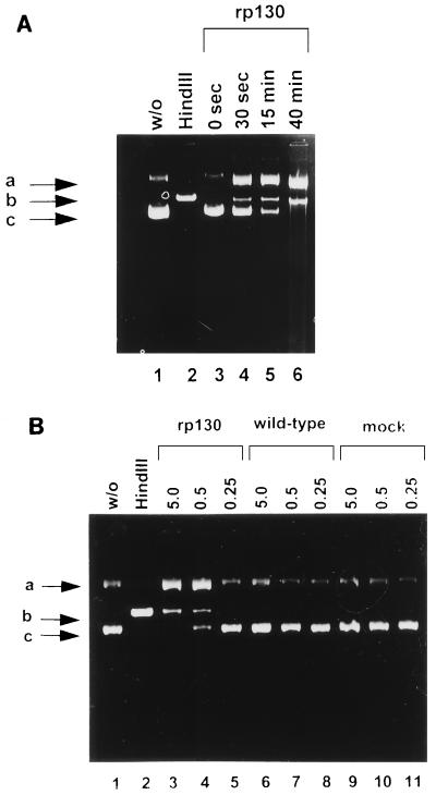
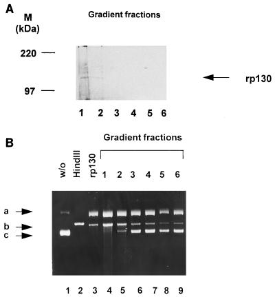
Using the cis-acting human cytomegalovirus (HCMV) packaging elements (pac 1 and pac 2) as DNA probes, specific DNA-protein complexes were detected by electrophoretic mobility shift assay (EMSA) in both HCMV-infected cell nuclear extracts and recombinant baculovirus-infected cell extracts containing the HCMV p130 (pUL56) protein. DNA-binding proteins, which were common in uninfected and infected cell extracts, were also detected. Mutational analysis showed that only the AT-rich core sequences in these cis-acting motifs, 5'-TAAAAA-3' (pac 1) and 5'-TTTTAT-3' (pac 2), were required for specific DNA-protein complex formation. The specificity of the DNA-protein complexes was confirmed by EMSA competition. Furthermore, a specific endonuclease activity was found to be associated with lysates of baculovirus-infected cells expressing recombinant p130 (rp130). This nuclease activity was time dependent, related to the amount of rp130 in the assay, and ATP independent. Nuclease activity remained associated with rp130 after partial purification by sucrose gradient centrifugation, suggesting that this activity is a property of HCMV p130. We propose a possible involvement of p130 in HCMV DNA packaging.


Figures

FIG. 1
FIG. 1
Effects of mutations in the packaging motifs on competition as well as on DNA-protein complex formation. EMSA was performed as described in Materials and Methods. (A) DNA sequences. pac 1, wild-type DNA containing the pac 1 motif; mut pac 1, DNA containing the mutagenized pac 1 motif; pac 2, wild-type DNA fragment containing the pac 2 motif; mut pac 2, DNA containing the mutated pac 2 motif. (B) Autoradiogram of EMSA competition with mut pac 1. Lanes: 1, DNA probe alone; 2, DNA probe plus mock-infected cell extracts; 3, DNA probe plus HCMV-infected cell nuclear extracts; 4, same as lane 3 plus 50-fold molar excess of unlabeled mut pac 1; 5, same as lane 3 plus 50-fold molar excess of unlabeled pac 1. (C) Autoradiogram of EMSA competition with mut pac 2. Lanes: 1, DNA probe alone; 2, DNA probe plus mock-infected cell nuclear extracts; 3, DNA probe plus HCMV-infected cell nuclear extracts; 4, same as lane 3 plus 50-fold molar excess of unlabeled wild-type pac 2; 5, same as lane 3 plus 50-fold molar excess of unlabeled mut pac 2. Arrows on the right indicate specific complexes.
FIG. 2
FIG. 2
Immunoblot of HCMV rp130 antigen for detection of the HCMV rp130 synthesized in insect cells infected with recombinant baculovirus-UL56. Infected cells were fractionated, and aliquots of each fraction were separated by SDS-PAGE followed by immunoblotting with a specific antiserum against p130. Lanes: 1, supernatant fraction from recombinant baculovirus-UL56-infected H5 cells; 2, sediment from recombinant baculovirus-infected H5 cells; 3, supernatant from wild-type baculovirus-infected H5 cells; 4, sediment from wild-type baculovirus-UL56-infected H5 cells. Molecular mass standards (M) are indicated on the left; the position of rp130 proteins is indicated by an arrow.
FIG. 3
FIG. 3
EMSA with baculovirus rp130-containing cell extract and pac DNA probe. (A) Lane 1, free pac 1 probe; lane 2, EMSA with extract from wild-type baculovirus-infected cells; lane 3, EMSA with extract containing rp130; lane 4, same as lane 3 plus 50-fold molar excess of unlabeled pac 1. (B) Lanes: 1, free pac 2 probe; 2, EMSA with extracts from wild-type baculovirus-infected cells; 3, EMSA with extract containing rp130; 4, same as lane 2 plus 50-fold molar excess of unlabeled pac 2. All the results represent an experiment on the same gel. Specific DNA-protein complexes are indicated by arrows.
FIG. 4
FIG. 4
Nicking activity of HCMV rp130. (A) Lane 1, plasmid pON205 in the absence of protein; lane 2, pON205 treated with restriction enzyme HindIII; lane 3, pON205 incubated with rp130; lane 4, pON205 incubated with wild-type-infected extracts; lane 5, pON205 incubated in the presence of mock-infected extracts; lane 6, same as lane 1 but treated with proteinase K; lane 7, same as lane 2 after treatment with proteinase K; lane 8, same as lane 3 plus treatment with proteinase K; lane 9, same as lane 4 treated with proteinase K; lane 10, same as lane 5 but incubated with proteinase K. (B) Lanes: 1, control plasmid pUC9 alone; 2, pUC9 treated with restriction enzyme HindIII; 3, pUC9 incubated with rp130; 4, pUC9 incubated with wild-type-infected extracts; 5, pUC9 plus mock-infected extracts; 6, same as lane 1 plus treatment with proteinase K; 7, same as lane 2 after proteinase K treatment; 8, same as lane 3 treated with proteinase K; 9, same as lane 4 incubated with proteinase K; 10, same as lane 5 after treatment with proteinase K. The arrows indicated four different plasmid DNA forms: open circular molecules (a), linear forms (b), supercoiled DNA (c), and additional fast-migrating molecules (d).
FIG. 5
FIG. 5
Effects of the incubation time and protein concentration on the enzyme activity. (A) Nuclease reaction mixtures were incubated at 37°C for the indicated time, and reactions were stopped by addition of proteinase K. Lanes: 1, pON205 alone; 2, pON205 treated with HindIII; 3, pON205 plus incubation of rp130 for 0 s; 4, incubation with rp130 for 30 s; 5, incubation for 15 min; 6, incubation for 40 min. (B) Nuclease reactions were performed for 40 min at 37°C prior to proteinase K treatment. Lanes: 1, pON205 alone; 2, pON205 treated with HindIII; 3, pON205 plus incubation with 5 μg of rp130; 4, incubation with 0.5 μg of rp130; 5, incubation with 0.25 μg of rp130; 6, pON205 plus incubation with 5 μg of extract of wild-type baculovirus; 7, incubation with 0.5 μg of wild-type extract; 8, incubation with 0.25 μg of wild-type extract; 9, pON205 plus 5 μg of mock-infected extract; 10, incubation with 0.5 μg of mock-infected extract; 11, incubation with 0.25 μg of mock-infected extract. Positions of open circular DNA molecules (a), linear molecules (b), and supercoiled molecules (c) are indicated.
FIG. 6
FIG. 6
Single-step purification of rp130 and direct association with enzyme activity. (A) Recombinant-infected cells were lysed and layered on a 5 to 20% sucrose gradient. Fractions were collected, separated by SDS-PAGE (8% gel) and transferred to a polyvinylidene difluoride membrane. Detection was performed with an anti-p130 antibody. Lane 1, fraction 1; lane 2, fraction 2; lane 3, fraction 3; lane 4, fraction 4; lane 5, fraction 5; lane 6, fraction 6. Molecular mass markers (M) are shown on the left; the position of rp130 is indicated by the arrow on the right. (B) Nuclease activity was assayed with an aliquot of each fraction. Lanes: 1, pON205 alone; 2, pON205 treated with HindIII; 3, pON205 plus extract containing rp130; 4, plasmid plus protein of fraction 1; 5, incubation with an aliquot of fraction 2; 6, incubation with an aliquot of fraction 3; 7, incubation with an aliquot of fraction 3; 8, incubation with an aliquot of fraction 4; 8, incubation with an aliquot of fraction 5; 9, incubation with an aliquot of fraction 6. The arrows indicate three different plasmid forms: open circular (a), linear (b), and supercoiled (c).

Similar articles

Cited by

References

    1. Addison C, Rixon F J, Preston V G. Herpes simplex virus type 1 UL28 gene product is important for the formation of mature capsids. J Virol. 1990;71:2377–2384. - PubMed
    1. Alford C A, Britt W J. Cytomegalovirus. In: Roizman B, Whiteley R J, Lopez C, et al., editors. The human herpesviruses. New York, N.Y: Raven Press, Ltd.; 1993. pp. 227–255.
    1. Bogner E, Reschke M, Reis B, Reis E, Britt W, Radsak K. Recognition of compartmentalized intracellular analogs of glycoprotein H of human cytomegalovirus. Arch Virol. 1992;126:67–80. - PubMed
    1. Bogner E, Reschke M, Reis B, Mockenhaupt T, Radsak K. Identification of the gene product encoded by ORF UL56 of human cytomegalovirus genome. Virology. 1993;196:290–293. - PubMed
    1. Chee M, Bankier A, Beck S, Bohni R, Brown C, Cerny R, Horsnell T, Huchison C, Kouzarides T, Martignetti E, Preddie E, Satchnell S, Tomlinson P, Weston K, Barell B. Analysis of the protein coding content of the sequence of human cytomegalovirus AD169. Curr Top Microbiol Immunol. 1990;154:125–169. - PubMed

Publication types

MeSH terms

LinkOut - more resources