. 1998 Mar;72(3):2280-8.
doi: 10.1128/JVI.72.3.2280-2288.1998.

CD4 glycoprotein degradation induced by human immunodeficiency virus type 1 Vpu protein requires the function of proteasomes and the ubiquitin-conjugating pathway

Affiliations

CD4 glycoprotein degradation induced by human immunodeficiency virus type 1 Vpu protein requires the function of proteasomes and the ubiquitin-conjugating pathway

U Schubert et al. J Virol. 1998 Mar.

Abstract

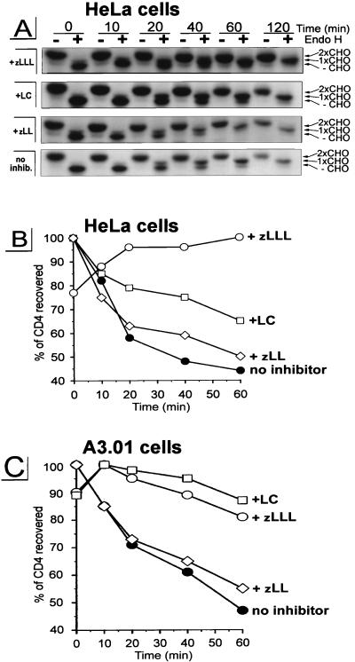
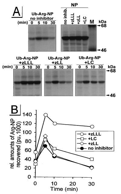
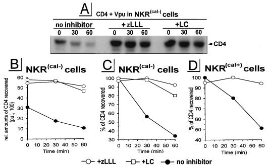
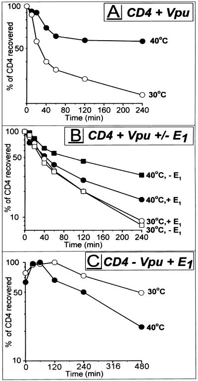
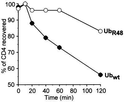
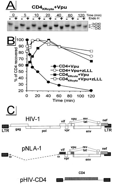
The human immunodeficiency virus type 1 (HIV-1) vpu gene encodes a type I anchored integral membrane phosphoprotein with two independent functions. First, it regulates virus release from a post-endoplasmic reticulum (ER) compartment by an ion channel activity mediated by its transmembrane anchor. Second, it induces the selective down regulation of host cell receptor proteins (CD4 and major histocompatibility complex class I molecules) in a process involving its phosphorylated cytoplasmic tail. In the present work, we show that the Vpu-induced proteolysis of nascent CD4 can be completely blocked by peptide aldehydes that act as competitive inhibitors of proteasome function and also by lactacystin, which blocks proteasome activity by covalently binding to the catalytic beta subunits of proteasomes. The sensitivity of Vpu-induced CD4 degradation to proteasome inhibitors paralleled the inhibition of proteasome degradation of a model ubiquitinated substrate. Characterization of CD4-associated oligosaccharides indicated that CD4 rescued from Vpu-induced degradation by proteasome inhibitors is exported from the ER to the Golgi complex. This finding suggests that retranslocation of CD4 from the ER to the cytosol may be coupled to its proteasomal degradation. CD4 degradation mediated by Vpu does not require the ER chaperone calnexin and is dependent on an intact ubiquitin-conjugating system. This was demonstrated by inhibition of CD4 degradation (i) in cells expressing a thermally inactivated form of the ubiquitin-activating enzyme E1 or (ii) following expression of a mutant form of ubiquitin (Lys48 mutated to Arg48) known to compromise ubiquitin targeting by interfering with the formation of polyubiquitin complexes. CD4 degradation was also prevented by altering the four Lys residues in its cytosolic domain to Arg, suggesting a role for ubiquitination of one or more of these residues in the process of degradation. The results clearly demonstrate a role for the cytosolic ubiquitin-proteasome pathway in the process of Vpu-induced CD4 degradation. In contrast to other viral proteins (human cytomegalovirus US2 and US11), however, whose translocation of host ER molecules into the cytosol occurs in the presence of proteasome inhibitors, Vpu-targeted CD4 remains in the ER in a transport-competent form when proteasome activity is blocked.


Figures

FIG. 1
FIG. 1
Treatment with proteasome-specific inhibitors rescues CD4 from Vpu-induced proteolysis. HeLa cells (A and B) or the CD4+ T-cell line A3.01 (C) were coinfected with rVVs expressing either human CD4 (v-CB3) at an MOI of 2 or Vpu (VV-Vpu) at an MOI of 5; 2.5 h p.i., the culture was split and cells were either pretreated with 5 μM zLLL, 5 μM zLL, or 10 μM LC or left untreated. After a 5-min pulse-labeling, cells were chased in the presence or absence of inhibitor for up to 2 h. CD4 molecules were recovered from detergent lysates by using a mixture of anti-CD4 antibodies. The immunoprecipitates recovered from HeLa cell lysates were split and either not treated (−) or treated with endo H (+), separated in a 10% AcrylAide gel, and analyzed by fluorography (A). Only parts of the fluorograms demonstrating CD4-specific bands are shown. The three CD4-specific bands in the endo H-treated samples represent CD4 molecules either partially resistant (1xCHO), completely resistant (2xCHO), or sensitive (−CHO) to endo H treatment as indicated on the right. (B) Relative amounts of CD4 detected in the untreated samples shown in panel A were quantitated with an Image Analyzer, and the stability of CD4 present at different times during the chase period was calculated, using as 100% the highest value for each treatment.
FIG. 2
FIG. 2
Effects of zLLL, zLL, and LC on proteasomal degradation of the model substrate Ub-Arg-NP in HeLa cells. HeLa cells that had been incubated for 45 min with or without the protease inhibitors were infected for 5 h with an rVV expressing either Ub-Arg-NP or wild-type NP (MOI = 10) in the presence or the absence of the appropriate inhibitor. Cells were pulse-labeled for 1 min and chased for up to 30 min. In panel NP, cells were lysed immediately after the pulse-labeling. Detergent lysates were immunoprecipitated with an anti-NP antibody and separated in a 9% acrylamide gel (A). Relative amounts of NP were quantitated with a PhosphorImager and plotted against time (pu, PhosphorImager units) (B). M, molecular mass standard protein.
FIG. 3
FIG. 3
Vpu-induced ERAD of CD4 does not require calnexin. Calnexin-deficient T-cell line NKR(cal−) (A to C) and calnexin-expressing T-cell line NKR(cal+) (D) were coinfected with VV-Vpu and v-CB3, treated with the inhibitors indicated, pulse-labeled for 5 min, and chased for up to 2 h. CD4 molecules recovered were analyzed by fluorography [shown only for NKR(cal−) cells (A)]. Relative amounts of CD4 established by PhosphorImager analysis (B) were used for quantitation of CD4 stability (C and D). pu, PhosphorImager units.
FIG. 4
FIG. 4
Ub-activating enzyme E1 is important for Vpu-induced CD4 degradation. tsA1S9 cells were coinfected with VV-Vpu (MOI = 2) and v-CB3 (MOI = 4) (A), in addition to either VV-E1 (MOI = 4) or VV-UDEL1 (MOI = 4) (B), or with v-CB3, VV-UDEL1, and VV-E1 (C). At 2.5 h p.i., cells were incubated at either permissive (30°C) or nonpermissive (40°C) temperature; 30 min later, standard pulse-chase experiments were conducted. Stability of CD4 recovered during 4 h (A and B) or 8 h (C) of chase periods (not shown) was calculated and plotted against time. Note the difference in time scales between panels A and B (0 to 240 min) and panel C (480 min).
FIG. 5
FIG. 5
Vpu-induced CD4 degradation is blocked by coexpression of transdominant mutant UbR48. Parallel HeLa cultures were cotransfected with VV-Vpu (MOI = 4), v-CB3 (MOI = 2), and either VV-UbR48 or VV-Ubwt (MOI = 10). At 10 h p.i., cells were pulse-labeled for 7.5 min, and CD4 molecules immunoprecipitated from chase samples were analyzed by fluorography (not shown). Stability of CD4 molecules recovered was calculated and plotted as a function of time. wt, wild type.
FIG. 6
FIG. 6
Potential ubiquitination sites within the CD4 cytoplasmic tail are required for Vpu-induced CD4 proteolysis. HeLa cells were cotransfected with the HIV-1 subgenomic expression vector pNLA-1, expressing wild-type Vpu, in combination with either pHIV-CD4, expressing wild-type CD4, or pHIV-CD4KRcyto, expressing mutant CD4KRcyto carrying Lys-to-Arg mutations within the cytoplasmic tail. At ∼24 h posttransfection, cells were preincubated with 5 μM zLLL or left untreated. Cells were pulse-labeled for 5 min and chased for up to 120 min in the absence or presence of zLLL. CD4 molecules were immunoprecipitated and analyzed by fluorography. Endo H analysis was performed as described for Fig. 1A. Only fluorograms demonstrating samples from experiment CD4KRcyto + Vpu are shown in panel A. Relative amounts of CD4 detected were quantitated and used to calculate the stability of CD4 molecules (B). (C) Schematic structure of HIV-1 subgenomic expression vectors pNLA-1 and pHIV-CD4. LTR, long terminal repeat; SD, splice donor; SA, splice acceptor.

Similar articles

Cited by

References

    1. Aiken C J, Konner J, Landau N R, Lenburg E, Trono D. Nef induces CD4 endocytosis: requirement of a critical dileucine motif in the membrane-proximal CD4 cytoplasmic domain. Cell. 1994;76:503–513. - PubMed
    1. Anderson S J, Lenburg M, Landau N R, Garcia V J. The cytoplasmic domain of CD4 is sufficient for its down-regulation from the cell surface by human immunodeficiency virus type 1 Nef. J Virol. 1994;68:3092–3101. - PMC - PubMed
    1. Bergeron J J M, Brenner M B, Thomas D Y, Williams D B. Calnexin: a membrane bound chaperone of the endoplasmic reticulum. Trends Biochem Sci. 1994;19:124–128. - PubMed
    1. Bonifacino J S, Klausner R D. Degradation of proteins in the endoplasmic reticulum. In: Ciechanover A, Schwartz A L, editors. Cellular proteolytic systems. Vol. 15. New York, N.Y: Wiley-Liss; 1994. pp. 137–160.
    1. Bour S, Boulerice F, Wainberg M A. Inhibition of gp160 and CD4 maturation in U937 cells after both defective and productive infections by human immunodeficiency virus type 1. J Virol. 1991;65:6387–6396. - PMC - PubMed

MeSH terms