Comparative Study
. 1998 Mar;72(3):2316-22.
doi: 10.1128/JVI.72.3.2316-2322.1998.

Distinct pathways for tumor necrosis factor alpha and ceramides in human cytomegalovirus infection

Affiliations
Comparative Study

Distinct pathways for tumor necrosis factor alpha and ceramides in human cytomegalovirus infection

J Allan-Yorke et al. J Virol. 1998 Mar.

Abstract

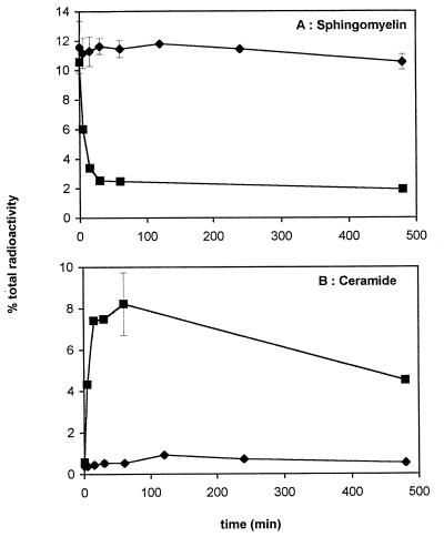
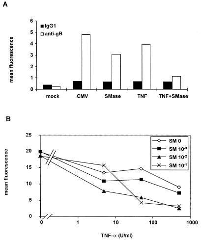
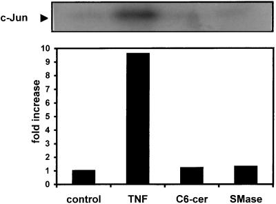
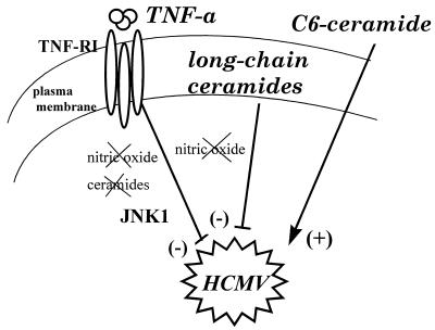
Human cytomegalovirus (HCMV) infection can be fatal to immunocompromised individuals. We have previously reported that gamma interferon and tumor necrosis factor alpha (TNF-alpha) synergistically inhibit HCMV replication in vitro. Ceramides have been described as second messengers induced by TNF-alpha. To investigate the mechanisms involved in the inhibition of HCMV by TNF-alpha, in the present study we have analyzed ceramide production by U373 MG astrocytoma cells and the effects of TNF-alpha versus ceramides on HCMV replication. Our results show that U373 MG cells did not produce ceramides upon incubation with TNF-alpha. Moreover, long-chain ceramides induced by treatment with exogenous bacterial sphingomyelinase inhibited HCMV replication in synergy with TNF-alpha. Surprisingly, short-chain permeant C6-ceramide increased viral replication. Our results show that the anti-HCMV activity of TNF-alpha is independent of ceramides. In addition, our results suggest that TNF-alpha and endogenous long-chain ceramides use separate pathways of cell signalling to inhibit HCMV replication, while permeant C6-ceramide appears to activate a third pathway leading to an opposite effect.


Figures

FIG. 1
FIG. 1
Effect of TNF-α on the cell cycle of infected U373 MG cells. U373 MG cells were incubated with 100 U of TNF-α per ml (b and d) or without TNF-α (a and c) for 24 h prior to HCMV infection (c and d) or mock infection (a and b). The medium was replaced 24 h later, and 6 days p.i. the nuclei were stained with propidium iodide and analyzed by flow cytometry.
FIG. 2
FIG. 2
Effect of C6-ceramide on the cell cycle of U373 MG cells. U373 MG cells were incubated for 24 h with 0 to 10 μM C6-ceramide prior to mock (A) or HCMV (B) infection. The inoculum was removed 24 h later, and the concentrations of ceramide were maintained throughout the experiment. At 6 days p.i., the nuclei were stained with propidium iodide and analyzed by flow cytometry.
FIG. 3
FIG. 3
Increase in gB expression and HCMV production by C6-ceramide. U373 MG cells were incubated with 0 to 10 μM C6-ceramide prior to HCMV infection. Medium was replaced after 24 h infection, maintaining the concentrations of ceramide throughout the experiment. (A) On day 6 p.i., the expression of gB was estimated by intracytoplasmic immunofluorescent staining followed by flow cytometry analysis. (B) Cells were harvested and sonicated at 6 days p.i. The sonicate was used to infect an MRC5 fibroblast monolayer. Infection was estimated by plaque assay.
FIG. 4
FIG. 4
Effect of TNF-α on ceramide and sphingomyelin levels. U373 MG cells were labelled with 1 μCi of [3H]palmitate (51 Ci/mmol) per dish for 48 h and then incubated for the indicated times with 2,000 U of TNF-α per ml (⧫) or 0.1 U of sphingomyelinase per ml (▪). The quantities of labelled sphingomyelin (A) and ceramide (B) were assessed, and the results are expressed as a percentage of total 3H-labelled lipids. Points on the TNF curve represent the mean ± standard deviation (n = 3).
FIG. 5
FIG. 5
Synergy between TNF-α and sphingomyelinase on HCMV inhibition. (A) gB expression. U373 MG cells were preincubated for 24 h with 100 U of TNF-α per ml and/or treated with 0.1 U of sphingomyelinase (SMase) per ml 1 h before HCMV infection. Expression of gB was assessed by intracellular immunofluorescent staining followed by flow cytometry analysis. (B) Viral production. U373 MG cells were pretreated or not treated for 24 h with the indicated concentrations of TNF-α and/or treated with the indicated concentrations of sphingomyelinase (SM) 1 h prior to HCMV infection at a MOI of 5. On day 6 p.i., the cells were sonicated and the sonicate was used at a dilution of 1/100 to infect a monolayer of FSF1 cells. Intracellular IE expression was estimated 4 days later by flow cytometry analysis. The results are expressed as mean fluorescence.
FIG. 6
FIG. 6
TNF-α alone increases JNK1 activity. U373 MG cells were either untreated (control) or treated with 500 U of TNF-α (TNF) per ml, 10 μM C6-ceramide (C6-cer), or 0.1 U of sphingomyelinase (SMase) per ml for 15 min prior to cell lysis. JNK1 was immunoprecipitated from cell lysates and incubated with recombinant c-Jun and [γ-32P]ATP in a 20-min assay at 30°C. Phosphorylation of c-Jun was determined by autoradiography following separation by sodium dodecyl sulfate-polyacrylamide gel electrophoresis (12.5% polyacrylamide). The bands were quantified by densitometry and expressed as fold increase compared to control. This data is from one of three experiments with similar results.
FIG. 7
FIG. 7
Summary of the pathways used by TNF-α and ceramides in the control of in vitro HCMV replication. TNF-α inhibits HCMV replication through a ceramide-independent and NO-independent pathway (see the text). Activation of JNK1 may be involved in this inhibition. Long-chain ceramides inhibit HCMV infection through another pathway, which is also independent of NO. Permeant C6-ceramide uses yet another pathway to activate HCMV replication.

References

    1. Bligh E G, Dyer W J. A rapid method of total lipid extraction and purification. Can J Biochem Physiol. 1959;37:911–918. - PubMed
    1. Boucher L-M, Wiegmann K, Fütterer A, Pfeffer K, Machleidt T, Schütze S, Mak T W, Krönke M. CD28 signals through acidic sphingomyelinase. J Exp Med. 1995;181:2059–2068. - PMC - PubMed
    1. Bresnahan W A, Boldogh I, Aubrey Thomson E, Albrecht T. Human cytomegalovirus inhibits cellular DNA synthesis and arrests productively infected cells in late G1. Virology. 1996;224:150–160. - PubMed
    1. Britt W J, Alford C A. Cytomegalovirus. In: Fields B N, editor. Virology. 3rd ed. Philadelphia, Pa: Lippincott-Raven Publishers; 1996. pp. 2493–2523.
    1. Brockhaus M, Schoenfeld H-J, Schlaeger E-J, Hunziker W, Lesslauer W, Loetscher H. Identification of two types of tumor necrosis factor receptors on human cell lines by monoclonal antibodies. Proc Natl Acad Sci USA. 1990;87:3127–3131. - PMC - PubMed

Publication types

MeSH terms

LinkOut - more resources