. 1998 Mar;72(3):2352-63.
doi: 10.1128/JVI.72.3.2352-2363.1998.

The R33 G protein-coupled receptor gene of rat cytomegalovirus plays an essential role in the pathogenesis of viral infection

Affiliations

The R33 G protein-coupled receptor gene of rat cytomegalovirus plays an essential role in the pathogenesis of viral infection

P S Beisser et al. J Virol. 1998 Mar.

Abstract

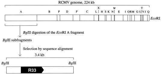
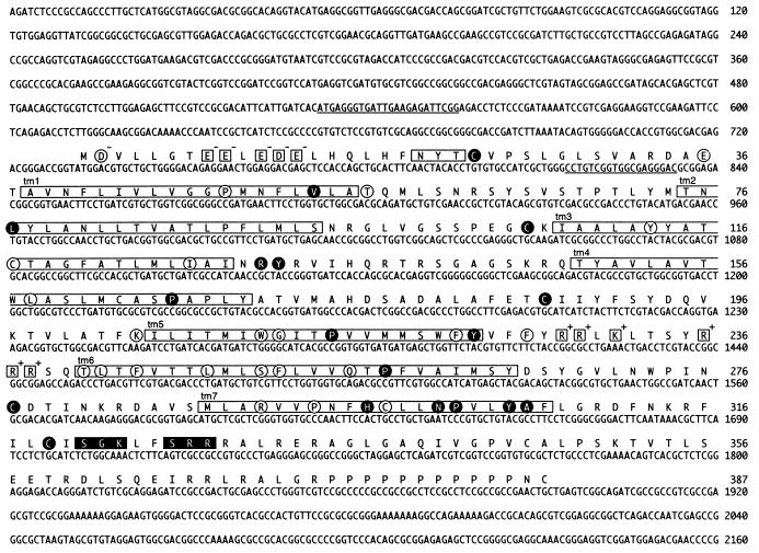
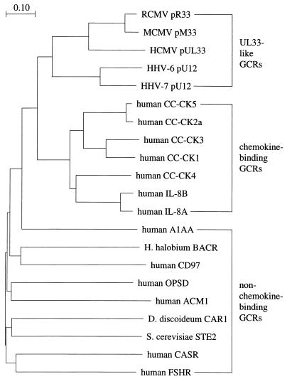
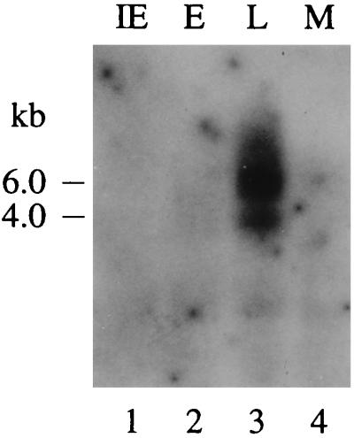
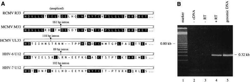
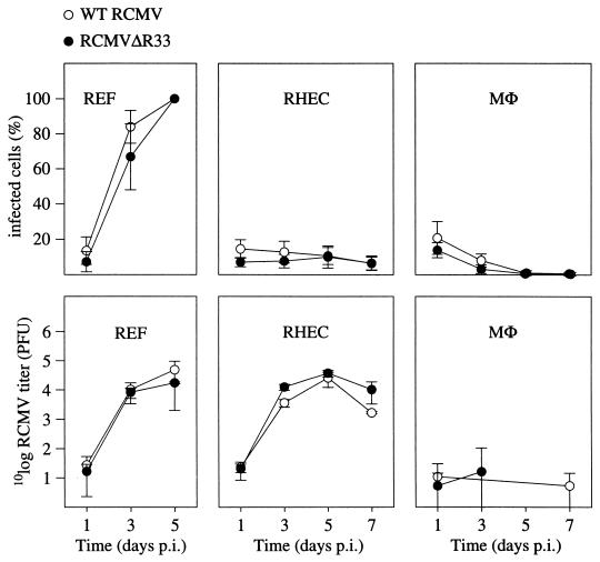
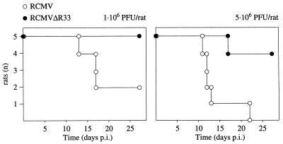
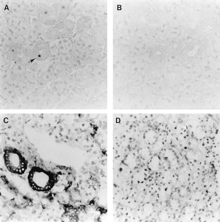
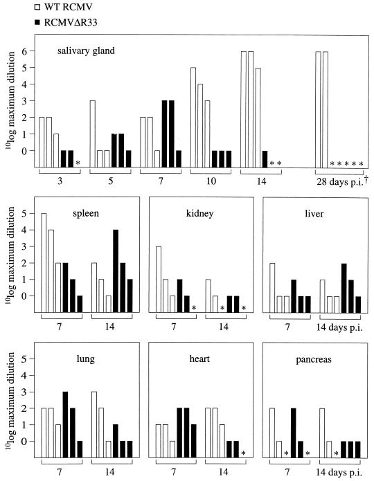
We have identified a rat cytomegalovirus (RCMV) gene that encodes a G-protein-coupled receptor (GCR) homolog. This gene (R33) belongs to a family that includes the human cytomegalovirus UL33 gene. R33 was found to be transcribed during the late phase of RCMV infection in rat embryo fibroblasts. Unlike the mRNAs from all the other members of the UL33 family that have been studied to date, the R33 mRNA is not spliced. To study the function of the R33 gene, we constructed an RCMV strain in which the R33 open reading frame is disrupted. The mutant strain (RCMV deltaR33) did not show differences in replication from wild-type RCMV upon infection of several rat cell types in vitro. However, marked differences were seen between the mutant and wild-type strain in the pathogenesis of infection in immunocompromised rats. First, the mutant strain induced a significantly lower mortality than the wild-type virus did. Second, in contrast to wild-type RCMV, the mutant strain did not efficiently replicate in the salivary gland epithelial cells of immunocompromised rats. Although viral DNA was detected in salivary glands of RCMV deltaR33-infected rats up to 14 days postinfection, it could not be detected at later time points. This indicates that although the strain with R33 deleted is probably transported to the salivary glands in a similar fashion to that for wild-type virus, the mutant virus is not able to either enter or replicate in salivary gland epithelial cells. We conclude that the RCMV R33 gene plays a vital role in the pathogenesis of infection.


Figures

FIG. 1
FIG. 1
Restriction map of the RCMV genome (36) and the relative position of the R33 gene, which encodes a putative G protein-coupled receptor (pR33). The 3.4-kb BglII fragment that contains the R33 ORF is indicated below the genome map.
FIG. 2
FIG. 2
Nucleotide sequence of the R33 gene, and predicted amino acid sequence of the pR33 peptide. The open boxes indicate seven putative transmembrane domains (tm1 to tm7) and a putative N-linked glycosylation site (NXT/S). Charged amino acid residues in the N-terminal (extracellular) region and the third intracellular region (between tm5 and tm6) are enclosed in open squares. The charges of these residues are printed at the top right of each square. Black boxes indicate consensus sequences [(S/T)X(K/R)], of which the S/T residue might be phosphorylated by protein kinase C. The amino acid residues that are conserved among all pUL33-like proteins (Fig. 3) are encircled. The residues that are conserved between chemokine receptors (Fig. 3) are enclosed in black circles. The underlined nucleotide sequences indicate sequences identical or complementary to the sequences of oligonucleotides that were used in RT-PCR (Fig. 5B).
FIG. 3
FIG. 3
pR33 is a member of the chemokine receptor-like GCR family. To compare the sequences of UL33-like GCRs, chemokine receptors, and non-chemokine-binding GCRs, a phylogenetic tree was calculated. The tree is based on a multiple alignment of amino acid sequences that are colinear with the putative seventh transmembrane region (amino acids 288 to 307) of pR33 (Fig. 2). CLUSTAL W pairwise alignment (48) was set to BLOSUM30 protein weight matrix, gap open penalty = 10, and gap extension penalty = 0.1. Multiple alignment was set to BLOSUM series, gap opening penalty = 10, gap extension penalty = 0.05, delay divergent sequences = 0.4. The virus-encoded GCRs are indicated analogous to pR33; i.e., the ORF designation is preceded by a ’p’. CC-CK, C-C chemokine receptor; IL-8, interleukin-8 receptor; A1AA, αA1-adrenergic receptor (13); H. halobium BACR, Halobacterium halobium bacteriorhodopsin (30); CD97, cluster designation 97 (28); OPSD, rhodopsin (39); ACM1, M1 muscarinic acetylcholine receptor (3); D. discoideum CAR1, Dictyostelium discoideum cyclic AMP receptor 1 (31); S. cerevisiae STE2, Saccharomyces cerevisiae pheromone α-factor receptor (14); CASR, extracellular calcium-sensing receptor (2); FSHR, follicle-stimulating hormone receptor (37).
FIG. 4
FIG. 4
The RCMV R33 gene is transcribed at late times of infection in REF. The figure shows an autoradiograph of a Northern blot that was hybridized with an R33-specific probe. Lanes 1, 2, and 3 represent the IE, E, and L phases of infection, respectively. In lane 4, mRNA from mock-infected (M) cells was separated. The estimated lengths of the transcripts are indicated on the left.
FIG. 5
FIG. 5
R33 transcripts are not spliced near the 5′ end. (A) Alignment of the N termini of UL33-like GCRs and the position of introns within the 5′ region of the corresponding genes. Amino acid residues that are conserved between pR33 and at least one of the other pUL33-like proteins are indicated as white letters in black boxes. (B) To identify a potential intron near the 5′ end of the R33 gene, an RT-PCR was performed on poly(A)+ RNA of RCMV-infected cells (lane 4, + RT). As negative controls, either target DNA (lane 2, − cDNA) or RT transcriptase (lane 3, − RT) was omitted. Genomic RCMV DNA was included as a positive control for the PCR (lane 5, genomic DNA). Lane 1 contains a molecular mass reference (100-bp marker). A photograph of an ethidium bromide-stained agarose gel is shown.
FIG. 6
FIG. 6
Generation of an RCMV R33 null mutant. (A) To determine the role of R33 in RCMV infection, a mutant RCMV strain was generated by replacing part of the R33 ORF by a neomycin resistance gene (neo). ORFs are indicated by black arrows. Black rectangles above and below each genome indicate the position of the DNA probes that were used for Southern blot hybridization. SV40 early, simian virus 40 early promoter; SV40 pA, polyadenylation signal. Note that the indicated MluNI sites are lost in the RCMVΔR33 genome. (B) Southern blot hybridization of RCMV and RCMVΔR33 virion DNA. A photograph of an ethidium bromide-stained gel containing BamHI-digested genomic DNA from RCMV (lane 1) and RCMVΔR33 (lane 2) is shown. After transfer to a nylon filter, the DNA from lanes 1 and 2 was hybridized to either the R33 probe (lanes 3 and 4, respectively) or the neo probe (lanes 5 and 6, respectively).
FIG. 7
FIG. 7
The expression of genes neighboring the R33 gene is not affected by disruption of R33. (A) To determine whether transcription of the R32 and R34 genes was affected by disruption of R33, transcription of the R33 region of the genomes of both RCMV and RCMVΔR33 was analyzed by Northern blot hybridization, using probes specific for R32, R33, R34, and neo. The figure shows autoradiographs in which lanes 1, 4, 7, and 10 represent poly(A)+ RNA from RCMV-infected REF, lanes 2, 5, 8, and 11 represent poly(A)+ RNA from RCMVΔR33-infected REF, and lanes 3, 6, 9, and 12 represent poly(A)+ RNA from mock-infected REF. The estimated lengths of the transcripts are indicated on the left in kilobases. (B) Estimated lengths and positions of transcripts from the RCMV R32, R33, and R34 genes, as derived from panel A. The lengths of the indicated R32 and R34 ORFs are estimated and based on the lengths of MCMV M32 and M34 (44); the complete DNA sequence of this region of the RCMV genome is not yet available. (C) Estimated lengths and positions of transcripts from the RCMVΔR33 R32, ’ΔR33’, R34, and neo genes, as derived from panel A. Both the 2.8-kb∗ and 4.2-kb RNA transcripts hybridize to the R33, R34 and neo probes. It is not known whether the 5′ ends of these transcripts map to the 5′ end of the R33 gene or the 5′ end of the neo gene.
FIG. 8
FIG. 8
The R33 gene is not essential for virus replication in vitro. REF, MHEC, and MΦ were infected with either RCMV or RCMVΔR33, and the replicative potential of these viruses was assessed by immunofluorescence and plaque assay. The upper graphs show the infected-cell/total-cell ratios at various time points p.i. The lower graphs show virus titers that were determined in culture medium up to 7 days p.i. Standard deviations are indicated by vertical bars. REF were monitored up to 5 days p.i., when 100% of the cells showed cytopathic effect. On days 5 and 7 p.i., virus could not be detected in medium samples that were taken from cultures of infected MΦ. Data from these time points are therefore not included in the graph.
FIG. 9
FIG. 9
Survival of two groups of immunocompromised rats after intraperitoneal inoculation with either wild-type RCMV or RCMVΔR33. Two groups of rats (five in each group) were infected with either 1 × 10⁶ or 5 × 10⁶ PFU of virus. Survival was recorded up to 28 days p.i.
FIG. 10
FIG. 10
Expression of RCMV (early) proteins and rat class II MHC proteins in salivary glands of RCMV- and RCMVΔR33-infected rats. The figure shows micrographs of 4-μm sections of rat salivary glands infected with either RCMV (A and C) or RCMVΔR33 (B and D). Tissue sections were stained with either MAb against viral (E) antigens (MAb RCMV8 [A and B]) or MAb against class II MHC proteins (OX-6 [C and D]). The tissue sections were photographed at a magnification of ×400.
FIG. 11
FIG. 11
PCR detection of RCMV DNA in several organs of RCMV- and RCMVΔR33-infected rats. To detect viral DNA in organs from either RCMV- or RCMVΔR33-infected rats, PCR was performed on serial dilutions of DNA that was purified from these organs. At each indicated time point, the results are shown for three RCMV-infected rats (open bars) and three RCMVDR33-infected rats (black bars). The height of each bar indicates the maximum dilution of a given DNA isolate with which a positive PCR result could still be obtained. ∗, below detection level (the number of virus genome copies in these samples is smaller than 300). We were able to detect a minimum of approximately 300 genome copies in 1 μg of tissue DNA (data not shown). †, The five samples indicated by ∗ on day 28 p.i. were infected with RCMVΔR33. The samples taken on day 28 p.i. were obtained from the experiment in Fig. 9, in which the rats were infected with 1 × 10⁶ PFU of virus. All the other samples were taken from rats that were infected with 5 × 10⁶ PFU of virus.

Similar articles

Cited by

References

    1. Ahuja S K, Murphy P M. Molecular piracy of mammalian interleukin-8 receptor type B by herpesvirus saimiri. J Biol Chem. 1993;268:20691–20694. - PubMed
    1. Aida K, Koishi S, Tawata M, Onaya T. Molecular cloning of a putative Ca(2+)-sensing receptor cDNA from human kidney. Biochem Biophys Res Commun. 1995;214:524–529. - PubMed
    1. Allard W J, Sigal I S, Dixon R A. Sequence of the gene encoding the human M1 muscarinic acetylcholine receptor. Nucleic Acids Res. 1987;15:10604. - PMC - PubMed
    1. Arvatakis L, Geras-Raaka E, Varma A, Gershengorn M C, Cesarman E. Human herpesvirus KSHV encodes a constitutively active G-protein-coupled receptor linked to cell proliferation. Nature. 1997;385:347–350. - PubMed
    1. Baer R J, Bankier A T, Biggin M D, Deininger P L, Farrell P J, Gibson T J, Hatful G F, Hudson G S, Satchwell S C, Sequin C, Tuffnell P S, Barrell B G. DNA sequence and expression of the B95-8 Epstein-Barr virus genome. Nature. 1984;310:207–211. - PubMed

MeSH terms

Associated data

LinkOut - more resources