. 1998 Mar;72(3):2373-87.
doi: 10.1128/JVI.72.3.2373-2387.1998.

African swine fever virus is wrapped by the endoplasmic reticulum

Affiliations
Free PMC article

African swine fever virus is wrapped by the endoplasmic reticulum

I Rouiller et al. J Virol. 1998 Mar.
Free PMC article

Abstract

African swine fever (ASF) virus is a large DNA virus that shares the striking icosahedral symmetry of iridoviruses and the genomic organization of poxviruses. Both groups of viruses have a complex envelope structure. In this study, the mechanism of formation of the inner envelope of ASF virus was investigated. Examination of thin cryosections by electron microscopy showed two internal membranes in mature intracellular virions and all structural intermediates. These membranes were in continuity with intracellular membrane compartments, suggesting that the virus gained two membranes from intracellular membrane cisternae. Immunogold electron microscopy showed the viral structural protein p17 and resident membrane proteins of the endoplasmic reticulum (ER) within virus assembly sites, virus assembly intermediates, and mature virions. Resident ER proteins were also detected by Western blotting of isolated virions. The data suggested the ASF virus was wrapped by the ER. Analysis of the published sequence of ASF virus (R. J. Yanez et al., Virology 208:249-278, 1995) revealed a reading frame, XP124L, that encoded a protein predicted to translocate into the lumen of the ER. Pulse-chase immunoprecipitation and glycosylation analysis of pXP124L, the product of the XP124L gene, showed that pXP124L was retained in the ER lumen after synthesis. When analyzed by immunogold electron microscopy, pXP124L localized to virus assembly intermediates and fully assembled virions. Western blot analysis detected pXP124L in virions isolated from Percoll gradients. The packaging of pXP124L from the lumen of the ER into the virion is consistent with ASF virus being wrapped by ER cisternae: a mechanism which explains the presence of two membranes in the viral envelope.


Figures

FIG. 1
FIG. 1
Schematic comparison of budding and wrapping mechanisms of virus envelopment. (a) Budding. Viral nucleoprotein complexes bind to the cytoplasmic domains of virally encoded integral membrane proteins (|formula image, membrane glycoproteins). Interactions between viral proteins lead to membrane curvature, and the virion gains a single membrane by budding into the lumen of the membrane compartment. When the virion is released from the cell, oligosaccharides (formula image) are exposed on the surface of the virus, and the cytoplasmic tail of the membrane glycoprotein is buried within the virion. (b) Wrapping. Viral nucleoprotein complexes bind to the cytoplasmic domains of virally encoded integral membrane proteins. The nucleoprotein complex is then wrapped by the membrane cisternae, and the virus gains two membranes. The particle remains in the cytosol. When the virion is released from the cell by cell lysis, oligosaccharides (formula image) are buried within the two membranes of the virion while the cytoplasmic tail of the membrane glycoprotein is exposed on the surface of the virus.
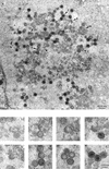
FIG. 2
FIG. 2
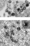
ASF virus assembly site examined by transmission electron microscopy of ultrathin Spurr resin sections. (A) A section, taken through an RS2 cell infected for 16 h with the Uganda isolate of ASF virus, shows a viral factory next to the nucleus (N). The micrograph shows the fully assembled intracellular forms of ASF virus visualized in cross-section as hexagons with electron-dense centers (F), empty particles seen as hexagons with electron-lucent cores (E), several assembly intermediates seen as one to six sided (1 to 6), and amorphous membrane material (m). (B) Selected images, from panel A, of the one- to six-sided intermediates (1 to 6) and of empty and full particles (E and F) are shown at higher magnification.
FIG. 3
FIG. 3
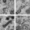
Intracellular ASF virus and all assembly intermediates have two membranes. RS2 cells were examined 16 h after infection with the Uganda isolate of ASF virus. The images were selected from thin cryosections taken through virus assembly sites. Note that the resolution of the membranes of the virions and assembly intermediates is improved in the cryosections compared with the resin sections presented in Fig. 2. (A to G) One- to six-sided assembly intermediates, indicating a possible assembly pathway. In several cases, continuity of the two viral membranes (im, inner membrane; om, outer membrane) with intracellular membranes was observed (arrowhead in panels C, E, and G). An increase of electron density on the concave surface of the virus (∗) paralleled the increase in complexity of the particles (A to H). (H and I) Fully assembled intracellular particles which appear empty and full, respectively. The different components of an ASF virus particle, referred to in the text, include a nucleoprotein core (n), a protein core shell (p), an inner membrane (im), and an outer membrane (om) as indicated in panel I.
FIG. 4
FIG. 4
The inner and outer membranes of ASF virus particles can be separated in damaged virions and when cells are treated with SLO. (A) Damaged intracellular virions observed in a Spurr resin section. In virion a, both the outer membrane (om) and the inner membrane (im) are broken and lost from two faces of the particle, exposing the protein core shell (p). In virion b, the outer membrane (om) is broken away from three faces of the particle, revealing an intact inner membrane (im), which is still covering the protein core shell (p). (B) These micrographs show virions present in cells treated with SLO. A dimple (arrowheads) is present in a single face of most of the fully assembled virions present in this virus factory. At a higher magnification (inset), the inner (im) and outer (om) membranes appear to be separated at the site of the dimple. The protein core shell (p) is clearly visible.
FIG. 5
FIG. 5
The membranes of ASF virus assembly intermediates are in continuity with host membrane compartments. RS2 cells were examined 16 h after infection with the Uganda isolate of ASF virus. (A and B) Thin sections of infected cells permeabilized by osmotic shock to increase the visualization of membranes embedded in Spurr resin. Note that the free edges of four- (A) and five- (B) sided assembly intermediates are seen attached to membranous material extending into the cytoplasm (arrows). Two membranes are clearly seen attached to the same free edge (upper edge on the micrographs) of the assembly intermediate. (C and D) Similar structures were observed in nontreated cells. The membranes (arrows) are however harder to visualize due to the compact nature of the assembly intermediate.
FIG. 6
FIG. 6
A spiculelike protein coat forms on the concave side of assembly intermediates. This micrograph shows a cryosection of a four-sided intermediate. The two membranes (im and om) are seen in the assembling virion and are extending from both edges into the cytosol (large arrowheads). Note the spiculelike protein coat on the concave face of the particle (small arrowheads).
FIG. 7
FIG. 7
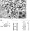
Marker proteins of the ER localize to ASF virus assembly sites and secreted virions. RS2 cells were examined 16 h after infection with the Uganda isolate of ASF virus. Cells were fixed and embedded in Lowicryl resin. (A) Immunogold labelling. Lowicryl sections were incubated with RxER, an antibody recognizing four ER membrane proteins, and visualized using goat anti-rabbit IgG coupled to 15-nm colloidal gold. Label (arrowheads) was observed within the virus factory on membranous material, virus assembly intermediates, and fully assembled virions. Note also the cigar-shaped structures (∗) occasionally seen in virus assembly sites. The inset shows labelling by the RxER antibody of secreted virions. The outer surface of the cell is indicated at the top right corner of the micrograph (large arrows). (B) Statistical analysis of immunolabelling. The specificity of the RxER labelling of virus assembly sites (VF) was assessed by comparison of immunolabelling of nuclei and cytoplasm in the same sections. The number of gold beads present per 7.5 μm2 are indicated. Preimmune sera were not available for the RxER antibody, a selection of 12 normal rabbit sera (NRS) were therefore used at the same dilution for control studies. The number of gold beads present per 7.5-μm2 area covering the virus factory are shown. The data represent the mean of the number of beads per 7.5-μm2 area. The error bars represent the standard deviation of the mean. (C) Biochemical analysis of virions isolated on Percoll gradients. (i) Virus isolation. Vero cells were infected with the BA71v strain of ASF virus. Virions were isolated using self-forming Percoll density gradients, and proteins present were analyzed by SDS-PAGE under reducing conditions. The left-hand panel shows a 12.5% gel after silver staining. The right-hand panel shows metabolically labelled proteins resolved by using a 10% gel and visualized by autoradiography. The major structural proteins of the virus described previously (4, 11) are indicated. (ii) Western blot analysis. Proteins present in Percoll-purified ASF virus were resolved by SDS-PAGE and analyzed by Western blotting. SDS-PAGE (7.5% polyacrylamide) was used to separate proteins for detection of the resident ER proteins ERP60, calnexin, and ERP72; 12.5% gels were used to detect pXP124L. The migration of molecular mass markers is shown.
FIG. 8
FIG. 8
Localization of ASF virus integral membrane protein, p17, within virus assembly sites and virions. RS2 cells were examined 16 h after infection with the Uganda isolate of ASF virus. The cells were fixed, embedded in Lowicryl, and immunolabelled with the 17KG12 antibody specific for p17, followed by goat anti-mouse IgG conjugated to 15-nm gold. As seen for the resident proteins of the ER (Fig. 7) p17 was localized to membranes, assembly intermediates, and fully assembled virions. The inset shows labelling of a secreted virion.
FIG. 9
FIG. 9
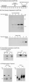
ASF virus-encoded protein pXP124L is a soluble protein retained within the lumen of the ER. (A) Predicted structure of pXP124L. The diagram indicates the structure of pXP124L predicted from the published DNA sequence of the XP124L gene (1, 51), showing the hydrophobic leader peptide, the predicted N-linked glycosylation site, and the synthetic peptide used to raise antiserum. (B) Time course of expression of pXP124L. Vero cells infected with the BA71v isolate of ASF virus were analyzed at increasing time after infection for expression of the XP124L gene product by quantitative Western blotting (bottom) or by metabolic labelling followed by immunoprecipitation (top). Proteins were resolved by SDS-PAGE (12.5% polyacrylamide). (C) (i) Chemical extraction. pXP124L is a soluble protein. Vero cells infected with the BA71v isolate of ASF virus for 16 h were pulse-labelled for 30 min. A crude membrane fraction prepared from the cells was extracted by using alkaline sodium carbonate or Triton X-114. Soluble (S) and membrane fractions (M) were immunoprecipitated with the antipeptide antibody recognizing pXP124L or 4H3 recognizing p73 and, analyzed using SDS-PAGE (12.5% polyacrylamide) followed by autoradiography. (ii) Endoglycosidase H digestion. pXP124L is retained in the ER. Vero cells infected with the BA71v isolate of ASF virus for 16 h or CHO-K1 cells infected with VSV for 5 h were pulse-labelled for 30 min (P) and then chased for 2 h (2HC). Cell lysates were immunoprecipitated by using antibodies specific for pXP124L or VSV G protein, and half of each precipitate was digested with endoglycosidase H (+ lanes). Proteins were resolved by using SDS-PAGE (12.5% polyacrylamide) followed by autoradiography.
FIG. 10
FIG. 10
pXP124L localizes to membranes of the viral factory and to assembled virions. RS2 cells were examined 16 h after infection with the Uganda isolate of ASF virus. The cells were fixed, embedded in Lowicryl, and immunolabelled with rabbit antipeptide antibody specific for pXP124L followed by goat anti-rabbit IgG conjugated to 15-nm gold. The section through the virus assembly site shows pXP124L localized to membranous structures, assembly intermediates, and fully assembled virions. The inset shows immunolabelling of secreted virions, with the plasma membrane being indicated with large arrows.
FIG. 11
FIG. 11
A model for the assembly and envelopment of the intracellular form of ASF virus. Viral structural proteins synthesized in the cytosol, for example p73 and the products of the pp220 and pp60 polyproteins, assemble on the ER membrane (a). Proteins present in the lumen of the ER and integral membrane proteins can also be packaged into assembly sites (b and c). Cooperative interactions between proteins on both sides of the ER cisternae constrict the ER lumen (d and e) and cause progressive bending of the ER cisternae into the one- to six-sided assembly intermediates seen in cross-sections of virus assembly sites (f and g). The genome may be packaged at a late stage during formation of the icosahedron (h). In the final steps, two membrane fusion events involving the inner and outer lipid bilayers allow release of the virion from the ER (i, j, and k). The outer (om) and inner membrane (im) layers of the intracellular form are indicated. Note that this mechanism allows the outer capsid (oc) and the inner core shell (p) to be assembled from the same pool of cytoplasmic structural proteins.

Similar articles

Cited by

References

    1. Almendral J M, Almazan F, Blasco R, Vinuela E. Multigene families in African swine fever virus: family 110. J Virol. 1990;64:2064–2072. - PMC - PubMed
    1. Alves De Matos A P, Marcal M R, Moura Nunes F J, Castro Portugal F L, Vigario J D. Ultrastructural and cytochemical aspects of the maturation of the ASFV particle. Rep Trab Inst Nac Vet. 1980;12:79–84.
    1. Andres G, Simon-Mateo C, Vinuela E. Assembly of African swine fever virus: role of polyprotein pp220. J Virol. 1997;71:2331–2341. - PMC - PubMed
    1. Arzuza O, Urzainqui A, Diaz-Ruiz J R, Tabares E. Morphogenesis of African swine fever virus in monkey kidney cells after reversible inhibition of replication by cycloheximide. Arch Virol. 1992;124:343–354. - PubMed
    1. Black D N, Brown F. Purification and physicochemical characteristics of ASFV. J Gen Virol. 1976;32:509–518. - PubMed

Publication types

MeSH terms

LinkOut - more resources