. 1998 Mar;72(3):2429-38.
doi: 10.1128/JVI.72.3.2429-2438.1998.

The extracellular domain of vaccinia virus protein B5R affects plaque phenotype, extracellular enveloped virus release, and intracellular actin tail formation

Affiliations

The extracellular domain of vaccinia virus protein B5R affects plaque phenotype, extracellular enveloped virus release, and intracellular actin tail formation

E Mathew et al. J Virol. 1998 Mar.

Abstract

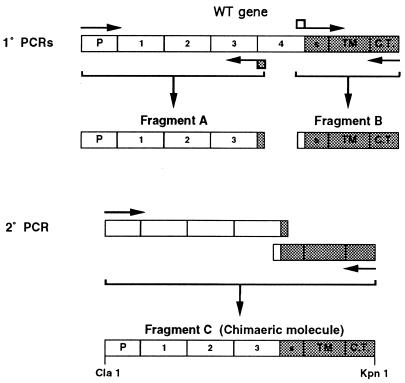
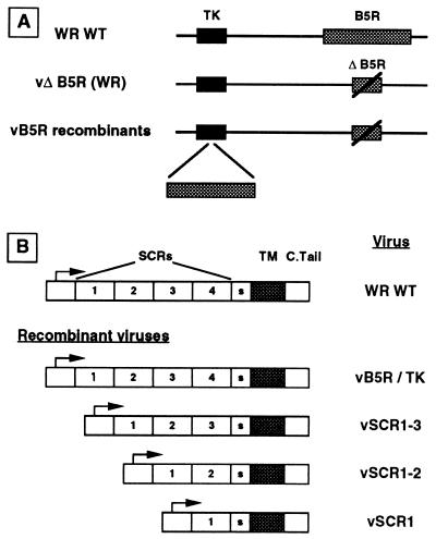
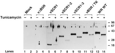
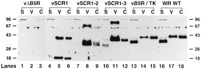
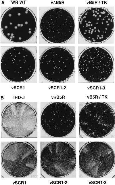
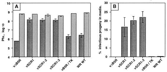
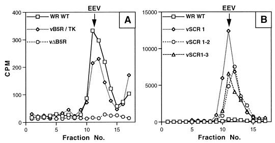
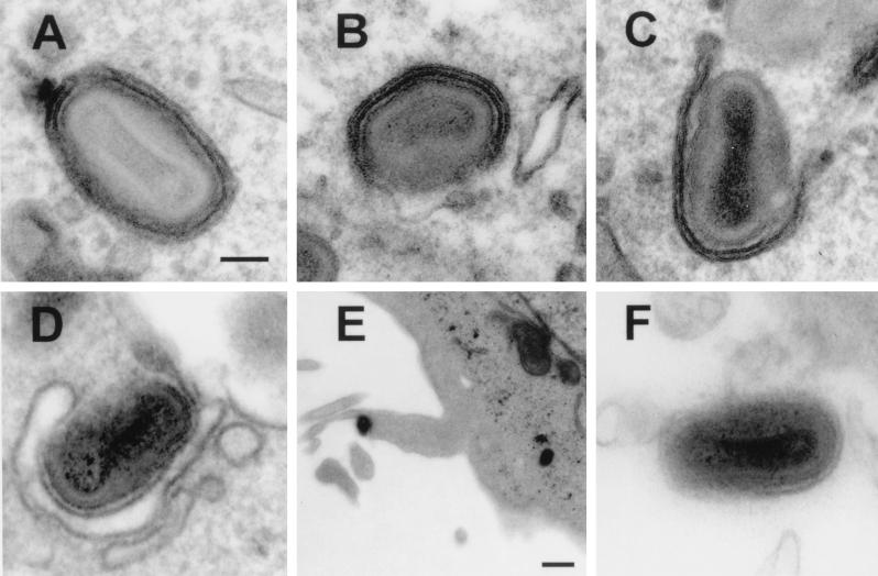
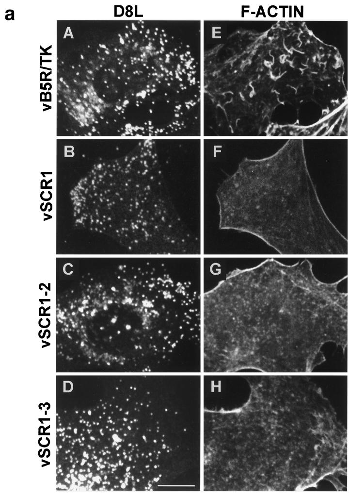
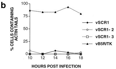
Vaccinia virus produces two morphologically distinct forms of infectious virus, termed intracellular mature virus (IMV) and extracellular enveloped virus (EEV). EEV is important for virus dissemination within a host and has different surface proteins which bind to cell receptors different from those used by IMV. Six genes are known to encode EEV-specific proteins. One of these, B5R, encodes a 42-kDa glycoprotein with amino acid similarity to members of the complement control protein superfamily and contains four copies of a 50- to 70-amino-acid repeat called the short consensus repeat (SCR). Deletion of B5R causes a small-plaque phenotype, a 10-fold reduction in EEV formation, and virus attenuation in vivo. In this study, we inserted mutated versions of the B5R gene lacking different combinations of the SCRs into a virus deletion mutant lacking the B5R gene. The resultant viruses each formed small plaques only slightly larger than those of the deletion mutant; however, the virus containing only SCR 1 formed plaques slightly larger than those of viruses with SCRs 1 and 2 or SCRs 1, 2, and 3. All of these viruses produced approximately 50-fold more infectious EEV than wild-type virus and formed comet-shaped plaques under liquid overlay. Despite producing more EEV, the mutant viruses were unable to induce the polymerization of actin on intracellular virus particles. The implications of these results for our understanding of EEV formation, release, and infectivity are discussed.


Figures

FIG. 1
FIG. 1
Schematic showing the formation of a mutant B5R gene lacking SCR 4 by SOE. Similar strategies were used to produce mutant B5R genes lacking SCRs 3 and 4 or SCRs 2, 3, and 4. P, promoter; S, spacer region between SCR 4 and transmembrane domain; TM, transmembrane domain; C.T., cytoplasmic tail.
FIG. 2
FIG. 2
Structures of recombinant VV genomes (A) and mutant B5R genes (B). TM, transmembrane domain; S, spacer; C.Tail, cytoplasmic tail.
FIG. 3
FIG. 3
Immunoblot showing the B5R proteins made in cells infected with the different viruses. BS-C-1 cells were infected with the indicated viruses at 10 PFU/cell in the presence or absence of tunicamycin (10 μg/ml), and extracts were prepared at 16 hpi. After electrophoresis through a 10% polyacrylamide gel, the proteins were transferred to nitrocellulose and B5R protein was detected by incubation with rabbit antibody against B5R (1:1,000 dilution) (10) followed by a horseradish peroxidase-conjugated species-specific secondary antibody and ECL reagents (Amersham). Molecular weight markers are shown in kilodaltons.
FIG. 4
FIG. 4
Immunoblot showing that mutant B5R proteins are released from infected cells. RK13 cells were infected with the indicated viruses at 10 PFU/cell and incubated in MEM with 2.5% FBS for 24 h. The supernatants of infected cells were clarified by low-speed centrifugation (2,000 rpm in a Beckman GPR benchtop centrifuge for 10 min) to pellet detached cells and cell debris and then high-speed centrifugation (14,000 rpm; SW28 rotor; Beckman ultracentrifuge; 80 min) to pellet virus particles (lanes V) from soluble protein (lanes S). An infected cell extract (C) was prepared as for Fig. 3. B5R proteins were resolved by electrophoresis and detected as described for Fig. 3. Molecular weight markers are shown in kilodaltons.
FIG. 5
FIG. 5
Plaque morphologies of virus mutants. (A) Monolayers of BS-C-1 cells were infected with the indicated viruses, incubated in 1.5% carboxymethyl cellulose in MEM with 2.5% FBS for 96 h, and then stained with crystal violet. (B) Monolayers of BS-C-1 cells were infected with the indicated viruses, incubated in MEM with 2.5% FBS for 96 h, and then stained with crystal violet.
FIG. 6
FIG. 6
B5R mutant viruses release enhanced levels of virus into the culture supernatant. RK13 cells were infected with the indicated viruses at 1 PFU/cell. At 24 hpi, the culture supernatant was collected and clarified by low-speed centrifugation (2,000 rpm in a Beckman GPR benchtop centrifuge for 10 min), and then the infectious virus present in this fraction was determined by duplicate plaque assay on monolayers of BS-C-1 cells. The infected cells were scraped from the monolayer into PBS, combined with the pellets derived from clarification of the culture supernatant, pelleted as described above, and lysed by freeze-thawing and sonication. Infectious virus present in the sonicate was determined by plaque assay on BS-C-1 cells. Plaque assays were stained between 48 and 120 hpi. (A) Data are presented as the titer of virus present in cells (hatched bars) or the culture supernatant (stippled bars) ± SEM (n = 3); (B) data are expressed as the percentage of total infectious virus that was released into the culture supernatant ± SEM (n = 3).
FIG. 7
FIG. 7
The virus released into the culture supernatant is resistant to neutralization by MAb 5B4/2F2. The experiment described in the legend to Fig. 6 was repeated, and this time the titer of infectious virus present in the culture supernatant was determined in the presence or absence of MAb 5B4/2F2 as described previously (45). (A) Stippled bars, total virus; open bars, virus resistant to neutralization by MAb 5B4/2F2; hatched bars, virus neutralized by MAb 5B4/2F2. Titers are shown ± SEM (n = 2). (B) Data expressed as the percentage of infectious virus present in the culture supernatant that is resistant to neutralization by MAb 5B4/2F2 ± SEM (n = 2).
FIG. 8
FIG. 8
CsCl density gradient contrifugation of EEV. RK13 cells were infected with the indicated virus and labeled with [3H]thymidine, and EEV was purified by CsCl density gradients as described in Materials and Methods. (A) EEV from cells infected with WT WR, vB5R/TK, and vΔB5R; (B) EEV from WT WR, vSCR1, vSCR1-2 and vSCR1-3. Note the different scales. The radioactivity from the peak fractions representing EEV was compared to the virus infectivity in the fresh culture supernatant to give a specific infectivity value for each virus. This value was compared to that of vB5R/TK or WT WR viruses, and ratio of the specific infectivity (mutant/WT) is given in the text.
FIG. 9
FIG. 9
Electron microscopy of cells infected with the indicated viruses. Note the formation of intracellular virus particles that have been, or are being, wrapped by intracellular membranes in cells infected with WR WT, vSCR1, vSCR1-2, and vSCR1-3 (A to D). (E) Enveloped particle leaving a B5R/TK-infected cell on an actin tail. (F) EEV particle produced from a vΔB5R-infected cell. Bars: (A to D and F) 100 nm; (E) 500 nm.
FIG. 10
FIG. 10
(a) Fluorescence microscopy of infected cells. BS-C-1 cells were infected with the indicated viruses at 10 PFU/cell. At 14 hpi, cells were permeabilized and stained with mouse MAb AB1.1 directed against the D8L gene product of IMV (25) and rhodamine isothiocyanate-phalloidin to detect F-actin. Note the actin tails in IEV particles in panel E. Bar = 10 μm. (b) Quantitation of actin tails made by the cells infected with the indicated viruses. Cells were infected and stained as for panel A at the indicated times. Between 30 and 40 cells containing approximately 300 D8L-positive particles were counted for each virus at each time point.
FIG. 10
FIG. 10
(a) Fluorescence microscopy of infected cells. BS-C-1 cells were infected with the indicated viruses at 10 PFU/cell. At 14 hpi, cells were permeabilized and stained with mouse MAb AB1.1 directed against the D8L gene product of IMV (25) and rhodamine isothiocyanate-phalloidin to detect F-actin. Note the actin tails in IEV particles in panel E. Bar = 10 μm. (b) Quantitation of actin tails made by the cells infected with the indicated viruses. Cells were infected and stained as for panel A at the indicated times. Between 30 and 40 cells containing approximately 300 D8L-positive particles were counted for each virus at each time point.

Similar articles

Cited by

References

    1. Appleyard G, Hapel A J, Boulter E A. An antigenic difference between intracellular and extracellular rabbitpox virus. J Gen Virol. 1971;13:9–17. - PubMed
    1. Blasco R, Moss B. Extracellular vaccinia virus formation and cell-to-cell virus transmission are prevented by deletion of the gene encoding the 37,000-dalton outer envelope protein. J Virol. 1991;65:5910–5920. - PMC - PubMed
    1. Blasco R, Moss B. Role of cell-associated enveloped vaccinia virus in cell-to-cell spread. J Virol. 1992;66:4170–4179. - PMC - PubMed
    1. Blasco R, Sisler J R, Moss B. Dissociation of progeny vaccinia virus from the cell membrane is regulated by a viral envelope glycoprotein: effect of a point mutation in the lectin homology domain of the A34R gene. J Virol. 1993;67:3319–3325. - PMC - PubMed
    1. Cudmore S, Cossart P, Griffiths G, Way M. Actin-based motility of vaccinia virus. Nature. 1995;378:636–638. - PubMed

Publication types

MeSH terms

LinkOut - more resources