. 1998 Mar;72(3):2449-55.
doi: 10.1128/JVI.72.3.2449-2455.1998.

The M2 ectodomain is important for its incorporation into influenza A virions

Affiliations

The M2 ectodomain is important for its incorporation into influenza A virions

E K Park et al. J Virol. 1998 Mar.

Abstract

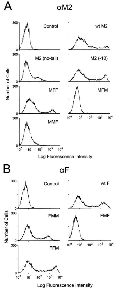
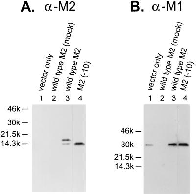
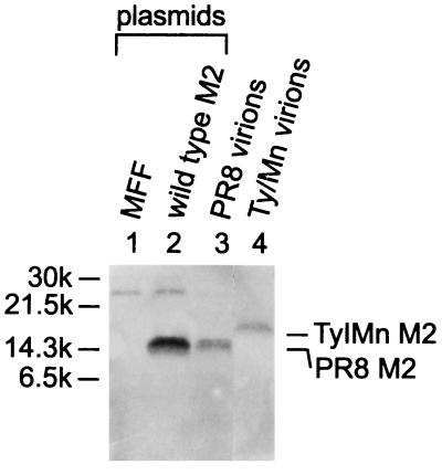
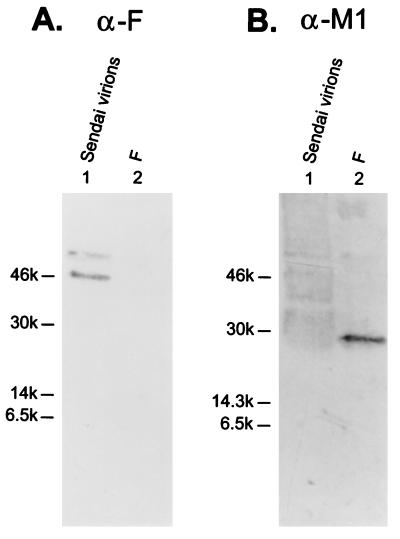
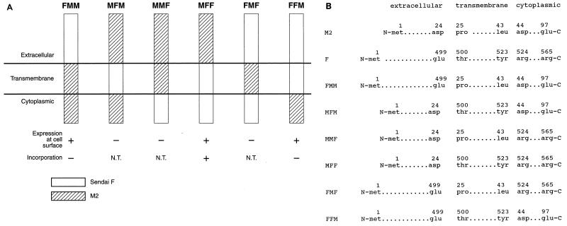
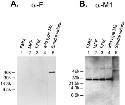
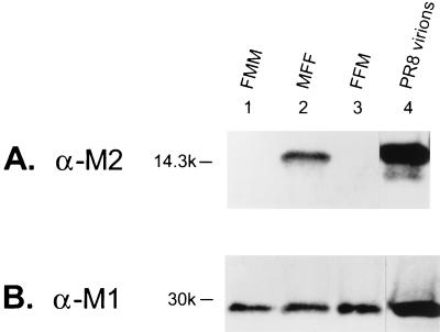
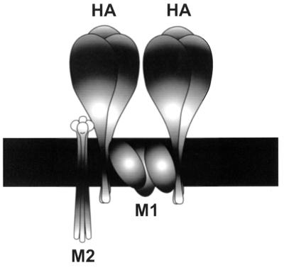
M2 is an integral protein of influenza A virus that functions as an ion channel. The ratio of M2 to HA in influenza A virions differs from that found on the cell surface, suggesting selective incorporation of M2 and HA into influenza virions. To examine the sequences that are important for M2 incorporation into virions, we used an incorporation assay that involves expressing M2 from a plasmid, transfecting the plasmid into recipient cells, and then infecting those cells with influenza virus. To test the importance of the different regions of the protein (extracellular, transmembrane, and cytoplasmic) in determining M2 incorporation, we created chimeric mutants of M2 and Sendai virus F proteins, exchanging corresponding extracellular, transmembrane, and cytoplasmic domains. Of the six possible chimeric mutants, only three were expressed on the cell surface. Of these three chimeric proteins, only one mutant (with the extracellular domain from M2 and the rest from F) was incorporated into influenza virions. These results suggest that the extracellular domain of M2 is important for its incorporation into virions.


Figures

FIG. 1
FIG. 1
Cell surface expression of wild-type and chimeric M2 and F mutants. The wild-type and chimeric constructs described in the text and in Fig. 6 were transfected into COS-1 cells and examined 24 h later by FACS analysis for cell surface expression, as described in Materials and Methods. Anti-M2 (αM2) monoclonal antibody 14C2 (A) and a pool of anti-F (αF) monoclonal antibodies (B) were used to label the cells.
FIG. 2
FIG. 2
Wild-type PR8 M2 is incorporated into Ty/MN virions. The incorporation assay was performed with a wild-type PR8 M2 plasmid (lanes 3) and vector alone (lanes 1) as described in Materials and Methods. Briefly, the plasmids were electroporated into COS-1 cells, which were then infected with Ty/MN virus. Virus in the culture supernatant was purified and examined by Western blot analysis. As another control for the assay, a wild-type M2 plasmid was transfected into cells, which were then subjected to a mock infection (lanes 2). Lanes 4 show the incorporation assay for the M2 mutant lacking the last 10 cytoplasmic residues [M2 (−10)]. The Western blot was probed with the anti-M2 (α-M2) monoclonal antibody 14C2 (A); the membrane was then stripped and reprobed with the anti-M1 (α-M1) monoclonal antibody pool (B).
FIG. 3
FIG. 3
Neither the wild-type PR8 M2 nor the chimeric protein between M2 and Sendai F proteins (MFF) hetero-oligomerizes with Ty/MN M2. (Lanes 1 and 2) Each plasmid indicated was transfected into COS-1 cells, which were then infected with Ty/MN. The viral lysate was immunoprecipitated with the anti-M2 monoclonal antibody 14C2 and then incubated with anti-mouse secondary antibody-coated protein A-Sepharose beads. The immunoprecipitated product was examined by a Western blot analysis. (Lanes 3 and 4) The blot was probed with the polyclonal antibody R3C, which recognizes the cytoplasmic tails of both PR8 M2 and Ty/MN M2. PR8 and Ty/MN virions were fractionated by SDS-PAGE without immunoprecipitation and then probed with the R3C antibody as controls.
FIG. 4
FIG. 4
M2 lacking its cytoplasmic tail is incorporated into virions at very low levels. (Lanes 1) The M2 (no-tail) mutant was tested for its incorporation into virions. (Lanes 2) Purified PR8 virions were analyzed as a control. (A) The blot was probed with the anti-M2 (α-M2) monoclonal antibody 14C2. (B) The M2 monoclonal antibody was stripped from the membrane, which was then reprobed with the anti-M1 (α-M1) antibody pool.
FIG. 5
FIG. 5
Sendai virus F protein is not incorporated into Ty/MN virions. The Sendai virus F gene in pCAGGS/MCS was transfected into COS-1 cells, and the incorporation assay was performed (lanes 2). Purified Sendai virions (lanes 1) were included as a control. (A) The Western blot was probed with the anti-F (α-F) monoclonal antibody pool. (B) The anti-F monoclonal antibodies were stripped from the membrane, which was then reprobed with a pool of anti-M1 (α-M1) monoclonal antibodies.
FIG. 6
FIG. 6
(A) Diagram of chimeric mutants of PR8 M2 and Sendai virus F. Chimeric mutants were constructed as shown, replacing a region (extracellular, transmembrane, or cytoplasmic) of one protein with its counterpart from the other protein. N.T., not tested. (B) Detailed description of chimeric mutants. The numbers above the amino acids indicate the positions of the amino acids in the wild-type protein.
FIG. 7
FIG. 7
Chimeric proteins containing the M2 transmembrane and cytoplasmic domains or only the cytoplasmic domain are not incorporated into Ty/MN virions. The three chimeric constructs found to be expressed on the cell surface were assayed for incorporation as described in Materials and Methods. (A) The viral lysates were probed on a Western blot with anti-F (α-F) monoclonal antibodies. (B) The anti-F monoclonal antibodies were stripped from the membrane, which was then reprobed with a pool of anti-M1 (α-M1) monoclonal antibodies. Purified Sendai virions were loaded in the right-hand lanes as a control.
FIG. 8
FIG. 8
The ectodomain of M2 contains its virion incorporation signal. The Western blot was probed with the anti-M2 (α-M2) monoclonal antibody 14C2 (A) and with anti-M1 (α-M1) antibodies (B). Purified PR8 virions were loaded in the right-hand lanes as a control.
FIG. 9
FIG. 9
Schematic model of M2 incorporation into influenza A virions. As a part of the postulated incorporation complex, the M2 proteins interact with HA (and possibly with NA as well) in the extracellular region, whereas HA trimers interact with each other in the transmembrane region, possibly through M1.

Similar articles

Cited by

References

    1. Arcus Y M, Knight C A. Properties of host components of PR8 influenza virus. Intervirology. 1981;15:171–176. - PubMed
    1. Ausubel F M, Brent R, Kingston R E, Moore D D, Seidman J G, Smith J A, Struhl K. Current protocols in molecular biology. New York, N.Y: John Wiley and Sons, Inc.; 1995.
    1. Blumberg B M, Giorgi C, Rose K, Kolakofsky D. Sequence determination of the Sendai virus fusion protein gene. J Gen Virol. 1985;66:317–331. - PubMed
    1. Bousse T, Takimoto T, Gorman W L, Takahashi T, Portner A. Regions on the hemagglutinin-neuraminidase proteins of human parainfluenza virus type-1 and Sendai virus important for membrane fusion. Virology. 1994;204:506–514. - PubMed
    1. Castrucci M R, Kawaoka Y. Reverse genetics system for generation of an influenza A virus mutant containing a deletion of the carboxyl-terminal residue of M2 protein. J Virol. 1995;69:2725–2728. - PMC - PubMed

Publication types

MeSH terms

LinkOut - more resources