. 1998 Mar;72(3):2509-15.
doi: 10.1128/JVI.72.3.2509-2515.1998.

Identification of determinants on a dualtropic human immunodeficiency virus type 1 envelope glycoprotein that confer usage of CXCR4

Affiliations

Identification of determinants on a dualtropic human immunodeficiency virus type 1 envelope glycoprotein that confer usage of CXCR4

M W Cho et al. J Virol. 1998 Mar.

Abstract

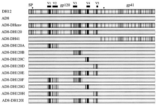
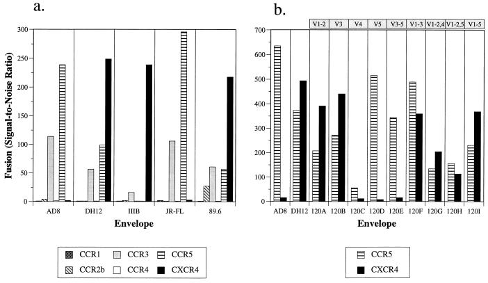
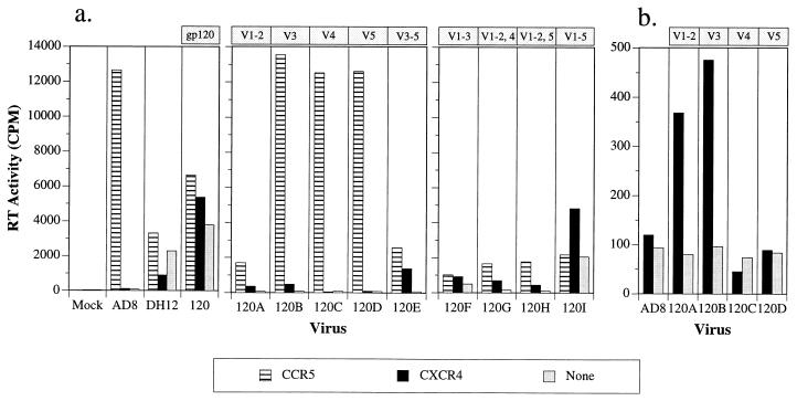
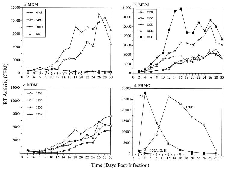
The chemokine receptors CCR5 and CXCR4, in combination with CD4, mediate cellular entry of macrophage-tropic (M-tropic) and T-cell-tropic strains of human immunodeficiency virus type 1 (HIV-1), respectively, while dualtropic viruses can use either receptor. We have constructed a panel of chimeric viruses and envelope glycoproteins in which various domains of the dualtropic HIV-1(DH12) gp160 were introduced into the genetic background of an M-tropic HIV-1 isolate, HIV-1(AD8). These constructs were employed in cell fusion and virus infectivity assays using peripheral blood mononuclear cells, MT4 T cells, primary monocyte-derived macrophages, or HOS-CD4 cell lines, expressing various chemokine receptors, to assess the contributions of different gp120 subdomains in coreceptor usage and cellular tropism. As expected, the dualtropic HIV-1(DH12) gp120 utilized either CCR3, CCR5, or CXCR4, whereas HIV-1(AD8) gp120 was able to use only CCR3 or CCR5. We found that either the V1/V2 or the V3 region of HIV-1(DH12) gp120 individually conferred on HIV-1(AD8) the ability to use CXCR4, while the combination of both the V1/V2 and V3 regions increased the efficiency of CXCR4 use. In addition, while the V4 or the V5 region of HIV-1(DH12) gp120 failed to confer the capacity to utilize CXCR4 on HIV-1(AD8), these regions were required in conjunction with regions V1 to V3 of HIV-1(DH12) gp120 for efficient utilization of CXCR4. Comparison of virus infectivity analyses with various cell types and cell fusion assays revealed assay-dependent discrepancies and indicated that events occurring at the cell surface during infection are complex and cannot always be predicted by any one assay.


Figures

FIG. 1
FIG. 1
Structure of the chimeric viruses generated between HIV-1DH12 and HIV-1AD8. Shown is a schematic diagram of the HIV-1 envelope, with processing sites (inverted triangles) and cleavage products (signal peptide [SP], gp120, and gp41) indicated. Vertical lines, individual amino acid residues of HIV-1DH12 that are divergent from those of HIV-1AD8; solid bars, variable regions of gp120 (V1 to V5).
FIG. 2
FIG. 2
Use of chemokine receptors in a cell fusion assay. (a) The abilities of the HIV-1DH12 (vvDHenv), HIV-1AD8 (vvADenv), HIV-1IIIB (BH8 clone, vSC60 [5]), HIV-1JR-FL (vCB28 [5]), and HIV-189.6 (vBD3 [18]) envelope glycoproteins to mediate fusion with quail QT6 cells expressing CD4 and the indicated coreceptors were determined by a gene reporter fusion assay. The extent of fusion was expressed (as signal-to-noise ratio) relative to background levels (i.e., the spontaneous fusion seen with QT6 cells expressing CD4 and CXCR2). (b) Fusion of the chimeric envelope proteins (expressed by transfecting pNVVDH120A to -I) with cells expressing CCR5 or CXCR4 in conjunction with CD4 was determined. The results were averaged for three experiments, each of which gave identical patterns of coreceptor usage for each envelope protein. Absolute numbers varied between experiments due to differences in cell numbers and transfection efficiencies in independent experiments. It also should be noted that the time elapsing between the addition of the substrate and the determination of luciferase activity strongly influences the absolute light units obtained, though not the pattern of coreceptor use. The variable regions of HIV-1DH12 gp120 transferred into the chimeric envelope are indicated at the top.
FIG. 3
FIG. 3
Infectivity of chimeric HIV-1 in HOS-CD4 cell lines expressing CCR5, CXCR4, or no chemokine coreceptor (HOS-CD4/pBABE-puro). Stocks of parental and chimeric HIV-1 were prepared as previously described (10). HOS cells were seeded (0.5 × 10⁴ to 1 × 10⁴ cells/well) in a 96-well plate 1 day prior to infection and were infected with equivalent amounts of viruses (normalized by virion-associated RT activity [42]). After an overnight incubation, the virus inoculum was removed and culture medium was added to a final volume of 250 μl. One hundred fifty microliters of culture medium was replaced on day 2 postinfection, and virus replication was monitored by measuring virion-associated RT activity on day 4 postinfection (a). The variable regions of HIV-1DH12 gp120 transferred into the chimeric envelope are indicated at the top. (b) Infectivities of HIV-1AD8 and the chimeric viruses AD8-DH120A to -D in HOS-CD4 cells expressing CXCR4 only. A smaller scale than that for panel a is shown here. The virion-associated RT activity in 0.8-μl aliquots of the culture supernatant is plotted.
FIG. 4
FIG. 4
Effects of β-chemokines on the infectivities of parental and chimeric viruses in human PBMC. Phytohemagglutinin-blasted PBMC (105 cells in 250 to 300 μl) were inoculated with equivalent amounts of virus (normalized by RT activity) in 96-well plates as previously described (10). One day postinfection, 200 μl of the inoculum was replaced with fresh medium. On the days indicated, 150 μl of the infection culture medium was harvested for RT assays and replaced with an equal volume of fresh medium. For infections with β-chemokines, PBMC were incubated with RANTES and MIP-1β (each at 200 ng/ml; R & D Systems) for 20 to 30 min at room temperature prior to the addition of virus. Subsequent to overnight adsorption, cells were cultured in medium containing β-chemokines at 100 ng/ml. Harvested samples were stored at −80°C and analyzed for virion-associated RT activity when samples had been collected at all the time points. Virion-associated RT activities in 0.8-μl aliquots of the culture supernatant are shown.
FIG. 5
FIG. 5
Replication of chimeric viruses in primary MDMs and PBMC. MDMs were prepared by differentiating freshly elutriated monocytes on Sterilin plates (Bibby Sterilin, Stone, Staffordshire, United Kingdom) over a 14-day period in macrophage medium (Dulbecco modified Eagle medium [DMEM], 10% pooled normal human serum, 1 mM sodium pyruvate, 2 mM glutamine, 25 U of penicillin/ml, and 25 μg of streptomycin/ml) as previously described (21). Differentiated MDMs were frozen in DMEM containing 20% human serum and 7.5% dimethyl sulfoxide. Cells were thawed and seeded 1 day prior to infection in 96-well plates (Nunc) (105 cells in 200 μl per well). MDMs (a through c) or PBMC (d) were infected with the parental virus or one of the chimeric viruses. Infection, sample collection, and analysis of progeny virus production were carried out as described for Fig. 4.

References

    1. Alkhatib G, Combadiere C, Broder C C, Feng Y, Kennedy P E, Murphy P M, Berger E A. CC CKR5: a RANTES, MIP-1α, MIP-1β receptor as a fusion cofactor for macrophage-tropic HIV-1. Science. 1996;272:1955–1958. - PubMed
    1. Andeweg A C, Leeflang P, Osterhaus A D M E, Bosch M L. Both the V2 and V3 regions of the human immunodeficiency virus type 1 surface glycoprotein functionally interact with other envelope regions in syncytium formation. J Virol. 1993;67:3232–3239. - PMC - PubMed
    1. Bleul C C, Farzan M, Choe H, Parolin C, Clark-Lewis I, Sodroski J, Springer T A. The lymphocyte chemoattractant SDF-1 is a ligand for LESTR/fusin and blocks HIV-1 entry. Nature. 1996;382:829–833. - PubMed
    1. Boyd M T, Simpson G R, Cann A J, Johnson M A, Weiss R A. A single amino acid substitution in the V1 loop of human immunodeficiency virus type 1 gp120 alters cellular tropism. J Virol. 1993;67:3649–3652. - PMC - PubMed
    1. Broder C C, Berger E A. Fusogenic selectivity of the envelope glycoprotein is a major determinant of human immunodeficiency virus type 1 tropism for CD4+ T-cell lines vs. primary macrophages. Proc Natl Acad Sci USA. 1995;92:9004–9008. - PMC - PubMed

Publication types

MeSH terms

Associated data

LinkOut - more resources