Lymantria dispar nucleopolyhedrovirus hrf-1 expands the larval host range of Autographa californica nucleopolyhedrovirus
- PMID: 9499118
- PMCID: PMC109557
- DOI: 10.1128/JVI.72.3.2526-2531.1998
Lymantria dispar nucleopolyhedrovirus hrf-1 expands the larval host range of Autographa californica nucleopolyhedrovirus
Abstract
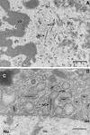
The gypsy moth (Lymantria dispar) is nonpermissive for Autographa californica nucleopolyhedrovirus (AcNPV) infection. We previously isolated a gene, host range factor 1 (hrf-1), from L. dispar nucleopolyhedrovirus that promotes AcNPV replication in Ld652Y cells, a nonpermissive L. dispar cell line (S. M. Thiem, X. Du, M. E. Quentin, and M. M. Berner, J. Virol. 70:2221-2229, 1996). In the present study, we investigated the ability of hrf-1 to alter the larval host range of AcNPV. Bioassays using recombinant AcNPV bearing hrf-1 were conducted with insect larvae by use of oral infection. AcNPV bearing hrf-1 was infectious for neonate L. dispar larvae, with a 50% lethal concentration of 1.2 x 10(5) polyhedral inclusion bodies/ml of diet, which is similar to that of wild-type AcNPV for permissive hosts. AcNPV can kill neonate L. dispar larvae at high doses, but it does not kill third-instar larvae. However, electron microscopy studies of AcNPV-inoculated third-instar larvae revealed virus replication in the midgut cells. PCR analyses indicated that the virus was AcNPV. These results suggest that the block for AcNPV infection of L. dispar larvae is its inability to spread systematically from primary infection sites in the midgut epithelium and that this barrier is leaky in neonates. hrf-1 allows AcNPV to overcome this barrier. AcNPV recombinants bearing hrf-1 were also significantly more infectious for Helicoverpa zea, a resistant species, suggesting that the blocks for AcNPV infection of L. dispar and H. zea larvae may be similar.
Figures


Similar articles
-
Responses of insect cells to baculovirus infection: protein synthesis shutdown and apoptosis.J Virol. 1997 Oct;71(10):7866-72. doi: 10.1128/JVI.71.10.7866-7872.1997. J Virol. 1997. PMID: 9311875 Free PMC article.
-
Host range factor 1 from Lymantria dispar Nucleopolyhedrovirus (NPV) is an essential viral factor required for productive infection of NPVs in IPLB-Ld652Y cells derived from L. dispar.J Virol. 2004 Nov;78(22):12703-8. doi: 10.1128/JVI.78.22.12703-12708.2004. J Virol. 2004. PMID: 15507661 Free PMC article.
-
Characterization of host range factor 1 (hrf-1) expression in Lymantria dispar M nucleopolyhedrovirus- and recombinant Autographa californica M nucleopolyhedrovirus-infected IPLB-Ld652Y cells.Virology. 1997 Jan 20;227(2):420-30. doi: 10.1006/viro.1996.8356. Virology. 1997. PMID: 9018141
-
Global protein synthesis shutdown in Autographa californica nucleopolyhedrovirus-infected Ld652Y cells is rescued by tRNA from uninfected cells.Virology. 1999 Aug 1;260(2):222-31. doi: 10.1006/viro.1999.9827. Virology. 1999. PMID: 10417257
-
Identification of a novel apoptosis suppressor gene from the baculovirus Lymantria dispar multicapsid nucleopolyhedrovirus.J Virol. 2011 May;85(10):5237-42. doi: 10.1128/JVI.00203-11. Epub 2011 Mar 16. J Virol. 2011. PMID: 21411519 Free PMC article.
Cited by
-
Analysis of the genomic sequence of Philosamia cynthia nucleopolyhedrin virus and comparison with Antheraea pernyi nucleopolyhedrin virus.BMC Genomics. 2013 Feb 20;14:115. doi: 10.1186/1471-2164-14-115. BMC Genomics. 2013. PMID: 23425301 Free PMC article.
-
Persistence of an occlusion-negative recombinant nucleopolyhedrovirus in Trichoplusia ni indicates high multiplicity of cellular infection.Appl Environ Microbiol. 2001 Nov;67(11):5204-9. doi: 10.1128/AEM.67.11.5204-5209.2001. Appl Environ Microbiol. 2001. PMID: 11679346 Free PMC article.
-
Expression and mutational analysis of Autographa californica nucleopolyhedrovirus HCF-1: functional requirements for cysteine residues.J Virol. 2005 Nov;79(22):13900-14. doi: 10.1128/JVI.79.22.13900-13914.2005. J Virol. 2005. PMID: 16254326 Free PMC article.
-
Genomic sequencing and analyses of Lymantria xylina multiple nucleopolyhedrovirus.BMC Genomics. 2010 Feb 18;11:116. doi: 10.1186/1471-2164-11-116. BMC Genomics. 2010. PMID: 20167051 Free PMC article.
-
Leucoma salicis nucleopolyhedrovirus (LesaNPV) genome sequence shed new light on the origin of the Alphabaculovirus orpseudotsugatae species.Virus Genes. 2024 Jun;60(3):275-286. doi: 10.1007/s11262-024-02062-x. Epub 2024 Apr 9. Virus Genes. 2024. PMID: 38594489 Free PMC article.
References
-
- Ayres M D, Howard S C, Kuzio J, Lopezferber M, Possee R D. The complete DNA sequence of Autographa californica nuclear polyhedrosis virus. Virology. 1994;202:586–605. - PubMed
-
- Cassar, S. S., and S. M. Thiem. Unpublished data.
-
- Cowan P, Bulach D, Goodge K, Robertson A, Tribe D E. Nucleotide sequence of the polyhedrin gene region of Helicoverpa zea single nucleocapsid nuclear polyhedrosis virus: placement of the virus in lepidopteran nuclear polyhedrosis virus group II. J Gen Virol. 1994;75:3211–3218. - PubMed
-
- Dall D, Sriskantha A, Vera A, Lai-Fook J, Symonds T. A gene encoding a highly expressed spindle body protein of Heliothis armigera entomopoxvirus. J Gen Virol. 1993;74:1811–1818. - PubMed
-
- Du X, Thiem S M. Characterization of host range factor 1 (hrf-1) expression in Lymantria dispar M nucleopolyhedrovirus- and recombinant Autographa californica M nucleopolyhedrovirus-infected IPLB-Ld652Y cells. Virology. 1997;227:420–430. - PubMed
Publication types
MeSH terms
Substances
Grants and funding
LinkOut - more resources
Full Text Sources

