. 1998 Mar 1;12(5):692-705.
doi: 10.1101/gad.12.5.692.

Cdc53 is a scaffold protein for multiple Cdc34/Skp1/F-box proteincomplexes that regulate cell division and methionine biosynthesis in yeast

Affiliations

Cdc53 is a scaffold protein for multiple Cdc34/Skp1/F-box proteincomplexes that regulate cell division and methionine biosynthesis in yeast

E E Patton et al. Genes Dev. .

Erratum in

  • Genes Dev 1998 Oct 1;12(19):3144

Abstract

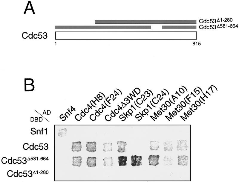
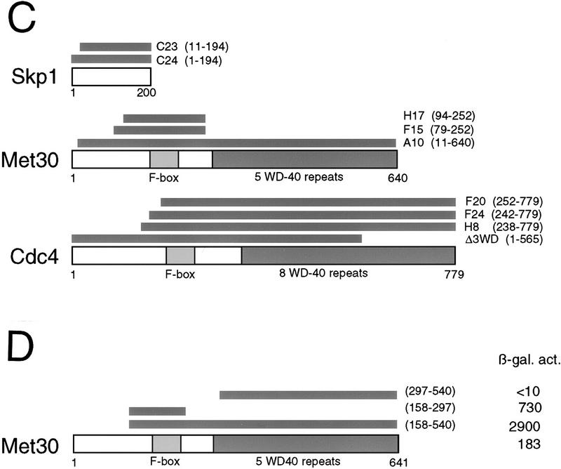
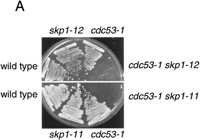
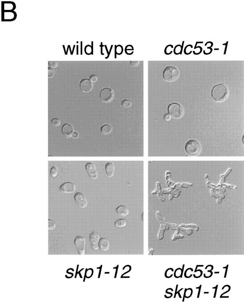
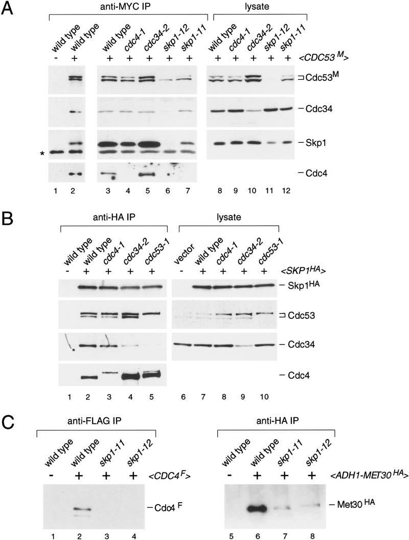
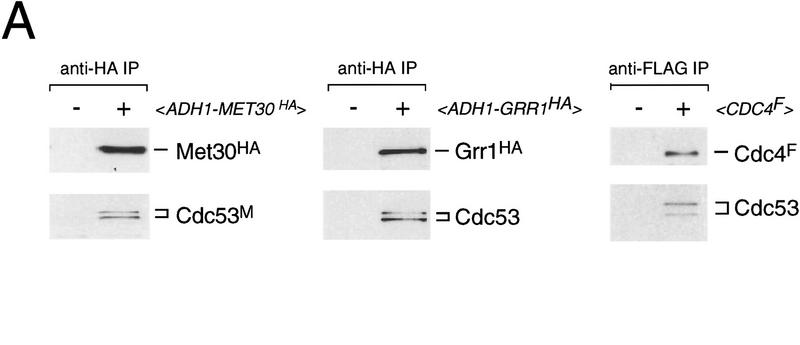
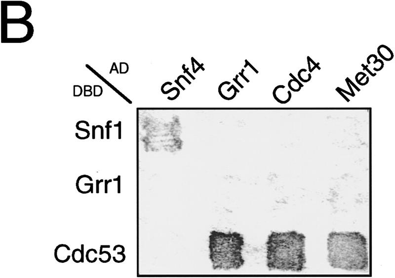
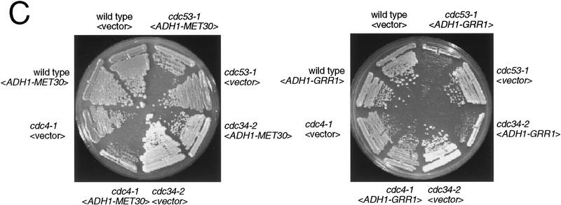
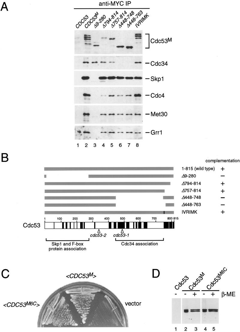
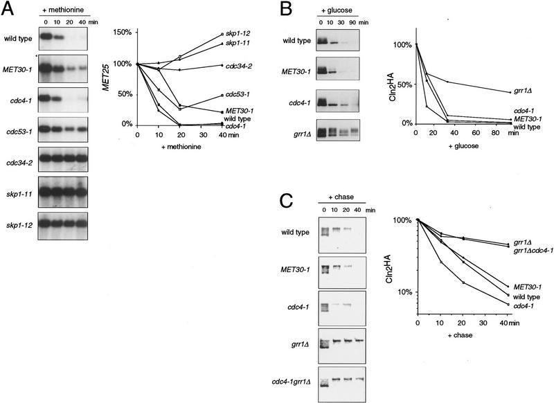
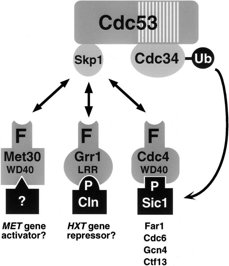
In budding yeast, ubiquitination of the cyclin-dependent kinase (Cdk) inhibitor Sic1 is catalyzed by the E2 ubiquitin conjugating enzyme Cdc34 in conjunction with an E3 ubiquitin ligase complex composed of Skp1, Cdc53 and the F-box protein, Cdc4 (the SCFCdc4 complex). Skp1 binds a motif called the F-box and in turn F-box proteins appear to recruit specific substrates for ubiquitination. We find that Skp1 interacts with Cdc53 in vivo, and that Skp1 bridges Cdc53 to three different F-box proteins, Cdc4, Met30, and Grr1. Cdc53 contains independent binding sites for Cdc34 and Skp1 suggesting it functions as a scaffold protein within an E2/E3 core complex. F-box proteins show remarkable functional specificity in vivo: Cdc4 is specific for degradation of Sic1, Grr1 is specific for degradation of the G1 cyclin Cln2, and Met30 is specific for repression of methionine biosynthesis genes. In contrast, the Cdc34-Cdc53-Skp1 E2/E3 core complex is required for all three functions. Combinatorial control of SCF complexes may provide a basis for the regulation of diverse cellular processes.


Figures

Figure 1
Figure 1
Cdc53 two-hybrid interactions. (A) Cdc53 two-hybrid screens were carried out with three Cdc53 fusion proteins: Gal4DBD–Cdc53, Gal4DBD–Cdc53Δ1–280, and Gal4DBD–Cdc53Δ581–664. (B) Interaction of two-hybrid Gal4AD isolates with Gal4DBD fusions in a β-galactosidase filter assay. (C) Schematic of Cdc53 interacting proteins and two-hybrid isolates. (D) Two-hybrid interactions of LexADBD–Met30 derivatives with VP16AD–Skp1. Interactions of the indicated constructs were quantitated by liquid β-galactosidase assays in Miller units.
Figure 1
Figure 1
Cdc53 two-hybrid interactions. (A) Cdc53 two-hybrid screens were carried out with three Cdc53 fusion proteins: Gal4DBD–Cdc53, Gal4DBD–Cdc53Δ1–280, and Gal4DBD–Cdc53Δ581–664. (B) Interaction of two-hybrid Gal4AD isolates with Gal4DBD fusions in a β-galactosidase filter assay. (C) Schematic of Cdc53 interacting proteins and two-hybrid isolates. (D) Two-hybrid interactions of LexADBD–Met30 derivatives with VP16AD–Skp1. Interactions of the indicated constructs were quantitated by liquid β-galactosidase assays in Miller units.
Figure 2
Figure 2
Genetic interaction between CDC53 and SKP1. (A) cdc53 skp1 double mutants are inviable at the semipermissive temperature. The indicated spore clones of a representative tetratype tetrad were grown at 30°C for 2 days. (B) Photomicrographs of cells from a representative tetratype tetrad grown at 25°C.
Figure 2
Figure 2
Genetic interaction between CDC53 and SKP1. (A) cdc53 skp1 double mutants are inviable at the semipermissive temperature. The indicated spore clones of a representative tetratype tetrad were grown at 30°C for 2 days. (B) Photomicrographs of cells from a representative tetratype tetrad grown at 25°C.
Figure 3
Figure 3
Characterization of Cdc53 complexes in yeast lysates. (A) Effects of temperature-sensitive mutations on the composition of Cdc53 immune complexes. The indicated strains containing <CDC53 TRP1 CEN> or <CDC53M TRP1 CEN> plasmids were arrested at 37°C for 2 hr. Anti-MYC immunoprecipitates from each strain were immunoblotted and sequentially probed with antibodies against Cdc4, Cdc34, Skp1, and the MYC epitope. The antibody against Cdc4 did not reliably detect Cdc4 in lysates and, therefore, these panels were omitted (see part C, below). (B) Effects of temperature-sensitive mutations on the composition of Skp1 immune complexes. Analysis was as above except that strains contained either empty vector or <SKP1HA LEU2 CEN> plasmids. Anti-HA immunoprecipitates were probed as in A. The cause of the lower mobility form of Cdc4 in lanes 3 and 5 has not been determined. (C) Abundance of Cdc4 and Met30 in skp1 mutants. Wild-type, skp1-11, skp1-12 strains containing either empty vector, <CDC4F TRP1 CEN> or <pADH1–MET30HA TRP1 2μ> plasmids, were analyzed as above. Anti-FLAG and anti-HA immunoprecipitates were probed with antibodies against Cdc4 and the HA epitope, respectively.
Figure 4
Figure 4
Interactions of different F-box proteins. (A) Interaction of Met30, Grr1, and Cdc4 with Cdc53 and Skp1. The indicated immunoprecipitates from wild-type cells containing either vector, <pADH1–MET30HA TRP1 2μ>, <pADH1–GRR1HA TRP1 2μ> and <CDC4F TRP1 CEN > plasmids were probed as indicated with antibodies against Cdc53, Skp1, Cdc4, and the HA epitope. IgG light chain is indicated by an asterisk. (B) Different F-box proteins do not interact with each other. Gal4AD–Met30 (A10), Gal4AD–Grr1 and Gal4AD–Cdc4 were tested against Gal4DBD–Grr1 and Gal4DBD–Cdc53 in the two-hybrid system by a β-galactosidase filter assay. (C) Effects of MET30 or GRR1 overexpression. Strains of the indicated genotype containing an empty vector plasmid <pADH1–MET30HA TRP1 2μ> (left) or <pADH1–GRR1HA TRP1 2μ> (right) plasmids were grown at 30°C for 3 days and photographed.
Figure 4
Figure 4
Interactions of different F-box proteins. (A) Interaction of Met30, Grr1, and Cdc4 with Cdc53 and Skp1. The indicated immunoprecipitates from wild-type cells containing either vector, <pADH1–MET30HA TRP1 2μ>, <pADH1–GRR1HA TRP1 2μ> and <CDC4F TRP1 CEN > plasmids were probed as indicated with antibodies against Cdc53, Skp1, Cdc4, and the HA epitope. IgG light chain is indicated by an asterisk. (B) Different F-box proteins do not interact with each other. Gal4AD–Met30 (A10), Gal4AD–Grr1 and Gal4AD–Cdc4 were tested against Gal4DBD–Grr1 and Gal4DBD–Cdc53 in the two-hybrid system by a β-galactosidase filter assay. (C) Effects of MET30 or GRR1 overexpression. Strains of the indicated genotype containing an empty vector plasmid <pADH1–MET30HA TRP1 2μ> (left) or <pADH1–GRR1HA TRP1 2μ> (right) plasmids were grown at 30°C for 3 days and photographed.
Figure 5
Figure 5
Mutational analysis of Cdc53. (A) Deletion analysis of Cdc53 protein–protein interaction domains. Cells were transformed with untagged Cdc53 (lane 1), MYC-tagged Cdc53 (lane 2) or MYC-tagged versions of the indicated Cdc53 mutants (lanes 3–8). All of the proteins were expressed from the wild-type CDC53 promoter on a CEN plasmid. Lysates from each strain were immunoprecipitated with anti-MYC antibody, immunoblotted, and probed with antibodies specific to each of the indicated proteins. (B) Schematic representation of Cdc53 mutant proteins and their ability to complement a cdc53 deletion strain. Regions of amino acid sequence conservation in the Cdc53 family are indicated in black (see Materials and Methods). The positions of the cdc53-1 (R488C) and cdc53-2 (G340D) point mutations, and the regions required for binding to Skp1/F-box proteins and Cdc34 are also indicated. (C) Cdc53 does not contain essential cysteine residues. A cdc53::ADE2 deletion strain containing a <CDC53HA URA3 CEN> plasmid was transformed with <CDC53M6C TRP1 CEN>, <CDC53M TRP1 CEN>, or an empty vector plasmid, plated on 5-FOA medium to select for Ura cells, and photographed after 2 days. (D) Cysteine residues in Cdc53 are not required for posttranslational modification of Cdc53. Lysates from strains containing a <CDC53M TRP1 CEN> or <CDC53M6C TRP1 CEN> plasmid were separated by SDS-PAGE in the absence or presence of reducing agent and immunoblotted with anti-MYC antibody.
Figure 6
Figure 6
Specificity of F-box protein function. (A) Methionine repression is mediated by Met30, Cdc34, Cdc53, and Skp1 but not Cdc4. The indicated strains were grown in methionine-free medium and then repressed with 1.0 mm methionine for the indicated times. MET25 expression was determined by Northern analysis and normalized to ACT1 expression. Values are expressed as percent of the signal at t = 0. (B) Grr1 specifically mediates Cln2 degradation. Cln2 stability was examined in the indicated strains carrying a <pGAL1–CLN2HA URA3 LEU2 CEN> plasmid by repression of the GAL1 promoter. Cln2HA was detected by immunoblotting with anti-HA antibody. Exposures were adjusted to give approximately equal Cln2HA signals at t = 0. Cln2HA was quantitated by densitometry and normalized to Cdc28 signals from the same blot probed with anti-Cdc28 antibody. Values are expressed as percent of the signal at t = 0. (C) Cdc4 does not overlap with Grr1 in Cln2 degradation. Cln2 stability was determined by [35S]methionine/cysteine pulse-chase analysis in the indicated strains. The 35S-labeled Cln2HA signal was quantitated by PhosphorImager and normalized to the 35S-labeled total lysate signal for each sample. Values are expressed as percent of the signal at t = 0.
Figure 7
Figure 7
Multiple F-box proteins recruit different substrates to an E2/E3 core ubiquitination complex. The hatched box in Cdc53 represents a conserved region found in all Cdc53 homologs that overlaps with the Cdc34 binding site in Cdc53. The F-box protein-substrate interactions are most firmly established for Cln1, Cln2, and Sic1. P indicates a phosphorylation dependent interaction, WD40 indicates WD40 repeats, and LRR indicates leucine rich repeats. See text for details.

References

    1. Amon A, Irniger S, Nasmyth K. Closing the cell cycle circle in yeast: G2 cyclin proteolysis initiated at mitosis persists until the activation of G1 cyclins in the next cycle. Cell. 1994;77:1037–1050. - PubMed
    1. Aso T, Haque D, Barstead RJ, Conaway RC, Conaway JW. The inducible elongin A elongation activation domain: Structure, function and interaction with the elongin BC complex. EMBO J. 1996;15:5557–5566. - PMC - PubMed
    1. Bai C, Sen P, Hofmann K, Ma L, Goebl M, Harper JW, Elledge SJ. SKP1 connects cell cycle regulators to the ubiquitin proteolysis machinery through a novel motif, the F-box. Cell. 1996;86:263–274. - PubMed
    1. Barral Y, Jentsch S, Mann C. G1 cyclin turnover and nutrient uptake are controlled by a common pathway in yeast. Genes & Dev. 1995;9:399–409. - PubMed
    1. Blondel M, Mann C. G2 cyclins are required for the degradation of G1 cyclins in yeast. Nature. 1996;384:279–282. - PubMed

Publication types

MeSH terms

Substances