. 1998 Mar 16;187(6):967-72.
doi: 10.1084/jem.187.6.967.

The transcription factor interferon regulatory factor 1 (IRF-1) is important during the maturation of natural killer 1.1+ T cell receptor-alpha/beta+ (NK1+ T) cells, natural killer cells, and intestinal intraepithelial T cells

Affiliations

The transcription factor interferon regulatory factor 1 (IRF-1) is important during the maturation of natural killer 1.1+ T cell receptor-alpha/beta+ (NK1+ T) cells, natural killer cells, and intestinal intraepithelial T cells

T Ohteki et al. J Exp Med. .

Abstract

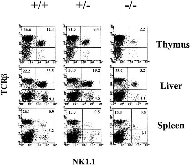
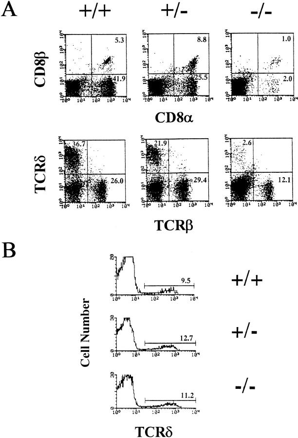
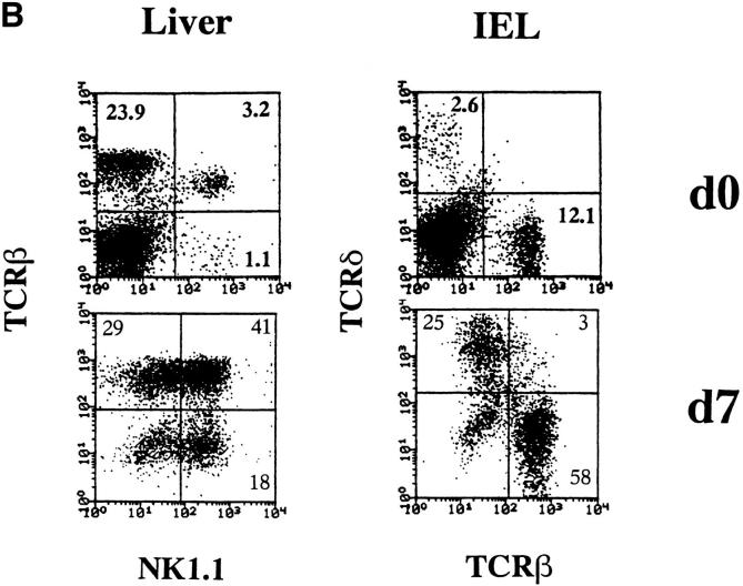
In contrast to conventional T cells, natural killer (NK) 1.1+ T cell receptor (TCR)-alpha/beta+ (NK1+T) cells, NK cells, and intestinal intraepithelial lymphocytes (IELs) bearing CD8-alpha/alpha chains constitutively express the interleukin (IL)-2 receptor (R)beta/15Rbeta chain. Recent studies have indicated that IL-2Rbeta/15Rbeta chain is required for the development of these lymphocyte subsets, outlining the importance of IL-15. In this study, we investigated the development of these lymphocyte subsets in interferon regulatory factor 1-deficient (IRF-1-/-) mice. Surprisingly, all of these lymphocyte subsets were severely reduced in IRF-1-/- mice. Within CD8-alpha/alpha+ intestinal IEL subset, TCR-gamma/delta+ cells and TCR-alpha/beta+ cells were equally affected by IRF gene disruption. In contrast to intestinal TCR-gamma/delta+ cells, thymic TCR-gamma/delta+ cells developed normally in IRF-1-/- mice. Northern blot analysis further revealed that the induction of IL-15 messenger RNA was impaired in IRF-1-/- bone marrow cells, and the recovery of these lymphocyte subsets was observed when IRF-1-/- cells were cultured with IL-15 in vitro. These data indicate that IRF-1 regulates IL-15 gene expression, which may control the development of NK1+T cells, NK cells, and CD8-alpha/alpha+ IELs.


Figures

Figure 1
Figure 1
IRF-1 is important for NK1+T cell and NK cell maturation. Thymocytes, liver, and spleen MNCs from indicated strains were stained with M1/69-FITC (anti-HSA), H57-597-PE (anti–TCR-β), and PK136-biotin (anti-NK1.1) plus streptavidin 670. HSA cells are shown.
Figure 2
Figure 2
Normal CD1 expression on IRF-1−/− thymocytes. Thymocytes from the indicated strains were stained with 1B1-FITC (anti-CD1), 57.6.7-PE (anti-CD8), and L3T4-biotin (anti-CD4) plus streptavidin 670, and double-positive CD4+8+ thymocytes were analyzed for CD1 expression. For H-2Db expression, total thymocytes were stained with B22 (anti–H-2Db) plus goat anti–mouse Ig-FITC.
Figure 3
Figure 3
IRF-1 controls intestinal IEL development. (A) Intestinal IELs were obtained from either IRF-1+/+ mice, IRF-1+/− mice, or IRF-1−/− mice and stained with H57-597 (anti–TCR-β) and GL-3-PE (anti– TCR-δ), or 53.6.7-FITC (anti–CD8α) and Lyt3-PE (anti-CD8β). (B) Thymocytes were stained with L3T4-FITC (anti-CD4), GL-3-PE (anti– TCR-δ), and 53.6.7-FITC (anti-CD8α). Histograms are gated on double-negative CD48 thymocytes and TCR-δ expression is shown.
Figure 4
Figure 4
Impaired lineage development correlates with the absence of IL-15. (A) Limited IL-15 expression in the absence of IRF-1. BM cells were isolated from IRF-1−/− mice or control wild-type (WT) mice. Total RNA was extracted from untreated BM cells or BM cells cultured for 6 h in the presence of LPS (30 μg/ml) and IFN-γ (100 U/ml). Northern blot analysis was performed using IL-15 cDNA and β-actin probes. (B) IL-15 induces the expansion of NK1+T cells, NK cells, and IEL subsets. Liver MNCs and intestinal IELs were isolated from IRF-1−/− mice and cultured with 100 ng/ml mouse IL-15 for 7 d.
Figure 4
Figure 4
Impaired lineage development correlates with the absence of IL-15. (A) Limited IL-15 expression in the absence of IRF-1. BM cells were isolated from IRF-1−/− mice or control wild-type (WT) mice. Total RNA was extracted from untreated BM cells or BM cells cultured for 6 h in the presence of LPS (30 μg/ml) and IFN-γ (100 U/ml). Northern blot analysis was performed using IL-15 cDNA and β-actin probes. (B) IL-15 induces the expansion of NK1+T cells, NK cells, and IEL subsets. Liver MNCs and intestinal IELs were isolated from IRF-1−/− mice and cultured with 100 ng/ml mouse IL-15 for 7 d.

References

    1. Bendelac A. Mouse NK1+T cells. Curr Opin Immunol. 1995;7:367–374. - PubMed
    1. MacDonald HR. NK1.1+ T cell receptor-α/β+cells: new clues to their origin, specificity, and function. J Exp Med. 1995;182:633–638. - PMC - PubMed
    1. Taniguchi M, Koseki H, Tokuhisa T, Masuda K, Sato H, Kondo E, Kawano T, Cui J, Perkes A, Koyasu S, Makino Y. Essential requirement of an invariant Vα14 T cell antigen receptor expression in the development of natural killer T cells. Proc Natl Acad Sci USA. 1996;93:11025–11028. - PMC - PubMed
    1. Ohteki T, MacDonald HR. Stringent Vβ requirement for the development of NK1.1+ T cell receptor-α/β+cells in mouse liver. J Exp Med. 1996;183:1277–1282. - PMC - PubMed
    1. Bendelac A, Lantz O, Quimby ME, Yewdell JW, Bennink JR, Brutkiewicz RR. CD1 recognition by mouse NK1+T lymphocytes. Science. 1995;268:863–865. - PubMed

Publication types

MeSH terms