

NADH-Monodehydroascorbate oxidoreductase is one of the redox enzymes in spinach leaf plasma membranes
- PMID: 9501135
- PMCID: PMC35072
- DOI: 10.1104/pp.116.3.1029
NADH-Monodehydroascorbate oxidoreductase is one of the redox enzymes in spinach leaf plasma membranes
Abstract
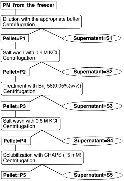
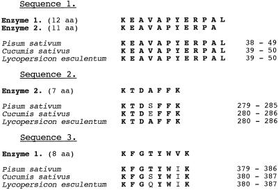
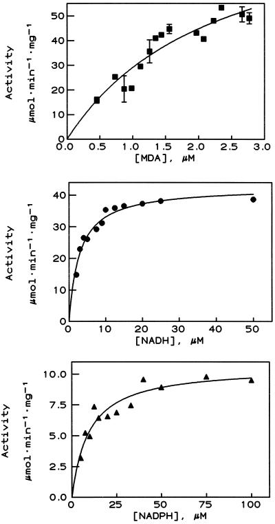
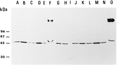
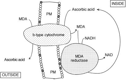
Amino acid analysis of internal sequences of purified NADH-hexacyanoferrate(III) oxidoreductase (NFORase), obtained from highly purified plasma membranes (PM) of spinach (Spinacia oleracea L.) leaves, showed 90 to 100% homology to internal amino acid sequences of monodehydroascorbate (MDA) reductases (EC 1.6.5.4) from three different plant species. Specificity, kinetics, inhibitor sensitivity, and cross-reactivity with anti-MDA reductase antibodies were all consistent with this identification. The right-side-out PM vesicles were subjected to consecutive salt washing and detergent (polyoxyethylene 20 dodecylether and 3-[(3-cholamido-propyl)-dimethylammonio]-1-propane sulfonate [CHAPS]) treatments, and the fractions were analyzed for NFORase and MDA reductase activities. Similar results were obtained when the 300 mm sucrose in the homogenization buffer and in all steps of the salt-washing and detergent treatments had been replaced by 150 mm KCl to mimic the conditions in the cytoplasm. We conclude that (a) MDA reductase is strongly associated with the inner (cytoplasmic) surface of the PM under in vivo conditions and requires washing with 1.0 m KCl or CHAPS treatment for removal, (b) the PM-bound MDA reductase activity is responsible for the majority of PM NFORase activity, and (c) there is another redox enzyme(s) in the spinach leaf PM that cannot be released from the PM by salt-washing and/or CHAPS treatment. The PM-associated MDA reductase may have a role in reduction of ascorbate in both the cytosol and the apoplast.
Figures
References
-
- Arrigoni O, Dipierro S, Borraccino G. Ascorbate free radical reductase, a key enzyme of the ascorbate acid system. FEBS Lett. 1981;125:242–245.
-
- Asard H, Horemans N, Caubergs RJ. Involvement of ascorbic acid and a b-type cytochrome in plant plasma membrane redox reactions. Protoplasma. 1995;184:36–41.
-
- Askerlund P, Larsson C, Widell S. Localization of donor and acceptor sites of NADH dehydrogenase activities using inside-out and right-side-out plasma membrane vesicles from plants. FEBS Lett. 1988;239:23–28.
-
- Askerlund P, Larsson C, Widell S. Cytochromes of plant plasma membranes. Characterization by absorbance difference spectrophotometry and redox titration. Physiol Plant. 1989;76:123–134.
LinkOut - more resources
Full Text Sources
