Comparative Study
. 1998 Mar 17;95(6):2868-73.
doi: 10.1073/pnas.95.6.2868.

Separation of inhibition and activation of the allosteric yeast chorismate mutase

Affiliations
Comparative Study

Separation of inhibition and activation of the allosteric yeast chorismate mutase

G Schnappauf et al. Proc Natl Acad Sci U S A. .

Abstract

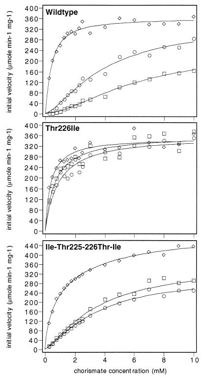
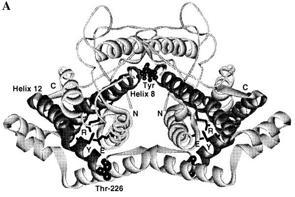
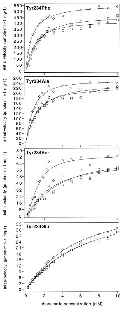
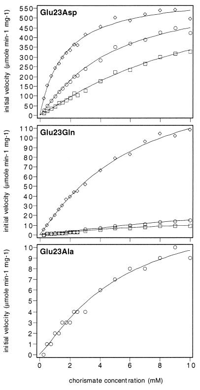
Yeast chorismate mutase (EC 5.4.99.5) shows homotropic activation by the substrate, allosteric activation by tryptophan, and allosteric inhibition by tyrosine. In this study mutants of chorismate mutase have been found that remain sensitive to one allosteric effector (tryptophan) but insensitive to the other (tyrosine). These mutations are located in the catalytic domain: loop 220s (212-226) and helix 12 (227-251). The first example starts with the Thr-266 --> Ile mutant that had previously been shown to be locked in the activated R state. The additional mutation Ile-225 --> Thr unlocks the R state and restores the activation by tryptophan but not the inhibition by tyrosine. The second example refers to a molecular trigger for the switch between the T and R state: a hydrogen-bonded system, which stabilizes only the T state, from Tyr-234 to Glu-23 to Arg-157. Various mutants of Tyr-234, especially Tyr-234 --> Phe, are unresponsive to tyrosine but are activated by tryptophan. This separation of activation from inhibition may indicate a pathway for activation that is independent of the allosteric transition and may also be consistent with an intermediate structure between T and R states.


Figures

Figure 1
Figure 1
Substrate saturation plots of wild-type, Thr-226 → Ile, and Ile-225 → Thr-Thr-226 → Ile mutant chorismate mutase. The enzymes were assayed with 10 μM tryptophan (diamonds), without effectors (circles), or in the presence of 100 μM tyrosine (squares). The data were fitted to functions describing cooperative or Michaelis–Menten-type saturation.
Figure 2
Figure 2
(A) Structure of the T state of chorismate mutase drawn in ribbon style. The N termini and C termini are labeled as N and C, respectively. The two tyrosine molecules located at the interface of the dimer are indicated as Tyr. Helix 8, which connects the allosteric and active sites, and helix 12 are drawn in a dark color. The side chains of Glu-23 (E) and Tyr-234 (Y), which are involved in the hydrogen-bonding interaction with the active residue Arg-157 (R), are drawn in stick model. Thr-226, which connects the 220s loop and helix 12, is indicated. (B) Residues Glu-23, Tyr-234, and Arg-157 in the crystal structures of the R and T states of yeast chorismate mutase. Hydrogen-bonding interactions are indicated by dotted lines. Glu-23 and Tyr-234 move toward the active site upon the transition from the R to the T state. In the T state, Glu-23 is in hydrogen-bonding distance to the active site residue Arg-157, and the OH of Tyr-234 is 2.7 Å from Oɛ2 of Glu-23; in the R state this distance increases to 4.7 Å.
Figure 2
Figure 2
(A) Structure of the T state of chorismate mutase drawn in ribbon style. The N termini and C termini are labeled as N and C, respectively. The two tyrosine molecules located at the interface of the dimer are indicated as Tyr. Helix 8, which connects the allosteric and active sites, and helix 12 are drawn in a dark color. The side chains of Glu-23 (E) and Tyr-234 (Y), which are involved in the hydrogen-bonding interaction with the active residue Arg-157 (R), are drawn in stick model. Thr-226, which connects the 220s loop and helix 12, is indicated. (B) Residues Glu-23, Tyr-234, and Arg-157 in the crystal structures of the R and T states of yeast chorismate mutase. Hydrogen-bonding interactions are indicated by dotted lines. Glu-23 and Tyr-234 move toward the active site upon the transition from the R to the T state. In the T state, Glu-23 is in hydrogen-bonding distance to the active site residue Arg-157, and the OH of Tyr-234 is 2.7 Å from Oɛ2 of Glu-23; in the R state this distance increases to 4.7 Å.
Figure 3
Figure 3
Substrate saturation plots of mutant chorismate mutases carrying amino acid substitutions at position Tyr-234. The enzymes were assayed with 10 μM tryptophan (diamonds), without effectors (circles), or in the presence of 100 μM tyrosine (squares). The data were fitted to functions describing cooperative or Michaelis–Menten-type saturation.
Figure 4
Figure 4
Substrate saturation plots of mutant chorismate mutases carrying amino acid substitutions at position Glu-23. The enzymes were assayed with 10 μM tryptophan (diamonds), without effectors (circles), or in the presence of 100 μM tyrosine (squares). The data were fitted to functions describing cooperative or Michaelis–Menten-type saturation.

References

    1. Bohr C, Hasselbalch K, Krough A. Scand Arch Physiol. 1904;16:402–412.
    1. Monod J, Changeux J P, Jacob F. J Mol Biol. 1963;6:306–329. - PubMed
    1. Monod J, Wyman J, Changeux J-P. J Mol Biol. 1965;12:88–118. - PubMed
    1. Hathaway J A, Atkinson D E. J Biol Chem. 1963;238:2875–2881. - PubMed
    1. Koshland D E, Nemethy G, Filmer D. Biochemistry. 1966;5:365–385. - PubMed

Publication types

LinkOut - more resources